. 2023 Oct 16:11:tkad041.
doi: 10.1093/burnst/tkad041. eCollection 2023.

A photoactivatable and phenylboronic acid-functionalized nanoassembly for combating multidrug-resistant gram-negative bacteria and their biofilms

Affiliations

A photoactivatable and phenylboronic acid-functionalized nanoassembly for combating multidrug-resistant gram-negative bacteria and their biofilms

Xiaoqing Zhou et al. Burns Trauma. .

Abstract

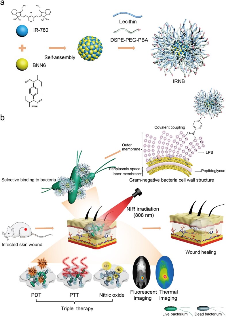
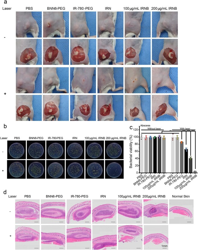
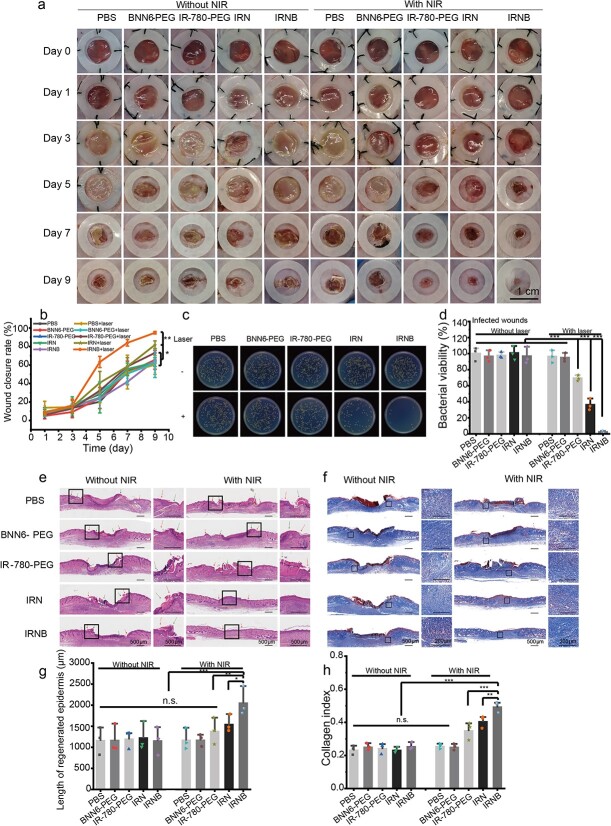
Background: Multidrug-resistant (MDR) gram-negative bacteria-related infectious diseases have caused an increase in the public health burden and mortality. Moreover, the formation of biofilms makes these bacteria difficult to control. Therefore, developing novel interventions to combat MDR gram-negative bacteria and their biofilms-related infections are urgently needed. The purpose of this study was to develop a multifunctional nanoassembly (IRNB) based on IR-780 and N, N'-di-sec-butyl-N, N'- dinitroso-1,4-phenylenediamine (BNN6) for synergistic effect on the infected wounds and subcutaneous abscesses caused by gram-negative bacteria.

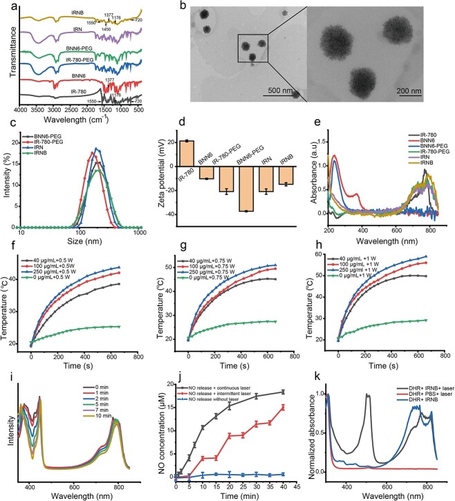
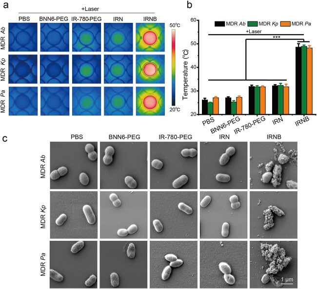
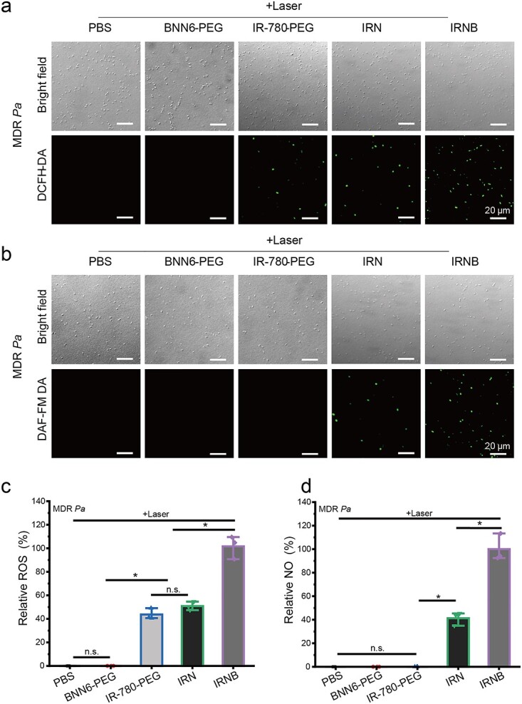
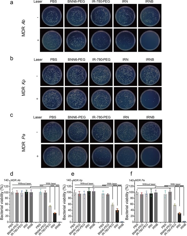
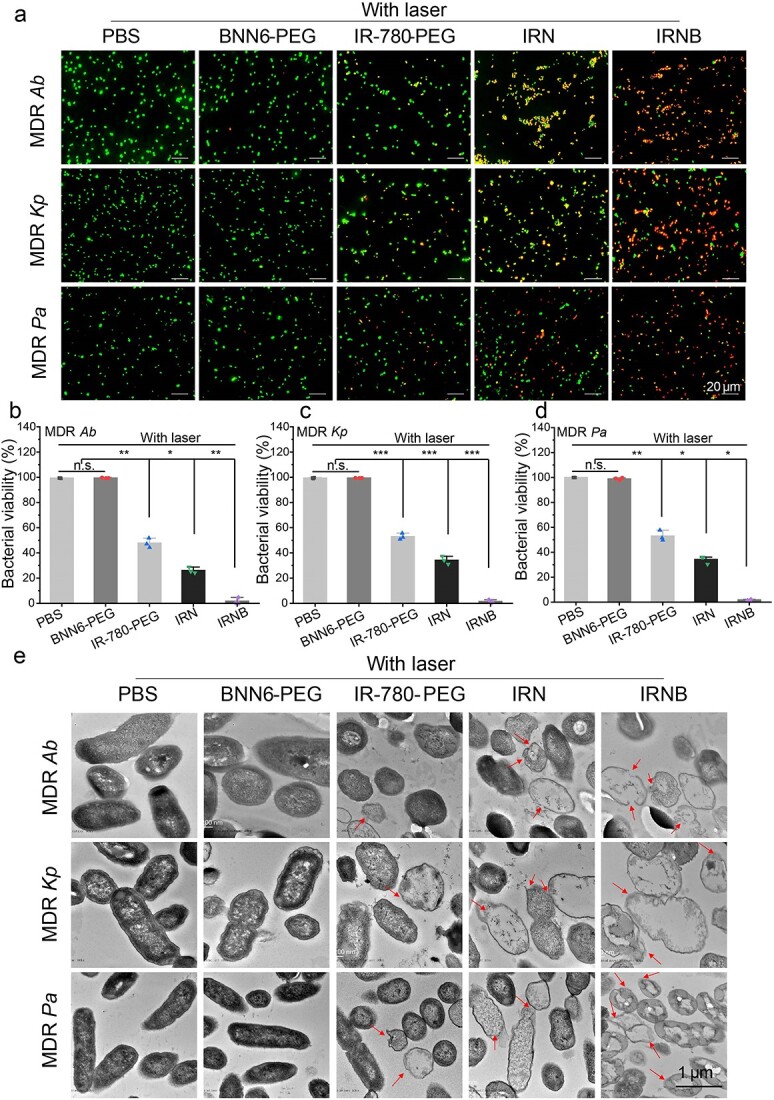
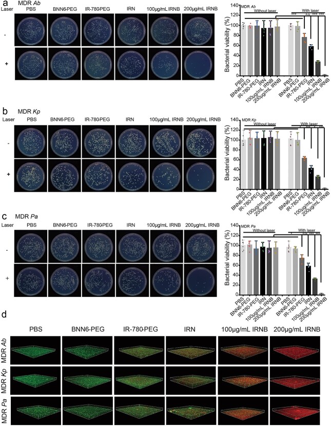
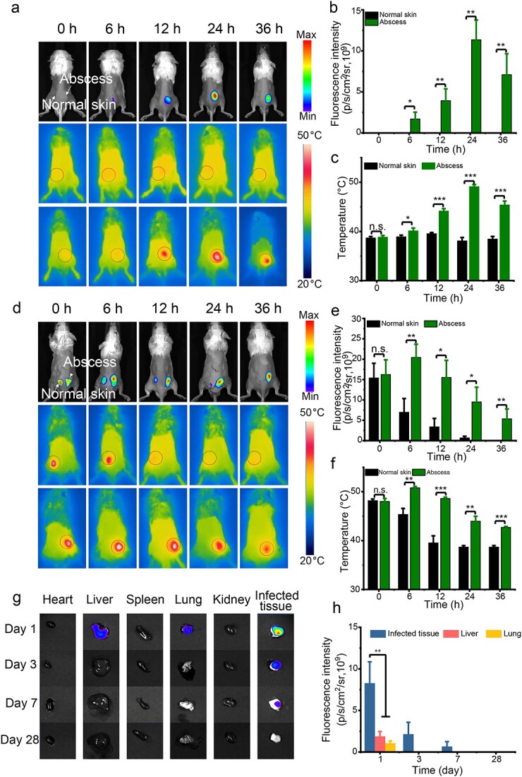
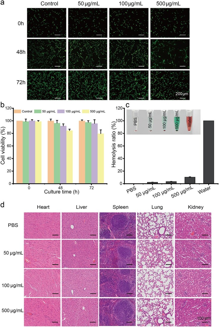
Methods: The characterization and bacteria-targeting ability of IRNB were investigated. The bactericidal efficacy of IRNB against gram-negative bacteria and their biofilms was demonstrated by crystal violet staining assay, plate counting method and live/dead staining in vitro. The antibacterial efficiency of IRNB was examined on a subcutaneous abscess and cutaneous infected wound model in vivo. A cell counting kit-8 assay, Calcein/PI cytotoxicity assay, hemolysis assay and intravenous injection assay were performed to detect the biocompatibility of IRNB in vitro and in vivo.

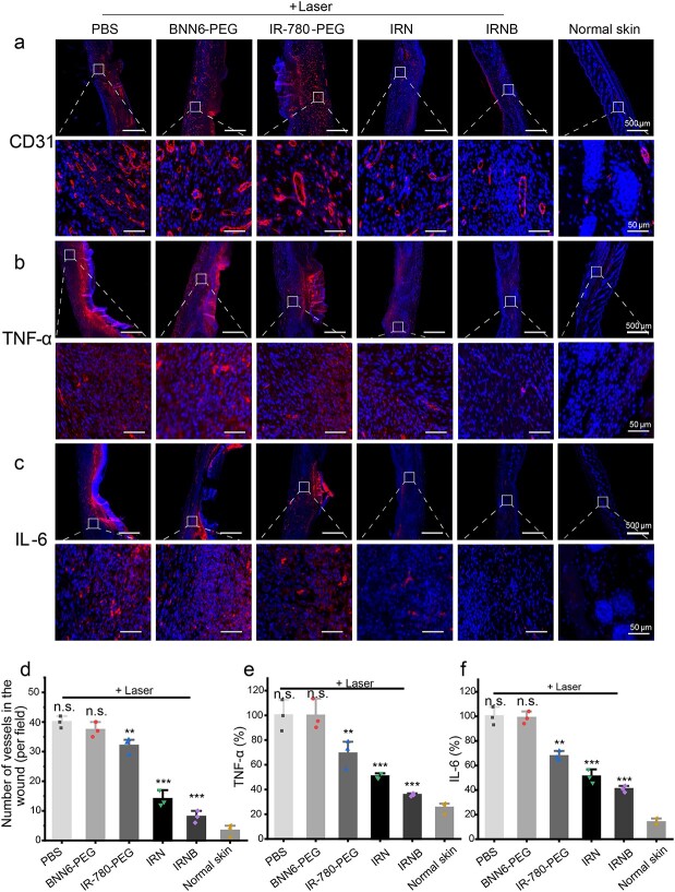
Results: Herein, we successfully developed a multifunctional nanoassembly IRNB based on IR-780 and BNN6 for synergistic photothermal therapy (PTT), photodynamic therapy (PDT) and nitric oxide (NO) effect triggered by an 808 nm laser. This nanoassembly could accumulate specifically at the infected sites of MDR gram-negative bacteria and their biofilms via the covalent coupling effect. Upon irradiation with an 808 nm laser, IRNB was activated and produced both reactive oxygen species (ROS) and hyperthermia. The local hyperthermia could induce NO generation, which further reacted with ROS to generate ONOO-, leading to the enhancement of bactericidal efficacy. Furthermore, NO and ONOO- could disrupt the cell membrane, which converts bacteria to an extremely susceptible state and further enhances the photothermal effect. In this study, IRNB showed a superior photothermal-photodynamic-chemo (NO) synergistic therapeutic effect on the infected wounds and subcutaneous abscesses caused by gram-negative bacteria. This resulted in effective control of associated infections, relief of inflammation, promotion of re-epithelization and collagen deposition, and regulation of angiogenesis during wound healing. Moreover, IRNB exhibited excellent biocompatibility, both in vitro and in vivo.

Conclusions: The present research suggests that IRNB can be considered a promising alternative for treating infections caused by MDR gram-negative bacteria and their biofilms.

Keywords: Boronic acid; Multidrug-resistant gram-negative bacteria; Nitric oxide; Photodynamic therapy; Photothermal therapy; Synergistic.


Conflict of interest statement

None declared.

Figures

Figure 1
Figure 1
Schematic diagram of the preparation and antimicrobial mechanism of IRNB. (a) Schematic illustration of the preparation of IRNB. (b) The antimicrobial mechanism of IRNB in vivo. IRNB IR-780/BNN6-PEG-PBA, BNN6 N,N′-di-sec-butyl-N,N′-dinitroso-1,4-phenylenediamine, DSPE-PEG-PBA 1,2-distearoyl-snglycero-3-phosphoethanolamine-N-[methoxy(polyethylene glycol)-2000]-phenylboronic acid, LPS lipopolysaccharide, NIR near infrared laser, PDT photodynamic therapy, PTT photothermal therapy
Figure 2
Figure 2
Characterization of IRNB. (a) FTIR spectra of free IR-780, free BNN6, synthesized IR-780-PEG, BNN6-PEG, IRN and IRNB. (b) TEM images of IRNB nanoparticles (Scale bar: 500 nm and 200 nm). (c) and (d) Hydrodynamic diameter and ζ potential of IR-780-PEG, BNN6-PEG, IRN and IRNB in PBS buffer measured by DLS. (e) UV–Vis absorption spectra of free IR-780, free BNN6, synthesized IR-780-PEG, BNN6-PEG, IRN and IRNB. (f), (g) and (h) Temperature evolution curves of IRNB with different concentrations (0–250 μg/mL) under 808 nm NIR irradiation at 0.5, 0.75 and 1 W/cm2. (i) UV–Vis absorbance spectrum of IRNB co-incubated with DPBF under 808 nm NIR irradiation at 0.75 W/cm2 for different time periods. (j) NO release profile of IRNB under different laser irradiation conditions. (k) DHR 123 UV–Vis absorbance spectrum after various treatments. FTIRfourier transform infrared spectroscopy, BNN6 N,N′-di-sec-butyl-N,N′-dinitroso-1,4-phenylenediamine, IRN IR-780/BNN6-PEG, IRNB IR-780/BNN6-PEG-PBA, TEM transmission electron microscopy, PBSphosphate-buffered saline, DLS dynamic light scattering, NIR near infrared laser, DPBF 1,3-diphenylisobenzofuran, NO nitric oxide, DHR 123 dihydrorhodamine 123, UV–Visultraviolet–visible
Figure 3
Figure 3
In vitro bacteria-targeting abilities of IRNB. (a) and (b) Thermographic images and the corresponding temperature measurements of MDR Ab, MDR Kp and MDR Pa suspensions after incubation with PBS, IR-780-PEG, BNN6-PEG, IRN and IRNB under laser irradiation (808 nm, 0.75 W/cm2,10 min). Statistical analysis: Mean ± SD; n = 3; ***p < 0.001, **p < 0.01, *p < 0.05. (c) Representative SEM images of MDR Ab, MDR Kp and MDR Pa after incubation with PBS, IR-780-PEG, BNN6-PEG, IRN and IRNB (Scale bar: 1 μm). BNN6 N,N′-di-sec-butyl-N,N′- dinitroso-1,4-phenylenediamine, IRN IR-780/BNN6-PEG, IRNB IR- 780/BNN6-PEG-PBA, MDR multidrug-resistant, Ab Acinetobacter baumannii, Kp Klebsiella pneumoniae, Pa Pseudomonas aeruginosa, SEMscanning electron microscopy, PBS phosphate-buffered saline
Figure 4
Figure 4
NIR-triggered ROS/NO generation in bacterial cells. (a) and (c) Confocal laser microscopy images and relative quantitative fluorescence intensity bar chart of ROS generation in MDR Pa after receiving various treatments (Scale bar: 20 μm). (b) and (d) Confocal laser microscopy images and relative quantitative fluorescence intensity bar chart of NO generation in MDR Pa after receiving various treatments (Scale bar: 20 μm). Statistical analysis: Mean ± SD; n = 3; ***p < 0.001, **p < 0.01, *p < 0.05. NIR near infrared laser, ROS reactive oxygen species, NO nitric oxide, MDR multidrug-resistant, Pa Pseudomonas aeruginosa, PBS phosphate-buffered saline, BNN6 N,N′-di-sec-butyl-N,N′- dinitroso-1,4-phenylenediamine, IRN IR-780/BNN6-PEG, IRNB IR-780/BNN6-PEG-PBA, DCFH-DA 2′,7’-Dichlorodihydrofluorescein diacetate, DAF-FM DA 4-Amino-5-methylamino-2′,7′-difluorofluorescein diacetate
Figure 5
Figure 5
In vitro antibacterial activities of IRNB. (a), (b) and (c) Representative images of bacterial CFUs of MDR Ab, MDR Kp and MDR Pa exposed to PBS, IR-780-PEG, BNN6-PEG, IRN and IRNB with or without laser irradiation. (d), (e) and (f) Relative bacterial viability analysis of MDR Ab, MDR Kp and MDR Pa after receiving different treatments. Statistical analysis: Mean ± SD; n = 6; ***p < 0.001, **p < 0.01, *p < 0.05. MDR multidrug-resistant, CFU colony forming unit, PBS phosphate-buffered saline, BNN6 N,N′-di-sec-butyl- N,N′- dinitroso-1,4-phenylenediamine, IRN IR-780/BNN6-PEG, IRNB IR-780/BNN6-PEG-PBA, Ab Acinetobacter baumannii, Kp Klebsiella pneumoniae, Pa Pseudomonas aeruginosa
Figure 6
Figure 6
In vitro antibacterial mechanisms of IRNB. (a) Live/dead staining of MDR Ab, MDR Kp and MDR Pa exposed to PBS, IR-780-PEG, BNN6-PEG, IRN and IRNB with laser irradiation (Scale bar: 20 μm). (b), (c) and (d) Relative bacterial viability analysis of MDR Ab, MDR Kp and MDR Pa after receiving different treatments. Statistical analysis: Mean ± SD; n = 3; ***p < 0.001, **p < 0.01, *p < 0.05. (e) Representative TEM images of MDR Ab, MDR Kp and MDR Pa after receiving different treatments (Scale bar: 1 μm). The arrows indicated the disrupted bacterial membranes. IRNB IR-780/BNN6-PEG-PBA, MDR multidrug-resistant, PBS phosphate-buffered saline, BNN6 N,N′-di-sec-butyl-N,N′- dinitroso-1,4-phenylenediamine, IRN IR- 780/BNN6-PEG, Ab Acinetobacter baumannii, Kp Klebsiella pneumoniae, Pa Pseudomonas aeruginosa, TEM transmission electron microscopy
Figure 7
Figure 7
In vitro antibiofilm activities of IRNB. (a–c) Representative images and their relative bacterial viability analysis of bacterial CFUs of MDR Ab, MDR Kp and MDR Pa biofilm exposed to PBS, IR-780-PEG, BNN6-PEG, IRN, 100 μg/mL IRNB and 200 μg/mL IRNB with or without laser irradiation. Statistical analysis: Mean ± SD; n = 6; ***p < 0.001, **p < 0.01, *p < 0.05. (d) Live/dead staining of MDR Ab, MDR Kp and MDR Pa biofilms after receiving different treatments. IRNB IR-780/BNN6-PEG-PBA, CFU colony forming unit, MDR multidrug-resistant, PBS phosphate-buffered saline, BNN6 N,N′-di-sec-butyl-N,N′- dinitroso-1,4-phenylenediamine, IRN IR-780/BNN6-PEG, Ab Acinetobacter baumannii, Kp Klebsiella pneumoniae, Pa Pseudomonas aeruginosa
Figure 8
Figure 8
In vivo biodistribution of IRNB. (a) Fluorescence images and thermographic images, (b) the corresponding fluorescence signal intensities and (c) temperature measurements of the mice with abscesses after intravenous injection with IRNB at 0, 6, 12, 24 and 36 h posttreatment. (d) Fluorescence images and thermographic images, (e) the corresponding fluorescence signal intensities and (f) temperature measurements of the mice with abscesses after local injection with IRNB at 0, 6, 12, 24 and 36 h posttreatment. (g) Fluorescence images and the corresponding (h) fluorescence signal intensities of the heart, liver, spleen, lung, kidney and the infected wound tissue extracted from the test mice intravenously injected with IRNB at the indicated time points (days 1, 3, 7, 28) post injection. Statistical analysis: Mean ± SD; n = 3; ***p < 0.001, **p < 0.01, *p < 0.05. IRNBIR-780/BNN6-PEG-PBA, Maxmaximal, Minminimal, h hour
Figure 9
Figure 9
Effect of IRNB on the healing of subcutaneous abscess model. (a) Representative macroscopic appearances and biopsied photographs of the abscesses from the PBS, IR-780-PEG, BNN6-PEG, IRN, 100 μg/mL IRNB and 200 μg/mL IRNB with or without laser irradiation groups after 10 days. (b) Representative photographs of bacterial CFUs and (c) corresponding quantitative results under various treatments. (d) Representative H&E staining images of abscesses that received various treatments (Scale bar:1 mm). Statistical analysis: Mean ± SD; n = 3; ***p < 0.001, **p < 0.01, *p < 0.05. IRNB IR-780/BNN6-PEG-PBA, PBS phosphate-buffered saline, BNN6 N,N′-di-sec-butyl-N,N′- dinitroso-1,4-phenylenediamine, IRNIR-780/BNN6-PEG, CFU colony forming unit, H&E hematoxylin and eosin
Figure 10
Figure 10
Effect of IRNB on the healing of cutaneous infected wound model. (a) Representative macroscopic appearances of the wounds from the PBS, IR-780-PEG, BNN6-PEG, IRN and IRNB with or without laser irradiation groups. (b) The wound closure rates at different time points. (c) Representative images of bacterial CFUs from the wounds and (d) the corresponding bacterial viabilities. (e) and (f) Representative H&E staining and Masson staining images of infected skin wounds that received various treatments on day 9 (red arrows indicate wound edges; green arrows indicate tips of epithelial tongues; yellow lines indicate the regenerated epidermis; Scale bar: 500 μm and 200 μm). (g) and (h) Quantitative determination of the length of regenerated epidermis and collagen index on day 9. Statistical analysis: Mean ± SD; n = 3; ***p < 0.001, **p < 0.01, *p < 0.05. IRNB IR-780/BNN6-PEG-PBA, PBS phosphate-buffered saline, BNN6 N,N′- di-sec-butyl-N,N′-dinitroso-1,4-phenylenediamine, IRN IR-780/BNN6-PEG, CFU colony forming unit, H&E hematoxylin and eosin
Figure 11
Figure 11
Evaluation of wound healing treated with IRNB by immunofluorescence. (a), (b) and (c) Representative confocal images of CD31, TNF-α and IL-6 in PBS, IR-780-PEG, BNN6-PEG, IRN and IRNB with laser irradiation treatment groups and normal skin on day 9 (Scale bar: 500 μm and 50 μm). (d), (e) and (f) Quantification of CD31, TNF-α and IL-6 in different treatment groups on day 9. Statistical analysis: Mean ± SD; n = 3; ***p < 0.001, **p < 0.01, *p < 0.05. IRNB IR-780/BNN6-PEG-PBA, PBS phosphate-buffered saline, BNN6N,N′-di-sec-butyl-N,N′- dinitroso-1,4-phenylenediamine, IRN IR-780/BNN6-PEG, CD31 platelet endothelial cell adhesion molecule-1, TNF-α tumor necrosis factor-α, IL-6 interleukin-6, NIR near infrared laser
Figure 12
Figure 12
In vitro and in vivo biocompatibility of IRNB. (a) Live/dead staining of 3T3 fibroblasts after incubation with various concentrations of IRNB at the indicated time points in vitro (Scale bar: 200 μm). (b) 3T3 fibroblast viabilities treated with various concentrations of IRNB at the indicated time points in vitro. (c) Hemocompatibilities of IRNB at different concentrations. (d) In vivo toxicity evaluation of IRNB in major organs (heart, liver, spleen, lung and kidney) on day 9. (Scale bar: 100 μm). IRNB IR-780/BNN6-PEGPBA, PBSphosphate-buffered saline, h hour

Similar articles

Cited by

References

    1. Mccann E, Srinivasan A, Deryke CA, Ye G, Depestel DD, Murray J, et al. . Carbapenem-nonsusceptible gram-negative pathogens in ICU and non-ICU settings in US hospitals in 2017: a Multicenter study. Open Forum Infect Di. 2018;5:ofy241. - PMC - PubMed
    1. Centers for Disease Control and Prevention (U.S.) . Antibiotic resistance threats in the United States. 2019;2019. 10.15620/cdc:82532. - DOI
    1. Falcone M, Paterson D. Spotlight on ceftazidime/avibactam: a new option for MDR gram-negative infections. J Antimicrob Chemoth. 2016;71:2713–22. - PubMed
    1. Corcione S, Lupia T, De Rosa FG. Microbiome in the setting of burn patients: implications for infections and clinical outcomes. Burns. Trauma. 2020;8:tkaa033. - PMC - PubMed
    1. Cook MA, Wright GD. The past, present, and future of antibiotics. Sci Transl Med. 2022;14:eabo7793. - PubMed