

A review of pulmonary neutrophilia and insights into the key role of neutrophils in particle-induced pathogenesis in the lung from animal studies of lunar dusts and other poorly soluble dust particles
- PMID: 37850621
- PMCID: PMC10872584
- DOI: 10.1080/10408444.2023.2258925
A review of pulmonary neutrophilia and insights into the key role of neutrophils in particle-induced pathogenesis in the lung from animal studies of lunar dusts and other poorly soluble dust particles
Abstract
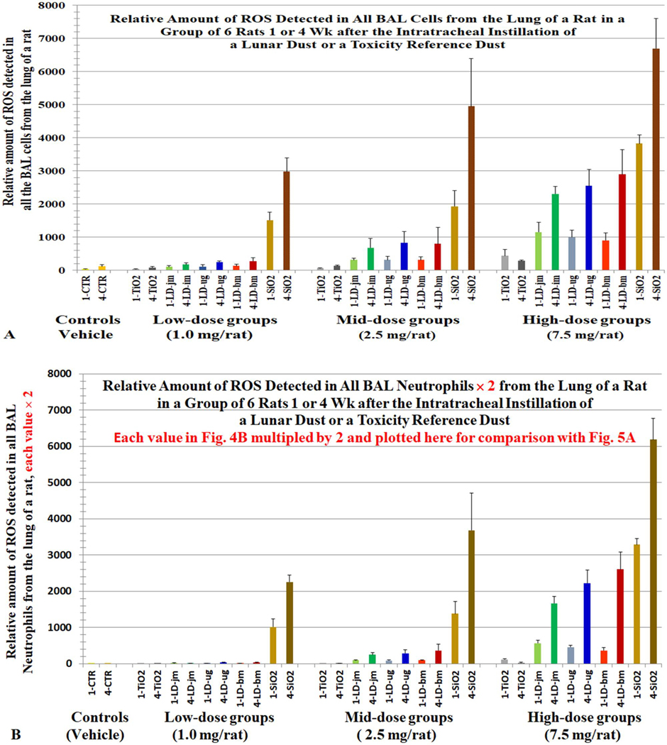
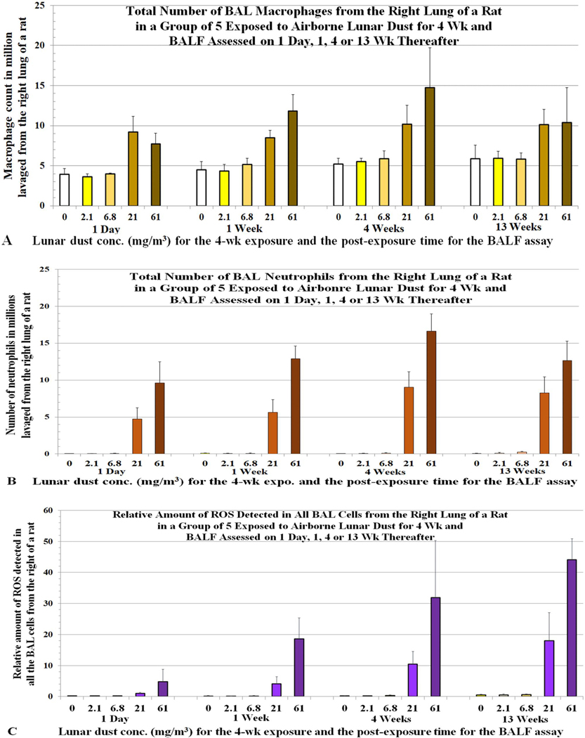
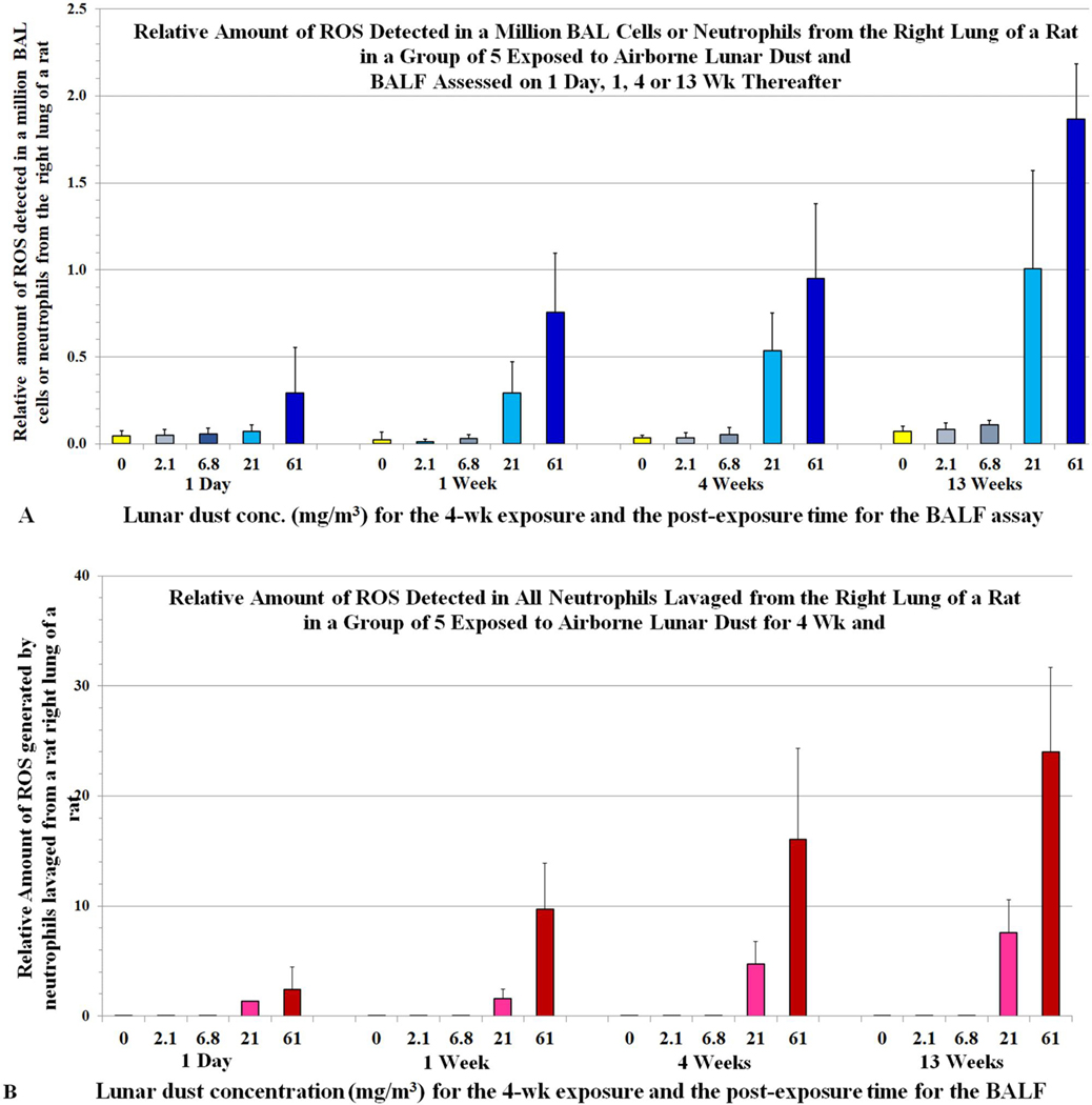
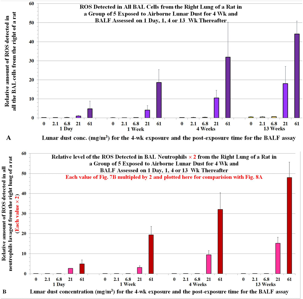
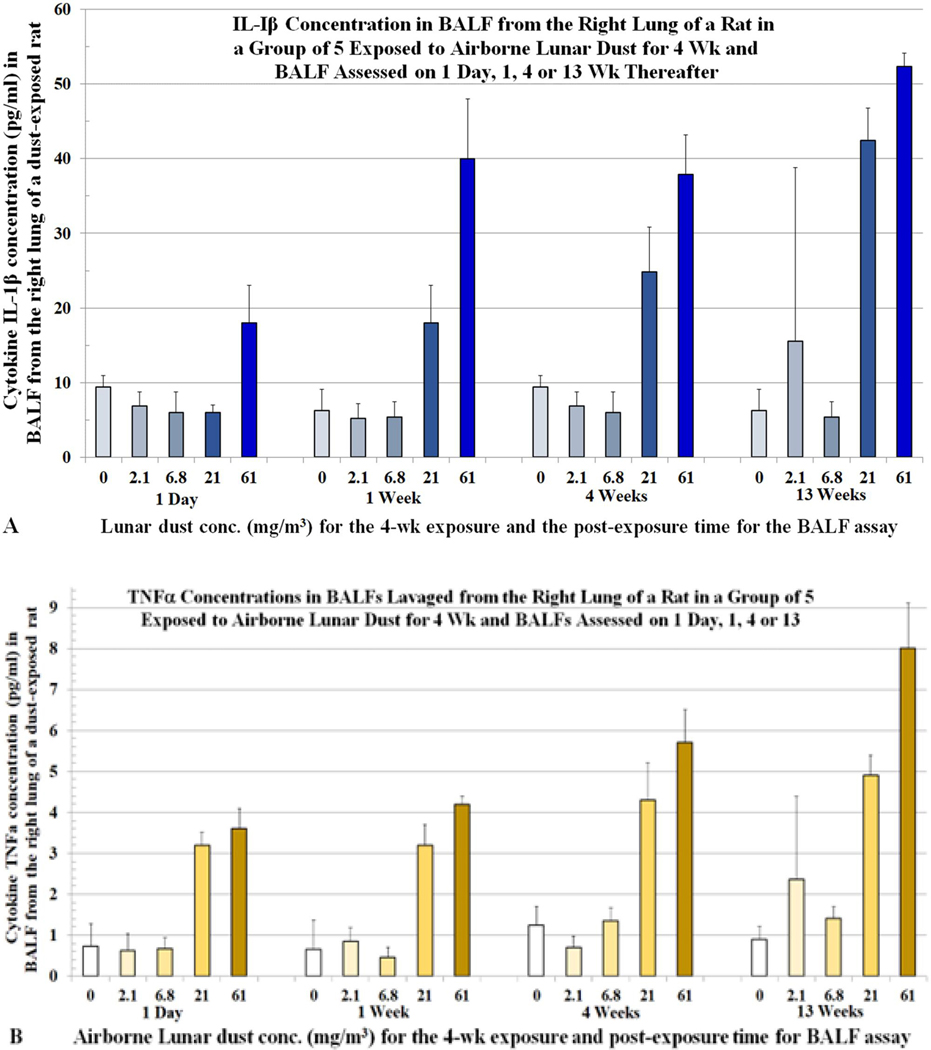
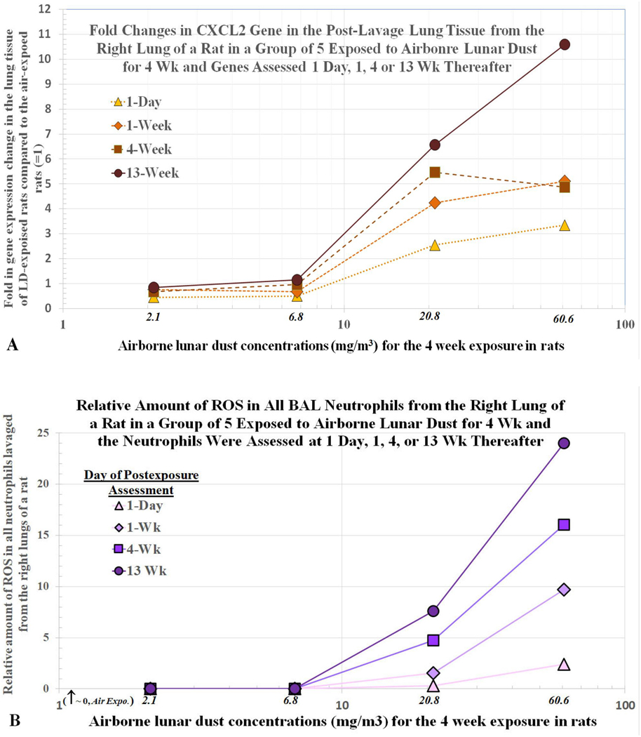
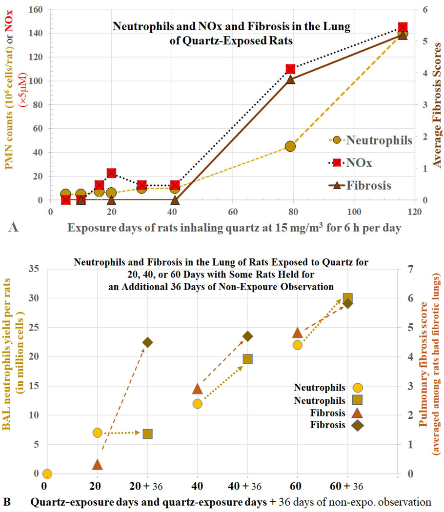
The mechanisms of particle-induced pathogenesis in the lung remain poorly understood. Neutrophilic inflammation and oxidative stress in the lung are hallmarks of toxicity. Some investigators have postulated that oxidative stress from particle surface reactive oxygen species (psROS) on the dust produces the toxicopathology in the lungs of dust-exposed animals. This postulate was tested concurrently with the studies to elucidate the toxicity of lunar dust (LD), which is believed to contain psROS due to high-speed micrometeoroid bombardment that fractured and pulverized lunar surface regolith. Results from studies of rats intratracheally instilled (ITI) with three LDs (prepared from an Apollo-14 lunar regolith), which differed 14-fold in levels of psROS, and two toxicity reference dusts (TiO2 and quartz) indicated that psROS had no significant contribution to the dusts' toxicity in the lung. Reported here are results of further investigations by the LD toxicity study team on the toxicological role of oxidants in alveolar neutrophils that were harvested from rats in the 5-dust ITI study and from rats that were exposed to airborne LD for 4 weeks. The oxidants per neutrophils and all neutrophils increased with dose, exposure time and dust's cytotoxicity. The results suggest that alveolar neutrophils play a critical role in particle-induced injury and toxicity in the lung of dust-exposed animals. Based on these results, we propose an adverse outcome pathway (AOP) for particle-associated lung disease that centers on the crucial role of alveolar neutrophil-derived oxidant species. A critical review of the toxicology literature on particle exposure and lung disease further supports a neutrophil-centric mechanism in the pathogenesis of lung disease and may explain previously reported animal species differences in responses to poorly soluble particles. Key findings from the toxicology literature indicate that (1) after exposures to the same dust at the same amount, rats have more alveolar neutrophils than hamsters; hamsters clear more particles from their lungs, consequently contributing to fewer neutrophils and less severe lung lesions; (2) rats exposed to nano-sized TiO2 have more neutrophils and more severe lesions in their lungs than rats exposed to the same mass-concentration of micron-sized TiO2; nano-sized dust has a greater number of particles and a larger total particle-cell contact surface area than the same mass of micron-sized dust, which triggers more alveolar epithelial cells (AECs) to synthesize and release more cytokines that recruit a greater number of neutrophils leading to more severe lesions. Thus, we postulate that, during chronic dust exposure, particle-inflicted AECs persistently release cytokines, which recruit neutrophils and activate them to produce oxidants resulting in a prolonged continuous source of endogenous oxidative stress that leads to lung toxicity. This neutrophil-driven lung pathogenesis explains why dust exposure induces more severe lesions in rats than hamsters; why, on a mass-dose basis, nano-sized dusts are more toxic than the micron-sized dusts; why lung lesions progress with time; and why dose-response curves of particle toxicity exhibit a hockey stick like shape with a threshold. The neutrophil centric AOP for particle-induced lung disease has implications for risk assessment of human exposures to dust particles and environmental particulate matter.
Keywords: Lunar dust; PM; PSP; ROS; diesel particles; inhalation toxicity; intratracheal instillation; lung pathogenesis; mechanism of toxicity; moon dust; neutrophilia; neutrophils; particulate matter.
Conflict of interest statement
Declaration of interest
This project was funded by the NASA Human Research Program. The authors participated in the development of the paper as individual professionals and have sole responsibility for the writing and content of the paper. None of the authors have been involved in the last 5 years with regulatory or legal proceedings related to the contents of the paper. The findings and conclusions in this report are those of the authors and do not necessarily represent the views of the NASA, NIOSH, the University of Texas Health Science Center at Houston or any other entities. No potential conflict of interest was reported by the author(s). SPECIAL DECLARATION OF INTERESTS DISCLOSURE STATEMENT by the Editor-in-Chief of CRT, Dr. Roger O. McClellan, was a member of the NASA-assembled Lunar Airborne Toxicity Assessment Group advising on the experimental design and the conduct of the LD toxicity studies in rodents. He is also a contributing coauthor of this paper. In the interest of transparency and to avoid any conflicts of interest, he recused himself from the review process for this paper following its submission to CRT for publication consideration. The review process for this paper was assigned to a member of the Journal’s Editorial Advisory Board, Samuel Cohen PhD, MD who is knowledgeable of the toxicology of respiratory toxicants and respiratory disease processes pathology. The publication fees and cost of open access for this article were paid by NASA.
Figures
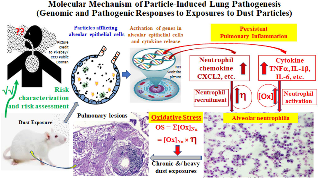
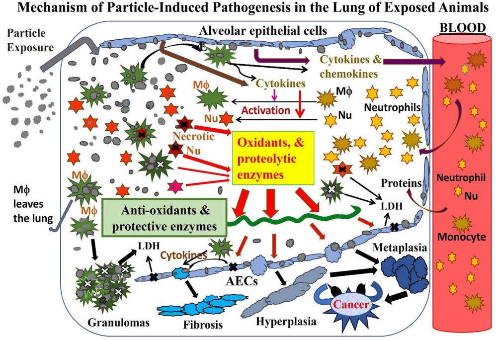
 ) and neutrophils (orangep) to the airspaces. Some of the cellular mediators are also released by resident alveolar macrophages (AM or Mu, green
 ). (3) The incoming leukocytes, especially neutrophils, are activated by cytokines to increase their oxidant levels; monocytes become activated macrophages (AM, green ) and neutrophils become activated neutrophils (redp). (4) Neutrophils are short-lived; apoptotic neutrophils and dust particles are phagocytized by AMs. (5) In animals exposed heavily to dust, AMs are overloaded with particles. Some of the dust-laden and dead AMs cannot leave the lung; they aggregate to produce granulomas. (6) The unphagocytized apoptotic neutrophils undergo necrosis (marked with x) and eventually release their oxidants and harmful enzymes onto the alveolar epithelium. (7) Some oxidants are destroyed by antioxidants and protective enzymes. (8) Oxidants and proteases released from neutrophils damage AECs. (9) If particles are below a threshold level, they can be removed by AMs and the inflammatory/damage cycle ends leading to repair. Above a threshold dust burden, persistent and chronic neutrophilic inflammation causes lung lesions to progress with time. (10) Fibroblasts proliferate on the wall of damaged epithelial areas, resulting in fibrosis. (11) Persistent and chronic oxidative damage to AECs can cause proliferation of type II AECs; some of these cells may eventually undergo hyperplasia, metaplasia, and, in the worst case, carcinogenesis.
Similar articles
-
Comparative pulmonary toxicities of lunar dusts and terrestrial dusts (TiO2 & SiO2) in rats and an assessment of the impact of particle-generated oxidants on the dusts' toxicities.Inhal Toxicol. 2022;34(3-4):51-67. doi: 10.1080/08958378.2022.2038736. Epub 2022 Mar 16. Inhal Toxicol. 2022. PMID: 35294311
-
NTP Toxicity Study Report on the atmospheric characterization, particle size, chemical composition, and workplace exposure assessment of cellulose insulation (CELLULOSEINS).Toxic Rep Ser. 2006 Aug;(74):1-62, A1-C2. Toxic Rep Ser. 2006. PMID: 17160106
-
Pulmonary responses of mice, rats, and hamsters to subchronic inhalation of ultrafine titanium dioxide particles.Toxicol Sci. 2004 Feb;77(2):347-57. doi: 10.1093/toxsci/kfh019. Epub 2003 Nov 4. Toxicol Sci. 2004. PMID: 14600271
-
Significance of particle parameters in the evaluation of exposure-dose-response relationships of inhaled particles.Inhal Toxicol. 1996;8 Suppl:73-89. Inhal Toxicol. 1996. PMID: 11542496 Review.
-
Toxicokinetics and effects of fibrous and nonfibrous particles.Inhal Toxicol. 2002 Jan;14(1):29-56. doi: 10.1080/089583701753338622. Inhal Toxicol. 2002. PMID: 12122559 Review.
Cited by
-
Pulmonary and systemic immune alterations in rats exposed to airborne lunar dust.Front Immunol. 2025 Feb 6;16:1538421. doi: 10.3389/fimmu.2025.1538421. eCollection 2025. Front Immunol. 2025. PMID: 39981230 Free PMC article.
-
Single-cell transcriptomic analysis of radiation-induced lung injury in rat.Biomol Biomed. 2024 Sep 6;24(5):1331-1349. doi: 10.17305/bb.2024.10357. Biomol Biomed. 2024. PMID: 38552230 Free PMC article.
-
Influence of macrophages and neutrophilic granulocyte-like cells on crystalline silica-induced toxicity in human lung epithelial cells.Toxicol Res (Camb). 2025 Jan 12;14(1):tfaf004. doi: 10.1093/toxres/tfaf004. eCollection 2025 Feb. Toxicol Res (Camb). 2025. PMID: 39822374
References
-
- Albrecht C, Knaapen AM, Becker A, Höhr D, Haberzettl P, van Schooten FJ, Borm PJ, Schins RP. 2005. The crucial role of particle surface reactivity in respirable quartz-induced reactive oxygen/nitrogen species formation and APE/Ref-1 induction in rat lung. Respir Res. 6(1):129–137. doi: 10.1186/1465-9921-6-129. - DOI - PMC - PubMed
Publication types
MeSH terms
Substances
Grants and funding
LinkOut - more resources
Full Text Sources
Medical
Research Materials
Miscellaneous