. 2023 Nov 27;51(21):11783-11796.
doi: 10.1093/nar/gkad804.

An archaeal virus-encoded anti-CRISPR protein inhibits type III-B immunity by inhibiting Cas RNP complex turnover

Affiliations

An archaeal virus-encoded anti-CRISPR protein inhibits type III-B immunity by inhibiting Cas RNP complex turnover

Jilin Liu et al. Nucleic Acids Res. .

Erratum in

Abstract

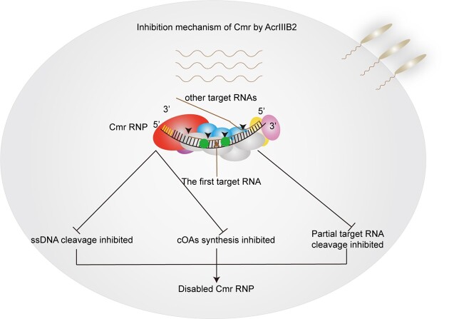
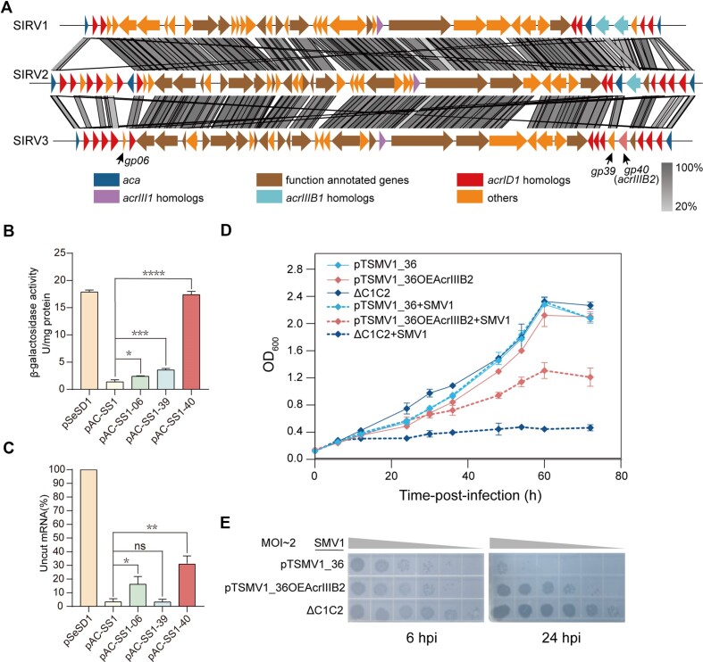
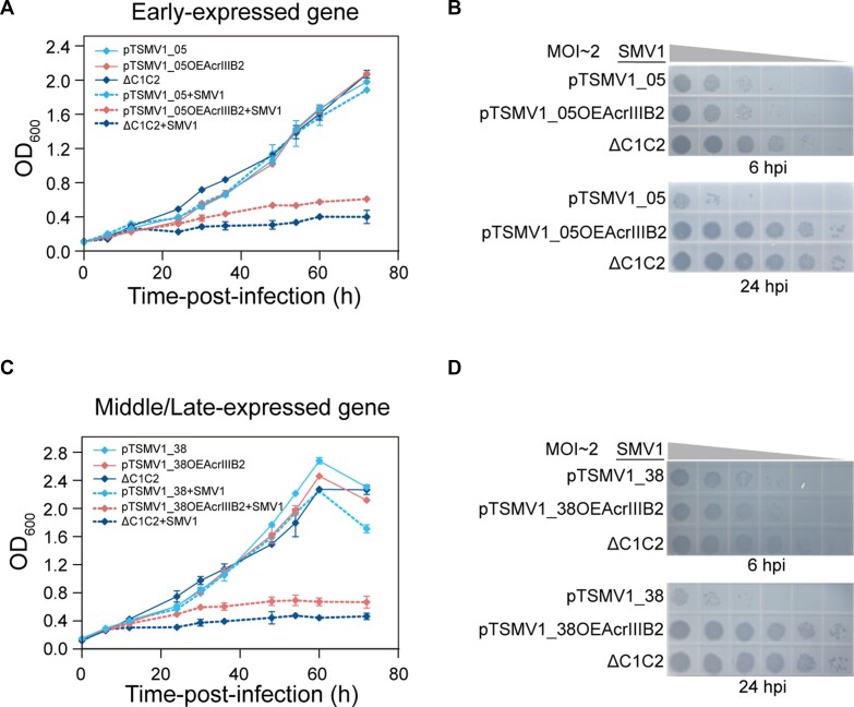
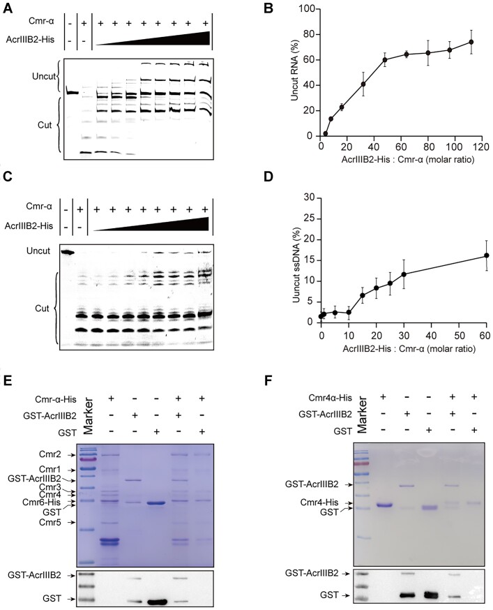
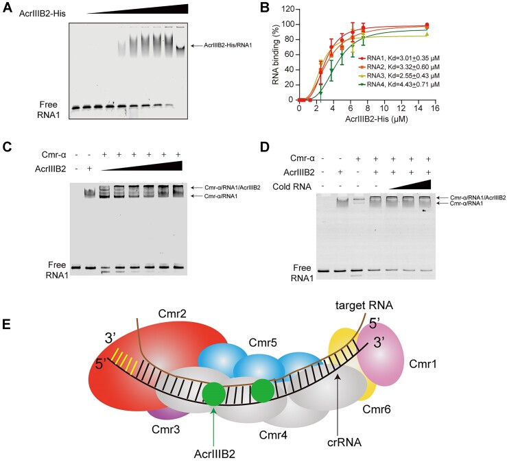
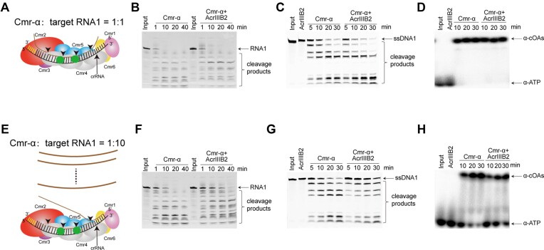
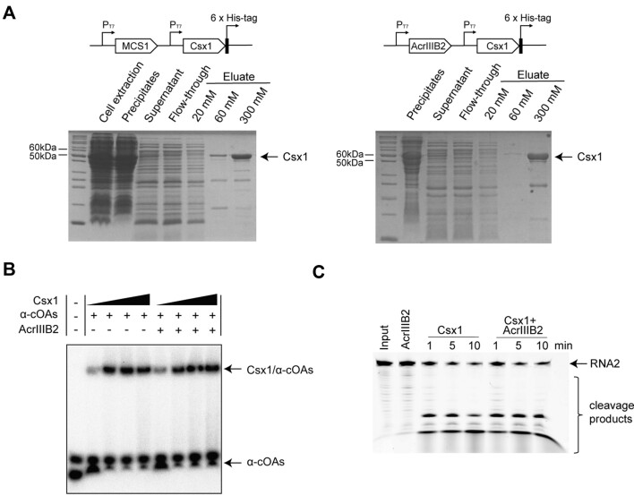
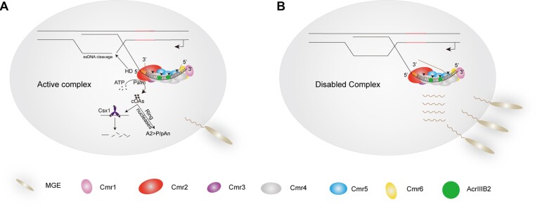
CRISPR-Cas systems are widespread in prokaryotes and provide adaptive immune against viral infection. Viruses encode a type of proteins called anti-CRISPR to evade the immunity. Here, we identify an archaeal virus-encoded anti-CRISPR protein, AcrIIIB2, that inhibits Type III-B immunity. We find that AcrIIIB2 inhibits Type III-B CRISPR-Cas immunity in vivo regardless of viral early or middle-/late-expressed genes to be targeted. We also demonstrate that AcrIIIB2 interacts with Cmr4α subunit, forming a complex with target RNA and Cmr-α ribonucleoprotein complex (RNP). Furtherly, we discover that AcrIIIB2 inhibits the RNase activity, ssDNase activity and cOA synthesis activity of Cmr-α RNP in vitro under a higher target RNA-to-Cmr-α RNP ratio and has no effect on Cmr-α activities at the target RNA-to-Cmr-α RNP ratio of 1. Our results suggest that once the target RNA is cleaved by Cmr-α RNP, AcrIIIB2 probably inhibits the disassociation of cleaved target RNA, therefore blocking the access of other target RNA substrates. Together, our findings highlight the multiple functions of a novel anti-CRISPR protein on inhibition of the most complicated CRISPR-Cas system targeting the genes involved in the whole life cycle of viruses.


Figures

Graphical Abstract
Graphical Abstract
Figure 1.
Figure 1.
AcrIIIB2 inhibits the immunity of Type III-B CRISPR–Cas systems in S. islandicus. (A) Genome comparison between Sulfolobus viruses SIRV1, SIRV2, and SIRV3. The acrID1, acrIIIB1, acrIII1 and their corresponding homologs are colored red, light blue and purple, respectively. The anti-CRISPR-associated protein genes (aca) are colored blue. The genes encoding function-annotated proteins are colored brown and other genes are colored orange. The black arrows point to BHS13_gp06, BSH13_gp39 and BHS13_gp40 genes. (B) Specific β-glycosidase activities of E233 transformants carrying empty vector (pSeSD1), a targeted plasmid (pAC-SS1) or pAC-SS1 overexpressing gp06, gp39 or gp40 genes, respectively. (C) Uncut lacS mRNA levels of E233 transformants carrying different plasmids. Three independent transformants of each construct were analysed for β-glycosidase assay as well as mRNA quantification (unpaired t-test; *P < 0.05; **P < 0.01; ***P < 0.001; ****P < 0.0001; Bars represent mean ± SD). (D) Growth curves of E233 strains containing plasmids and ΔC1C2 strain with or without SMV1 infection. pTSMV1_36: plasmid containing spacer targeting the early gene CF87_gp36 gene of SMV1; pTSMV1_36OEgp40: plasmid pTSMV1_36 cloned with the SIRV3 BHS13_gp40 gene expression cassette. The data show means of three replicates. Error bars indicate the standard deviations. (E) Plaques of the supernatant of SMV1-infected-cultures at 6 hpi and 24 hpi from (D) on the plates spread with ΔC1C2 cells.
Figure 2.
Figure 2.
AcrIIIB2 inhibits Cmr-α immunity targeting early-expressed or middle/late-expressed viral genes. Targeting of SMV1 early-expressed gene (A) or middle/late-expressed gene (C) by Cmr-α strain carrying the plasmid pTSMV1_05 or pTSMV1_38 is inhibited by AcrIIIB2 at MOI ∼2. (B) Plaques of the supernatant of SMV1-infected-cultures sampled at 6 and 24 hpi from (A) or (D) 6 and 24 hpi from (C) on the plates spread with ΔC1C2 cells. The data show means of three replicates. Error bars indicate the standard deviations.
Figure 3.
Figure 3.
AcrIIIB2 interacts with Cmr4α to inhibit the RNase and ssDNase activities of Cmr-α RNP. (A) AcrIIIB2 inhibits cis RNase activity of Cmr-α RNP. Cmr-α RNP (100 nM), AcrIIIB2 (at different concentrations), 250 nM labeled target RNA were co-incubated for 5 minutes at 70°C at AcrIIB2 to Cmr-α RNP molar ratio of 0, 1, 5, 10, 15, 20, 25 and 30. (B) Dose response of Cmr-α RNP cis-RNase inhibition by AcrIIIB2. (C) AcrIIIB2 inhibits trans ssDNase activity. Cmr-α RNP (100nM), AcrIIIB2 (at different concentrations), unlabeled target RNA (300 nM) and 5′-FAM-labeled ssDNA (300 nM) were co-incubated for 5min at 70°C at AcrIIIB2 to Cmr-α RNP molar ratios of 0, 1, 5, 10, 15, 20, 25 and 30. (D) Dose response of Cmr-α RNP trans-ssDNase inhibition by AcrIIIB2. (E) His-tag pull-down assays of GST-AcrIIIB2 with Cmr-α RNP complex. GST-AcrIIIB2, GST and Cmr-α subunit proteins are indicated by black arrows (top). Western blot assays were performed to show GST-tagged AcrIIIB2 using the anti-GST antibody. (F) His-tag pull-down assays of GST-AcrIIIB2 with Cmr4α subunit. GST-AcrIIIB2, GST and Cmr4α-His proteins are indicated by black arrows (top). Western blot assays were performed to show GST-tagged AcrIIIB2 using the anti-GST antibody.
Figure 4.
Figure 4.
AcrIIIB2 bound with Cmr-α and target RNA to form a super-complex. (A) EMSA analysis of 5′-FAM labeled RNA substrate 1 with increasing amount of AcrIIIB2. 250 nM labeled target RNA were co-incubated for 5 min at 70°C with AcrIIIB2 (0, 0.25, 0.5, 1.25, 2.5, 3.75, 5, 6.25, 7.5 and 15 μM). (B) Quantified fractions of RNA substrates bound by AcrIIIB2 as determined by EMSA (mean ± SD, n = 3 independent experiments). (C) EMSA analysis of 5′-FAM labeled RNA substrate 1 with Cmr-α RNP in the presence of increasing amount of AcrIIIB2. Cmr-α RNP (250 nM), AcrIIIB2 (0, 0.25, 0.5, 1.25, 2.5 and 3.75 μM), 250 nM labeled target RNA were co-incubated for 5 min at 70°C at AcrIIIB2 to Cmr-α RNP molar ratio of 0, 1, 2, 5, 10 and 15. (D) EMSA analysis of 5′-FAM labeled RNA substrate 1 with Cmr-α RNP in the presence of increasing amount of cold non-target RNA substrates (RNA5). Cmr-α RNP (250 nM), AcrIIIB2 (250 nM), labeled target RNA (100 nM) and cold non-target RNA (100, 500 and 1000 nM) were co-incubated for 5 min at 70°C. (E) Model for AcrIIIB2- target RNA-Cmr-α super complex. AcrIIIB2 interact with intermediate Cmr4α and probably with target RNA.
Figure 5.
Figure 5.
AcrIIIB2 inhibits Cmr-α turnover to reduce Cmr-α activities. (A) Diagram of target RNA cleavage when molar ratio of Cmr-α to target RNA equal to 1. (B) Target RNA cleavage by Cmr-α, and AcrIIIB2-bound Cmr-α complex at molar ratio of proteins to target RNA equal to 1. Cmr-α RNP (100nM) or Cmr-α-AcrIIIB2 (100 nM) were incubated with 100 nM 5′ FAM-labeled target RNA at 70°C for 1, 10, 20 and 40 min. Samples were separated by a 18% denaturing PAGE gel. (C) ssDNA cleavage by Cmr-α, or AcrIIIB2-bound Cmr-α complex at molar ratio of proteins to target RNA equal to 1. Cmr-α RNP (100 nM) or Cmr-α-AcrIIIB2 (100 nM) were incubated with 100 nM cold target RNA and 100 nM 5′ FAM-labeled ssDNA substrates at 70°C for 5, 10, 20 and 30 min. Samples were separated by a 18% denaturing PAGE gel. (D) COAs synthesis by Cmr-α, or AcrIIIB2-bound Cmr-α complex at molar ratio of proteins to target RNA equal to 1. Cmr-α RNP (100 nM) or Cmr-α-AcrIIIB2 (100 nM) were incubated with 100 nM cold target RNA and 100 μM cold ATP and radiolabeled α-ATP at 70°C for 10, 20 and 30 min. Samples were separated by a 24% denaturing PAGE gel. (E) Diagram of target RNA cleavage when molar ratio of Cmr-α to target RNA equal to 10. (F) Target RNA cleavage by Cmr-α, and AcrIIIB2-bound Cmr-α complex at molar ratio of proteins to target RNA equal to 10. Cmr-α RNP (25 nM) or Cmr-α-AcrIIIB2 (25 nM) were incubated with 250 nM 5′ FAM-labeled target RNA at 70°C for 1, 10, 20 and 40 min. Samples were separated by a 18% denaturing PAGE gel. (G) DNA cleavage by Cmr-α, or AcrIIIB2-bound Cmr-α complex at molar ratio of proteins to target RNA equal to 10. Cmr-α RNP (25 nM) or Cmr-α-AcrIIIB2 (25 nM) were incubated with 250 nM cold target RNA and 500 nM 5′ FAM-labeled ssDNA substrates at 70°C for 5, 10, 20 and 30 min. Samples were separated by a 18% denaturing PAGE gel. (H) COAs synthesis by Cmr-α, or AcrIIIB2-bound Cmr-α complex at molar ratio of proteins to target RNA equal to 10. Cmr-α RNP (25 nM) or Cmr-α-AcrIIIB2 (25 nM) were incubated with 250 nM cold target RNA and 1000 μM cold ATP and radiolabeled α-ATP at 70°C for 10, 20 and 30 min. Samples were separated by a 24% denaturing PAGE gel.
Figure 6.
Figure 6.
AcrIIIB2 has no direct effect on Csx1. (A) Co-purification of His-tagged Csx1 and non-tagged AcrIIIB2. Constructs were transformed into E. coli BL21(DE3) cells, which were cultured in 500 ml of LB medium containing 25 μg/ml Chloramphenicol at 37°C for 3 h to an OD600 of 0.6–0.7, and then induced with 0.1 mM IPTG for 16 h at 12°C. The cells were harvested and resuspended, then lysed by ultrasonication. Supernatant was collected after centrifugation and filtrated with a 0.22-μm filter. The filtered supernatant was loaded onto a Ni-NTA agarose column. The column was washed with 10 volumes of elution buffer (50 mM Tris-HCl pH 8.0, 300 mM NaCl and 20 mM Imidazole) and protein eluted by gradient imidazole solution. Samples collected at each step were analysis by SDS–PAGE. (B) Binding of Csx1 and α 32 P-cOAs. The mixture of Csx1 (250 nM, 500 nM, 1 μM and 2 μM), AcrIIIB2 (2 μM) and α 32 P-cOAs (500 nM) were incubated at 70°C for 5 min. (C) AcrIIIB2 could not inhibit Csx1 RNA cleavage activity. Csx1 (50 nM) or Csx1 (50 nM) with AcrIIIB2 (250 nM) were incubated with 1 μM cold cOAs at 70°C for 1, 5 and 10 min. Samples were separated by a 18% denaturing PAGE gel.
Figure 7.
Figure 7.
A model for the inhibitory effects of AcrIIIB2 on Type III-B CRISPR–Cas system. (A) When virus expresses AcrIIIB2, once the target RNA is cleaved by Cmr-α RNP, AcrIIIB2 binds with intermediate Cmr4α and probably grabs the cleaved target RNA fragments, inhibiting the dissociation of the fragments. Cmr-α is allosterically activated at this step. When the amount of target RNA is less than or equal to that of Cmr-α, it can sustain allosteric activation of Cmr-α and viruses will be eliminated. (B) When target RNA is more than the amount of Cmr-α, the undisassociated RNA block the access of other target RNA substrates to inhibit Cmr-α activities. As Cmr-α is activated for a short time, a few cOAs are produced and will probably be degraded by host encoded ring nucleases. Under this condition, viruses propagate successfully.

Similar articles

Cited by

References

    1. Makarova K.S., Wolf Y.I., Iranzo J., Shmakov S.A., Alkhnbashi O.S., Brouns S.J.J., Charpentier E., Cheng D., Haft D.H., Horvath P.et al. .. Evolutionary classification of CRISPR-Cas systems: a burst of class 2 and derived variants. Nat. Rev. Microbiol. 2020; 18:67–83. - PMC - PubMed
    1. Wang J.Y., Pausch P., Doudna J.A.. Structural biology of CRISPR-Cas immunity and genome editing enzymes. Nat. Rev. Microbiol. 2022; 20:641–656. - PubMed
    1. Dimitriu T., Szczelkun M.D., Westra E.R.. Evolutionary ecology and interplay of prokaryotic innate and adaptive immune systems. Curr. Biol. 2020; 30:R1189–R1202. - PMC - PubMed
    1. Barrangou R., Fremaux C., Deveau H., Richards M., Boyaval P., Moineau S., Romero D.A., Horvath P.. CRISPR provides acquired resistance against viruses in prokaryotes. Science. 2007; 315:1709–1712. - PubMed
    1. Marraffini L.A., Sontheimer E.J.. CRISPR interference limits horizontal gene transfer in staphylococci by targeting DNA. Science. 2008; 322:1843–1845. - PMC - PubMed