. 2023 Dec;32(6):575-582.
doi: 10.1007/s11248-023-00373-7. Epub 2023 Oct 18.

Knockout of M-LP/Mpv17L, a newly identified atypical PDE, induces physiological afferent cardiac hypertrophy in mice

Affiliations

Knockout of M-LP/Mpv17L, a newly identified atypical PDE, induces physiological afferent cardiac hypertrophy in mice

Reiko Iida et al. Transgenic Res. 2023 Dec.

Abstract

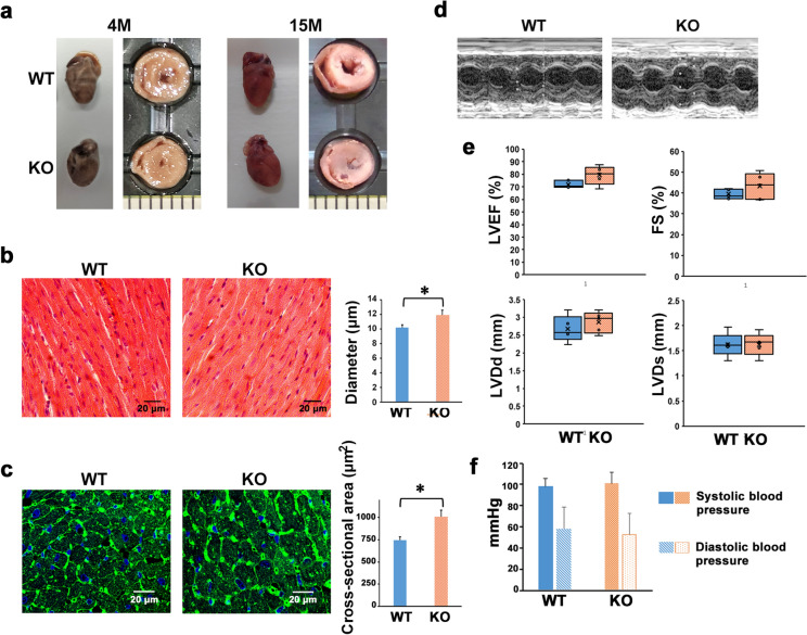
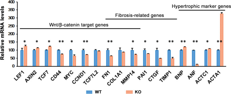
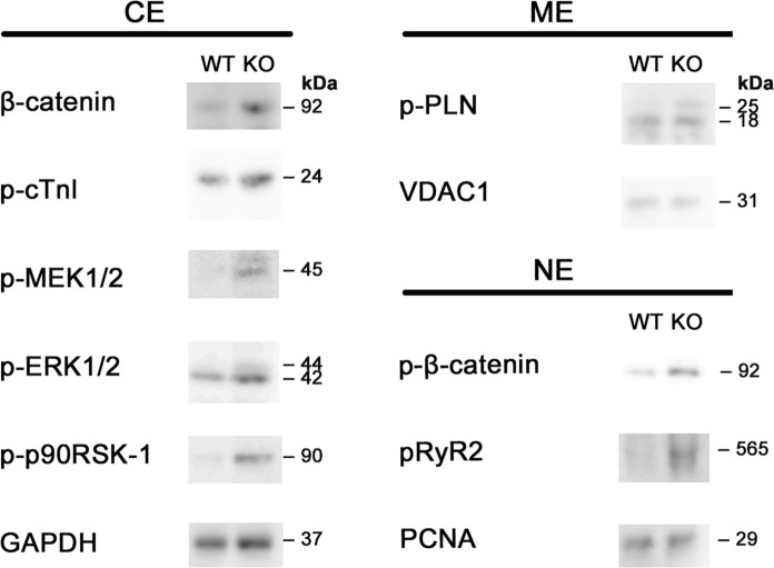
M-LP/Mpv17L (Mpv17-like protein) is an atypical cyclic nucleotide phosphodiesterase (PDE) without the molecular structure characteristic of the PDE family. Deficiency of M-LP/Mpv17L in mice has been found to result in development of β-cell hyperplasia and improved glucose tolerance. Here, we report another phenotype observed in M-LP/Mpv17L-knockout (KO) mice: afferent cardiac hypertrophy. Although the hearts of M-LP/Mpv17L-KO mice did not differ in size from those of wild-type mice, there was marked narrowing of the left ventricular lumen and thickening of the ventricular wall. The diameter and cross-sectional area of cardiomyocytes in 8-month-old M-LP/Mpv17L-KO mice were increased 1.16-fold and 1.35-fold, respectively, relative to control mice, but showed no obvious abnormalities of cell structure, fibrosis or impaired cardiac function. In 80-day-old KO mice, the expression of hypertrophic marker genes, brain natriuretic peptide (BNF), actin alpha cardiac muscle 1 (ACTC1) and actin alpha 1 skeletal muscle (ACTA1), as well as the Wnt/β-catenin pathway target genes, lymphoid enhancer-binding factor-1 (LEF1), axis inhibition protein 2 (AXIN2) and transcription factor 7 (TCF7), was significantly up-regulated relative to control mice, whereas fibrosis-related genes such as fibronectin 1 (FN1) and connective tissue growth factor (CTGF) were down-regulated. Western blot analysis revealed increased phosphorylation of molecules downstream of the cAMP/PKA signaling pathway, such as β-catenin, ryanodine receptor 2 (RyR2), phospholamban (PLN) and troponin I (cTnI), as well as members of the MEK1-ERK1/2 signaling pathway, which is strongly involved in afferent cardiac hypertrophy. Taken together, these findings indicate that M-LP/Mpv17L is one of the PDEs actively functioning in the heart and that deficiency of M-LP/Mpv17L in mice promotes physiological cardiac hypertrophy.

Keywords: Cardiac hypertrophy; Cyclic nucleotide phosphodiesterase; Mpv17-like protein; cAMP/PKA signaling.


Conflict of interest statement

The authors declare no competing interests.

Figures

Fig. 1
Fig. 1
Levels of M-LP/Mpv17L mRNA in mouse hearts at various ages. mRNA expression was determined by Q-PCR using β-actin as an internal control. The results are expressed as ratios relative to the value for 10-day-old mice (n = 3)
Fig. 2
Fig. 2
M-LP/Mpv17L deficiency leads to physiological afferent cardiac hypertrophy. a Gross morphology and cross-sections of representative hearts obtained from M-LP/Mpv17L-WT and -KO mice. b H&E staining of heart tissues from 8-month-old M-LP/Mpv17L-WT and -KO mice. Scale bars: 20 μm. Diameters of cardiomyocytes are shown as means ± SD (n = 10). *p < 0.05 c WGA staining of heart tissues from 8-month-old M-LP/Mpv17L-WT and -KO mice. Scale bars: 20 μm. Cross-sectional areas of cardiomyocytes are shown as means ± SD (n = 10). *p < 0.05 d Representative M-mode echocardiographic images of 14-month-old M-LP/Mpv17L-WT and -KO mice. e The echocardiographic parameters LVEF, FS, LVDd and LVDs of 14-month-old M-LP/Mpv17L-WT and -KO mice. Data are shown as means ± SD (n = 5). f Systolic and diastolic blood pressure of 16-month-old M-LP/Mpv17L-WT and -KO mice. Data are shown as means ± SD (n = 7)
Fig. 3
Fig. 3
Transcriptomic analysis of hearts obtained from 80-day-old M-LP/Mpv17L-WT and -KO mice. Expression of mRNA was determined by Q-PCR using GAPDH as an internal control. The results are expressed as ratios relative to the value for M-LP/Mpv17L-WT mice (n = 3). *p < 0.05, **p < 0.005
Fig. 4
Fig. 4
Western blot analysis of cytosolic, membrane and nuclear soluble extracts from 80-day-old M-LP/Mpv17L-WT and -KO mice. GAPDH, VDAC1 and PCNA were used as loading controls for cytosolic, membrane and nuclear protein, respectively. CE: cytosolic extract; ME: membrane extract; NE: nuclear soluble extract

References

    1. Alkass K, Panula J, Westman M, et al. No evidence for cardiomyocyte number expansion in preadolescent mice. Cell. 2015;163:1026–1036. doi: 10.1016/j.cell.2015.10.035. - DOI - PubMed
    1. Bobin P, Belacel-Ouari M, Bedioune I, et al. Cyclic nucleotide phosphodiesterases in heart and vessels: a therapeutic perspective. Arch Cardiovasc Dis. 2016;109:431–443. doi: 10.1016/j.acvd.2016.02.004. - DOI - PubMed
    1. Bueno OF, De Windt LJ, Tymitz KM, et al. The MEK1-ERK1/2 signaling pathway promotes compensated cardiac hypertrophy in transgenic mice. EMBO J. 2000;19:6341–6350. doi: 10.1093/emboj/19.23.6341. - DOI - PMC - PubMed
    1. Haq S, Michael A, Andreucci M, et al. Stabilization of β-catenin by a wnt-independent mechanism regulates cardiomyocyte growth. Proc Natl Acad Sci USA. 2003;100:4610–4615. doi: 10.1073/pnas.0835895100. - DOI - PMC - PubMed
    1. Heallen TR, Kadow ZA, Wang J, et al. Determinants of cardiac growth and size. Cold Spring Harb Perspect Biol. 2020;12:a037150. doi: 10.1101/cshperspect.a037150. - DOI - PMC - PubMed