. 2023 Oct 13:56:e12391.
doi: 10.1590/1414-431X2023e12391. eCollection 2023.

Total rupture of Achilles tendon induces inflammatory response and glial activation on the spinal cord of mice

Affiliations

Total rupture of Achilles tendon induces inflammatory response and glial activation on the spinal cord of mice

D R De Paula et al. Braz J Med Biol Res. .

Abstract

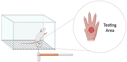
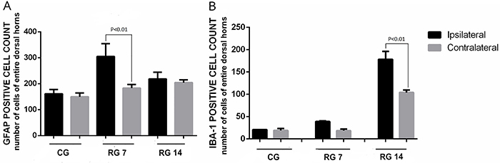
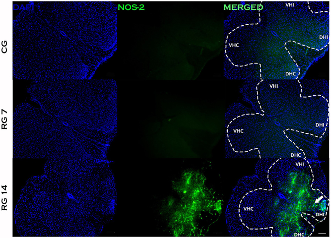
Rupture of Achilles tendon is a common accident affecting professional and recreational athletes. Acute and chronic pain are symptoms commonly observed in patients with rupture. However, few studies have investigated whether Achilles tendon rupture is able to promote disorders in the central nervous system (CNS). Therefore, the current study aimed to evaluate nociceptive alterations and inflammatory response in the L5 lumbar segment of Balb/c mice spinal cord after Achilles tendon rupture. We found increased algesia in the paw of the ruptured group on the 7th and 14th days post-tenotomy compared with the control group. This phenomenon was accompanied by overexpression of cyclooxygenase-2 (COX-2) and inducible nitric oxide synthase-2 (NOS-2) as well as hyperactivation of astrocytes and microglia in nociceptive areas of L5 spinal cord as evidenced by intense GFAP and IBA-1 immunostaining, respectively. Biochemical studies also demonstrated increased levels of nitrite in the L5 spinal cord of tenotomized animals compared with the control group. Thus, we have demonstrated for the first time that total rupture of the Achilles tendon induced inflammatory response and nitrergic and glial activation in the CNS in the L5 spinal cord region.


Figures

Figure 1
Figure 1. Image of the von Frey apparatus. The von Frey filaments are inserted through the elevated mesh platform to press the paw of the mouse. The animal is repeatedly stimulated with increasingly strong filaments to determine the threshold at which the paw withdrawal response is reliably elicited.
Figure 2
Figure 2. Effects of Achilles tendon rupture in the mechanical nociceptive threshold. CG (control group) and RG (rupture group) were analyzed before surgery and 7 and 14 days post-Achilles tendon tenotomy. Data are reported as means±SD. ***P<0.01 CG vs RG, n=6/group (two-way ANOVA followed by Tukey’s post hoc comparisons).
Figure 3
Figure 3. Effects of Achilles tendon rupture on astrocyte reactivity in the spinal cord. A, GFAP immunostaining of transversal sections of mice spinal cord (segment L5) on the 7th and 14th days post-Achilles tendon rupture. B, GFAP-positive cell count in the L5 spinal cord of the groups. Data are reported as means±SD, n=6/group (one-way ANOVA followed by Tukey's post hoc comparisons). VHC: ventral horn contralateral; VHI: ventral horn ipsilateral; DHC: dorsal horn contralateral; DHI: dorsal horn ipsilateral. Black arrows in the enlarged images show strong astrogliosis in the dorsal horn of the ipsilateral side to injury (scale bar=100 μm). CG: control group; RG 7: rupture group analyzed on the 7th day after tenotomy; RG 14: rupture group analyzed on the 14th day after tenotomy.
Figure 4
Figure 4. Effects of Achilles tendon rupture on microglial reactivity in the spinal cord. A, IBA-1 immunostaining of transversal sections of mice spinal cord (segment L5) on the 7th and 14th days post-Achilles tendon rupture. B, IBA-1-positive cell count in L5 spinal cord. Data are reported as means±SD, n=6/group (one-way ANOVA followed by Tukey's post hoc comparisons). VHC: ventral horn contralateral; VHI: ventral horn ipsilateral; DHC: dorsal horn contralateral; DHI: dorsal horn ipsilateral. Black arrows in the enlarged images show strong microgliosis in the dorsal horn of ipsilateral side to injury, mainly in RG 14. Scale bar=100 μm. CG: control group; RG 7: rupture group analyzed on the 7th day after tenotomy; RG 14: rupture group analyzed on the 14th day after tenotomy.
Figure 5
Figure 5. Effects of Achilles tendon rupture on glial reactivity in dorsal horns. A, GFAP-positive cell count in dorsal horns of L5 spinal cord. B, IBA-1-positive cell count in dorsal horns of L5 spinal cord. Data are reported as means±SD, n=6/group assessed (ANOVA followed by Tukey's post hoc comparisons). CG: control group; RG 7: rupture group analyzed on the 7th day after tenotomy; RG 14: rupture group analyzed on the 14th day after tenotomy.
Figure 6
Figure 6. Effects of Achilles tendon rupture on glial reactivity between the ipsi- and contralateral sides. A, GFAP-positive cell count in dorsal horns of L5 spinal cord. B, IBA-1-positive cell count of only dorsal horns of L5 spinal cord. Data are reported as means±SD, n=6/group (one-way ANOVA followed by Tukey's post hoc comparisons). CG: control group; RG 7: rupture group analyzed on the 7th day after tenotomy; RG 14: rupture group analyzed 14th day after tenotomy.
Figure 7
Figure 7. Inflammatory response of the spinal cord induced by Achilles tendon rupture. Immunofluorescence to COX-2 of transversal sections of mice spinal cord (segment L5) on the 7th and 14th days post-Achilles tendon rupture. The nuclei of cells were stained with DAPI. VHC: ventral horn contralateral; VHI: ventral horn ipsilateral; DHC: dorsal horn contralateral; DHI: dorsal horn ipsilateral. The white arrow shows strong reactivity in the dorsal horn of ipsilateral side to injury. n=6/group. Scale bar=100 μm. CG: control group; RG 7: rupture group analyzed on the 7th day after tenotomy; RG 14: rupture group analyzed 14th day after tenotomy.
Figure 8
Figure 8. Inflammatory response of the spinal cord induced by Achilles tendon rupture. Immunofluorescence to NOS-2 of transversal sections of mice spinal cord (segment L5) on the 7th and 14th days post-Achilles tendon rupture. The nuclei of cells were stained with DAPI. VHC: ventral horn contralateral; VHI: ventral horn ipsilateral; DHC: dorsal horn contralateral; DHI: dorsal horn ipsilateral. White arrow shows strong reactivity in the dorsal horn of ipsilateral side to injury. n=6/group. Scale bar=100 μm. CG: control group; RG 7: rupture group analyzed on the 7th day after tenotomy; RG 14: rupture group analyzed 14th day after tenotomy.
Figure 9
Figure 9. Nitrite quantification in the spinal cord at the 7th and 14th days post-Achilles tendon rupture. The lumbar intumescence was removed to assess nitrite levels in the tissue by the Griess method. Data are reported as means±SD, n=5-6/group (ANOVA followed by Tukey's post hoc comparisons). CG: control group; RG 7: rupture group analyzed on the 7th day after tenotomy; RG 14: rupture group analyzed 14th day after tenotomy.
Figure 10
Figure 10. Representative image of inflammatory response in the spinal cord after total Achilles tendon rupture.

References

    1. Padanilam TG. Chronic Achilles tendon ruptures. Foot Ankle Clin. 2009;14:711–728. doi: 10.1016/j.fcl.2009.08.001. - DOI - PubMed
    1. Gajhede-Knudsen M, Ekstrand J, Magnusson H, Maffulli N. Recurrence of Achilles tendon injuries in elite male football players is more common after early return to play: an 11-year follow-up of the UEFA Champions League injury study. Br J Sports Med. 2013;47:763–768. doi: 10.1136/bjsports-2013-092271. - DOI - PubMed
    1. Docking S, Rio E, Fortington L, Orchard J, Cook J. Prevalence and impact of Achilles and patellar tendinopathy in the Australian Football League: The role of imaging in the diagnosis and prediction of symptoms. J Sci Med Sport. 2017;20:e106–e128. doi: 10.1016/j.jsams.2017.01.188. - DOI
    1. Manegold S, Tsitsilonis S, Gehlen T, Kopf S, Duda GN, Agres AN. Alterations in structure of the muscle-tendon unit and gait pattern after percutaneous repair of Achilles tendon rupture with the Dresden instrument. Foot Ankle Surg. 2019;25:529–533. doi: 10.1016/j.fas.2018.04.004. - DOI - PubMed
    1. Riley G. Tendinopathy - from basic science to treatment. Nat Clin Pract Rheumatol. 2008;4:82–89. doi: 10.1038/ncprheum0700. - DOI - PubMed