. 2023 Nov 9;186(23):5028-5040.e14.
doi: 10.1016/j.cell.2023.09.021. Epub 2023 Oct 17.

Molecular basis of Wnt biogenesis, secretion, and Wnt7-specific signaling

Affiliations

Molecular basis of Wnt biogenesis, secretion, and Wnt7-specific signaling

Xiaofeng Qi et al. Cell. .

Abstract

Wnt proteins are enzymatically lipidated by Porcupine (PORCN) in the ER and bind to Wntless (WLS) for intracellular transport and secretion. Mechanisms governing the transfer of these low-solubility Wnts from the ER to the extracellular space remain unclear. Through structural and functional analyses of Wnt7a, a crucial Wnt involved in central nervous system angiogenesis and blood-brain barrier maintenance, we have elucidated the principles of Wnt biogenesis and Wnt7-specific signaling. The Wnt7a-WLS complex binds to calreticulin (CALR), revealing that CALR functions as a chaperone to facilitate Wnt transfer from PORCN to WLS during Wnt biogenesis. Our structures, functional analyses, and molecular dynamics simulations demonstrate that a phospholipid in the core of Wnt-bound WLS regulates the association and dissociation between Wnt and WLS, suggesting a lipid-mediated Wnt secretion mechanism. Finally, the structure of Wnt7a bound to RECK, a cell-surface Wnt7 co-receptor, reveals how RECKCC4 engages the N-terminal domain of Wnt7a to activate Wnt7-specific signaling.

Keywords: N-glycan; Porcupine; RECK; Wnt; Wntless; calreticulin; cryo-EM; phospholipid.


Conflict of interest statement

Declaration of interests The authors declare no competing interests.

Figures

Figure 1
Figure 1. Structural analysis of Wnt7a-WLS-CALR complex.
(A) The general production pathway of Wnt ligands. In the ER, Wnts (purple) are modified by PORCN (green). Secretion of the modified Wnts is mediated by WLS (blue). It remains unclear how Wnts are transferred from PORCN to WLS and how Wnts are released from WLS. Created with BioRender.com. (B) Overall structure of Wnt7a-WLS-CALR complex. The interaction details between PAM and WLS are indicated. The interaction area between the Wnt7a and CALR is shown in the right panel. (C) Cryo-EM map of N-glycan of Wnt7a and its linked loop (contour level: 0.44). (D) Interaction details between CALR and hairpin3, N-glycan of Wnt7a. The hydrophilic interactions are indicated by dashed lines. (E) Structural comparison of Wnt7a-WLS-CALR complex with the PLC complex (PDB: 7QPD). The clash between Wnt7a and ERp57 is zoomed-in in the right box.
Figure 2
Figure 2. Structure of Wnt3a-WLS-CALR complex.
(A) Anti-Flag coimmunoprecipitation (co-IP) assays in total HEK293 cell lysates of cells which expressed C-terminal Flag-tagged WLS with C-terminal Strep-HA-tagged Wnt proteins. Endogenous CALR is associated with WLS in the presence of Wnt1/2b/3/3a/5a/7a/11, but not Wnt8a/8b/9a/9b. (B) Overall structure of Wnt3a-WLS-CALR complex viewed from the side of the membrane and cryo-EM map of its N-glycan at Asn298 (contour level: 0.285). (C) The comparison of the overall structures of Wnt3a-WLS-CALR complex and Wnt7a-WLS-CALR complex. (D) The comparison of the CALR structures of Wnt3a-WLS-CALR and Wnt7a-WLS-CALR complexes. The shifts of the CALR structural elements and the N-glycans are indicated by arrows.
Figure 3
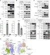
Figure 3. Validation of the roles of CALR and the Wnt glycosylation site in Wnt production and signaling.
(A) and (B) Anti-Flag co-IP assays in total HEK293 cell lysates of cells which expressed C-terminal Flag-tagged WLS with or without C-terminal Strep-HA-tagged Wnt3a (A) or Strep-tagged Wnt7a (B). Endogenous CALR is coimmunoprecipitated with WLS in the presence of Wnt3aWT and Wnt7aWT but not Wnt3aN298T and Wnt7aN295T. (C) The secreted level of Wnt3aWT, Wnt7aWT, Wnt3aN298T and Wnt7aN295T. (D) Signaling activity for different Wnt variants. Activity was measured by TOPFlash luciferase reporter assay. Normalized activity for wild-type Wnt (WT) is taken as 100%, and activity of Wnt mutants is shown as percentage activity compared to WT. n=4 biological repeats. Data are mean ± s.d. Two-sided t-test was performed between the wild-type and mutant groups by GraphPad Prism 9. ***, p<0.001; ****, p<0.0001. (E) and (F) Anti-Flag co-IP assays in total HEK293 cell lysates of cells which expressed N-terminal Flag-tagged PORCNL335A with or without C-terminal Strep-HA-tagged Wnt3a (E) and Strep-tagged Wnt7a (F). Endogenous CALR and WLS were coimmunoprecipitated with PORCNL335A in the presence of Wnt3a and Wnt7a. (G) The hypothetical model of CALR in Wnt transfer from PORCN to WLS. While the hairpin 2 is inserted into PORCN for lipidation, the rest of Wnt binds to WLS under the facilitation of CALR.
Figure 4
Figure 4. Structural analysis of Wnt7a-WLS complex and the apo WLS.
(A) Overall structure of Wnt7a-WLS complex. Electrostatic surface representation of WLS is shown. The cryo-EM map of putative POPC in the central cavity is shown. The central cavity opens to the extracellular/luminal leaflet and is indicated by a dashed rectangle. (B) The interaction details between POPC and WLS. The residue Ser206 of Wnt7a is underlined. (C) Electrostatic surface representation of the apo WLS. The closed central cavity is indicated by a red oval. A potential drug binding pocket is indicated by a dashed rectangle and zoomed-in on the right. (D) Anti-Flag co-IP assays in total HEK293 cell lysates of cells which expressed C-terminal Strep-tagged Wnt7a with C-terminal Flag-tagged WLS. 3M: F230A, W234A and F474A (colored in olive green in panel B). (E) Structural comparison between Wnt7a bound WLS (blue) and the apo WLS (gray). (F) The conformational changes of TM6 and TM7 in the apo state disrupt the POPC and PAM binding.
Figure 5
Figure 5. The analysis of the interaction energy between WLS and Wnt7a by MD simulations at 150 ns scale.
(A) The interaction energy between WLS and Wnt7a at the hairpin 3 and hairpin 2-PAM binding sites. The energy from the complex with the POPC is colored in orange and the energy from the complex without the POPC is colored in gray. (B) The conformational change of TM5 without POPC at 150 ns scale. F347 and F352 rotate towards the edge of WLS and trigger the opening of cavity which accommodates the PAM. The surface representations of the PAM binding cavity from the side of the membrane are shown. (C) The conformational change of TM5 with POPC at 150 ns scale. F347 and F352 retain a similar position in the presence of the POPC. The surface representations of the PAM binding cavity from the side of the membrane are shown.
Figure 6
Figure 6. Structural and functional analysis of the RECKCC4-Wnt7a complex.
(A) Overall structure of RECKCC4-Wnt7a-WLS complex. (B) Structural comparison of RECKCC4 in the complex with Wnt7a to crystal structure of mRECKCC4 in the apo state (PDB: 6WBJ). The conformational change of L3 loop which binds to Wnt7a is indicated by a red arrow. (C) Interactions between RECKCC4 and Wnt7a. The disulfide bonds in RECKCC4 are indicated. The hydrophilic interactions are indicated by dashed lines. (D) and (E) Signaling activity for Wnt7a variants (D) and RECK variants (E). Activity was measured by TOPFlash luciferase reporter assay. Normalized activity for wild-type protein (WT) is taken as 100%, and activity of mutants are shown as percentage activity compared to WT. n=3-4 biological repeats. Data are mean ± s.d. Two-sided t-test was performed between the wild-type and mutant groups by GraphPad Prism 9. **, p<0.01; ****, p<0.0001. (F) A structural model of RECKCC4-Wnt7a-FZD8CRD-LRP6E1E2 complex. The structure of RECKCC4-Wnt7a was docked to the structure of Wnt8-FZD8CRD-LRP6E1E2 (PDB: 8CTG).
Figure 7
Figure 7. Working model of Wnt production and Wnt7 specific signaling.
The hairpin 2 of Wnt interacts with the lumen cavity of PORCN (light green) to undergo lipidation. Then, hairpins 1 and/or 3 of Wnt bind to WLS (blue), with the assistance of CALR (wheat). Once the lipidation process is complete, the hairpin 2 of Wnt dissociates from PORCN and the TMs of WLS recruit a phospholipid (yellow), which facilitates the opening of the hairpin 2 binding cavity in WLS. Then the hairpin 2 of Wnt binds to the TMs of WLS, forming a complete Wnt complex. ER-resident glucosidases will trim the sugar moiety on Wnt. At this point, CALR can disengage from the Wnt-WLS complex, allowing the complex to traffic to the Golgi apparatus. Upon reaching the cell surface, the central phospholipid may dissociate from WLS, resulting in the secretion of Wnt into the extracellular space. Outside the cell, Wnt7 can bind to FZD, and interact with the CC4 domain of RECK (green) along with other co-receptors. This interaction triggers the Wnt ligand specific signaling. Created with BioRender.com.

References

    1. Nusse R, and Clevers H (2017). Wnt/beta-Catenin Signaling, Disease, and Emerging Therapeutic Modalities. Cell 169, 985–999. 10.1016/j.cell.2017.05.016. - DOI - PubMed
    1. Willert K, Brown JD, Danenberg E, Duncan AW, Weissman IL, Reya T, Yates JR 3rd, and Nusse R (2003). Wnt proteins are lipid-modified and can act as stem cell growth factors. Nature 423, 448–452. 10.1038/nature01611. - DOI - PubMed
    1. Taipale J, and Beachy PA (2001). The Hedgehog and Wnt signalling pathways in cancer. Nature 411, 349–354. 10.1038/35077219. - DOI - PubMed
    1. MacDonald BT, Tamai K, and He X (2009). Wnt/beta-catenin signaling: components, mechanisms, and diseases. Developmental cell 17, 9–26. 10.1016/j.devcel.2009.06.016. - DOI - PMC - PubMed
    1. Zhan T, Rindtorff N, and Boutros M (2017). Wnt signaling in cancer. Oncogene 36, 1461–1473. 10.1038/onc.2016.304. - DOI - PMC - PubMed

Publication types

LinkOut - more resources