. 2023 Dec 31;253(Pt 7):127486.
doi: 10.1016/j.ijbiomac.2023.127486. Epub 2023 Oct 16.

DnaJs are enriched in tau regulators

Affiliations

DnaJs are enriched in tau regulators

Abigail R Esquivel et al. Int J Biol Macromol. .

Abstract

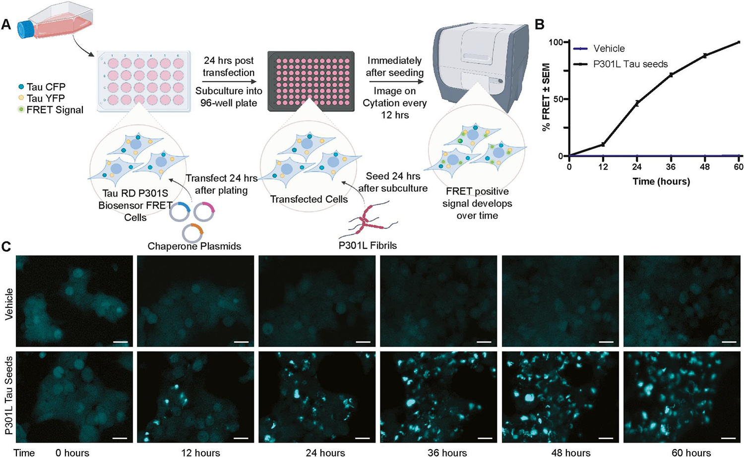
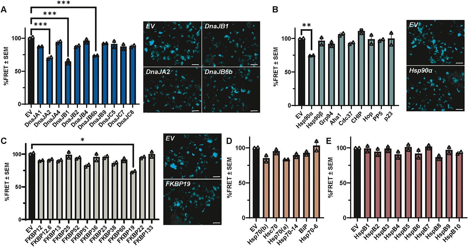
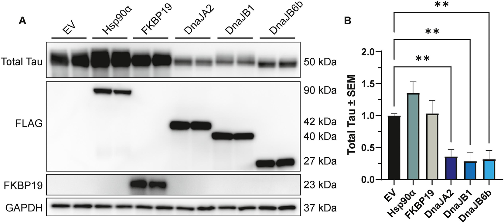
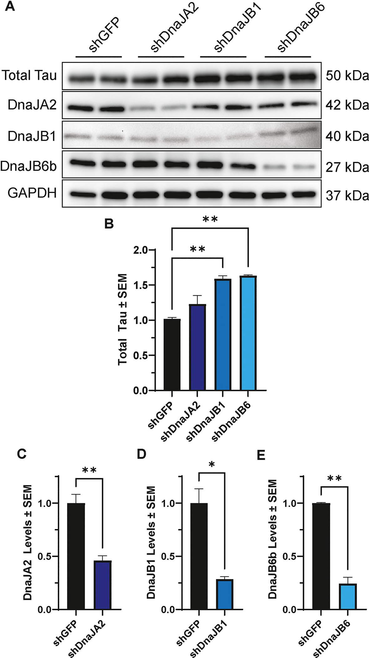
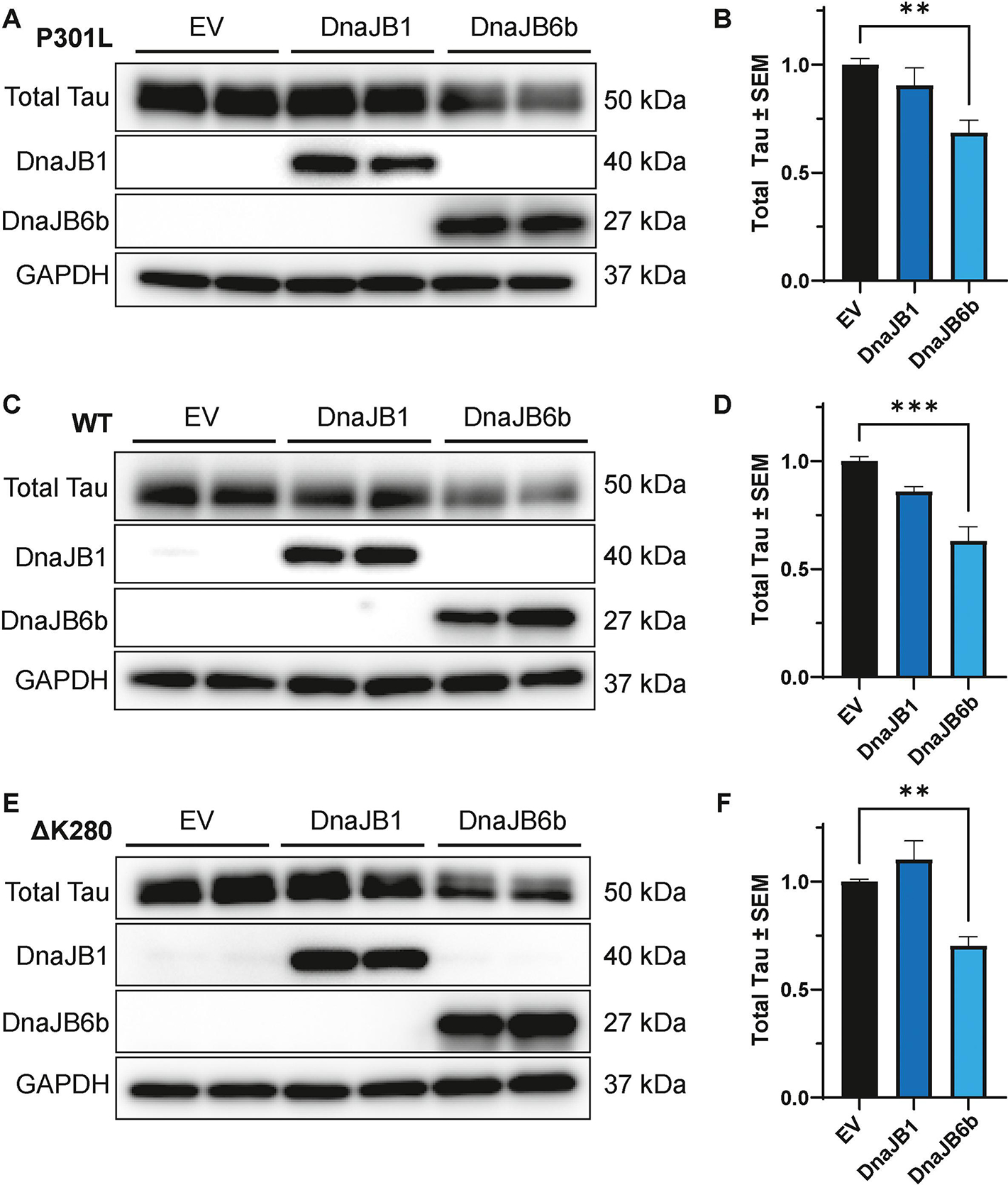
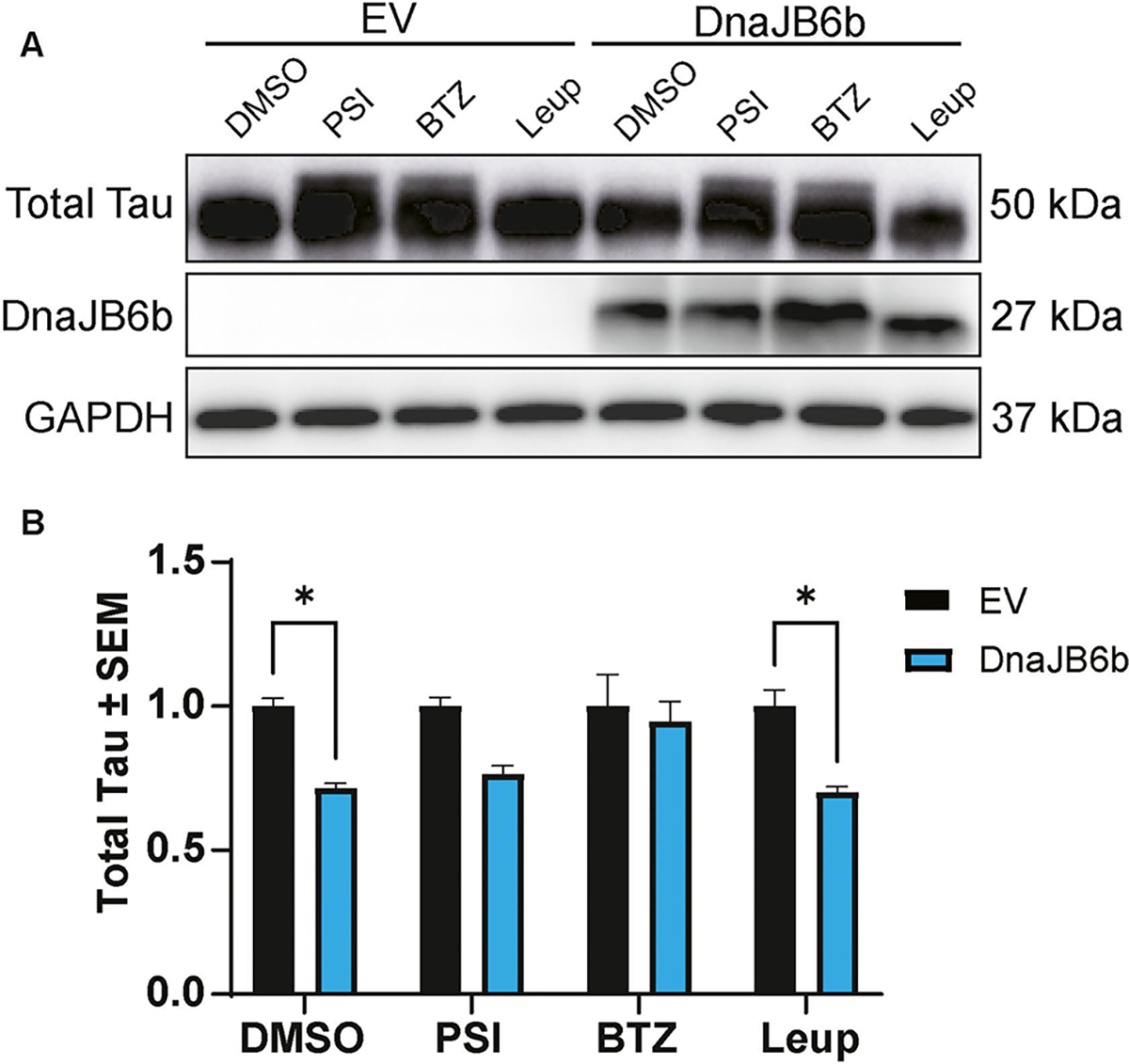
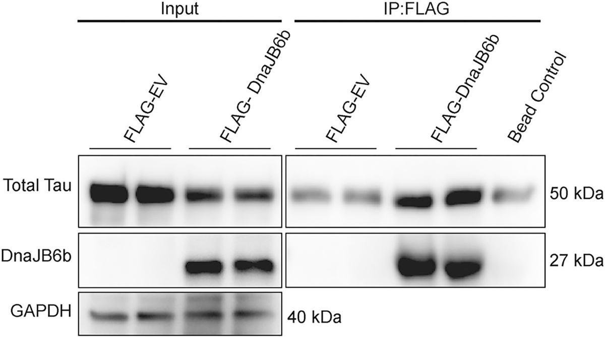
The aberrant accumulation of tau protein is implicated as a pathogenic factor in many neurodegenerative diseases. Tau seeding may underlie its predictable spread in these diseases. Molecular chaperones can modulate tau pathology, but their effects have mainly been studied in isolation. This study employed a semi-high throughput assay to identify molecular chaperones influencing tau seeding using Tau RD P301S FRET Biosensor cells, which express a portion of tau containing the frontotemporal dementia-related P301S tau mutation fused to a FRET biosensor. Approximately fifty chaperones from five major families were screened using live cell imaging to monitor FRET-positive tau seeding. Among the tested chaperones, five exhibited significant effects on tau in the primary screen. Notably, three of these were from the DnaJ family. In subsequent studies, overexpression of DnaJA2, DnaJB1, and DnaJB6b resulted in significant reductions in tau levels. Knockdown experiments by shRNA revealed an inverse correlation between DnaJB1 and DnaJB6b with tau levels. DnaJB6b overexpression, specifically, reduced total tau levels in a cellular model with a pre-existing pool of tau, partially through enhanced proteasomal degradation. Further, DnaJB6b interacted with tau complexes. These findings highlight the potent chaperone activity within the DnaJ family, particularly DnaJB6b, towards tau.

Keywords: DnaJ; Molecular chaperone; Tau.


Conflict of interest statement

Declaration of competing interest The authors declare that they have no known competing financial interests or personal relationships that could have appeared to influence the work reported in this paper.

Figures

Figure 1.
Figure 1.. Schematic of semi-high throughput chaperone screen.
(A) Workflow of the semi-high throughput assay developed to identify molecular chaperones that alter tau seeding. In short, Tau RD P301S FRET Biosensor cells were transfected with chaperone plasmids, subcultured into 96-well plates with 3 wells per chaperone and seeded with sonicated recombinant P301L tau fibrils or vehicle control (100 mM sodium acetate pH 7.0) 48 hours after transfection. (B) Cells were imaged every 12 hours for 60 hours with 4 non-overlapping images per well. Threshold masking was used to measure the FRET intensity within the total cell area at each time point and then normalized to the final time point to calculate %FRET signal. (C) Representative images of FRET signal at each time point are shown from cells treated with vehicle or P301L tau seed. Scale bar = 20 μm. Image created with Biorender.com
Figure 2.
Figure 2.. Discrete molecular chaperones alter tau seeding in vitro.
Using the semi-high throughput Tau RD P301S FRET biosensor assay, 49 molecular chaperones from five chaperone families were screened for their effects on tau seeding. For each chaperone, FRET intensity within the total cell area at 60 hours was normalized to EV control to calculate the relative %FRET signal. Bar graphs from the 60-hour timepoint show the average of 2 independent experiments as %FRET ± S.E.M. for chaperone members of the (A) DnaJ family, (B) Hsp90 and Hsp90 cochaperone families, (C) FKBP family, (D) Hsp70 family, and (E) sHsp family compared to their EV control, respectively. Data analysis was performed by repeated measures ANOVA with a Greenhouse-Geisser correction over the course of the experiment (Fig. S1) across each family of chaperones, except for Hsp90 and Hsp90 cochaperones, which were combined. The 60-hour timepoint from Fig. S1 is displayed here for simplicity. Significance indicated as follows: *p<0.05, **p<0.01, ***p<0.001. Representative images of select chaperones found to be significant in the post hoc analysis are shown. Scale bar = 20 μm.
Figure 3.
Figure 3.. Overexpression of DnaJs significantly affect intracellular tau levels.
(A) Representative western blot images of cell lysates from HEK293T cells co-transfected with P301L tau and Hsp90α, FKBP19, DnaJA2, DnaJB1, DnaJB6b, or EV control for 48 hours, prior to harvesting for western blot analysis. (B) Quantification of total tau relative to GAPDH normalized to EV control is shown from three independent experiments ± S.E.M. A one-way ANOVA with Dunnett’s post hoc test was used to identify significance. Significance indicated as follows: **p<0.01.
Figure 4.
Figure 4.. Knockdown of DnaJB1 or DnaJB6b significantly increases total tau levels.
(A) Representative western blot of cell lysates from HEK293T cells transfected with P301L tau and shDnaJA2, shDnaJB1, shDnaJB6, or shGFP control, as indicated. (B) Quantification of total tau relative to GAPDH normalized to shGFP control is shown from two independent experiments ± S.E.M. A one-way ANOVA with Dunnett’s post hoc test was used to identify significance. Quantification of knockdown was confirmed for (C) DnaJA2, (D) DnaJB1 and (E) DnaJB6b by unpaired t-test. Significance indicated as follows: *p<0.05, **p<0.01.
Figure 5.
Figure 5.. DnaJB6b overexpression mitigates tau levels in iHEK P301L, WT, and ΔK280 tau cells.
Tau expression was induced by tetracycline for 48 hours in iHEK P301L, WT, and ΔK280 tau cells, followed by transfection of DnaJB1, DnaJB6b, or EV for 48 hours, prior to harvesting for western blot analysis. Representative western blot and corresponding quantification of cell lysates from iHEK (A-B) P301L, (C-D) WT and (D-E) ΔK280 tau cells expressing DnaJB1, DnaJB6b, or EV control. Quantification was performed for total tau relative to GAPDH normalized to EV control from two independent experiments ± S.E.M. One-way ANOVA with Dunnett’s post hoc test was used to identify significance. Significance indicated as follows: **p<0.01, ***p<0.001.
Figure 6.
Figure 6.. Inhibition of the proteasome counteracts the reduction of tau by DnaJB6b.
(A) Representative western blot from iHEK P301L tau cells induced by tetracycline for 48 hours, followed by transfection of DnaJB6b or EV. Twenty-four hours after transfection, cells were treated with DMSO control, PSI, Bortezomib (BTZ), or Leupeptin (Leup) for 16 hours prior to harvesting cells for western blot analysis. (B) Quantification of total tau relative to GAPDH normalized to respective EV treatment controls is shown from two independent experiments ± S.E.M. Two-way ANOVA with Šídák’s post hoc test was used to identify significance. Significance indicated as follows: *p<0.05.
Figure 7.
Figure 7.. DnaJB6b complexes with P301L tau.
Tau expression was induced by tetracycline for 48 hours in iHEK P301L tau cells, followed by transfection of FLAG-DnaJB6b, FLAG-EV, or EV for 48 hours, prior to harvesting. DnaJB6b and DnaJB6b interacting proteins were immunoprecipitated using FLAG-tag conjugated beads and analyzed by western blot as indicated. Representative blot represents results from two independent experiments.

References

    1. Narasimhan S, Guo JL, Changolkar L, Stieber A, McBride JD, Silva LV, He Z, Zhang B, Gathagan RJ, Trojanowski JQ, Lee VMY, Pathological Tau Strains from Human Brains Recapitulate the Diversity of Tauopathies in Nontransgenic Mouse Brain, J. Neurosci. 37(47) (2017) 11406–11423. - PMC - PubMed
    1. He Z, McBride JD, Xu H, Changolkar L, Kim S.-j., Zhang B, Narasimhan S, Gibbons GS, Guo JL, Kozak M, Schellenberg GD, Trojanowski JQ, Lee VMY, Transmission of tauopathy strains is independent of their isoform composition, Nature Communications 11(1) (2020) 7. - PMC - PubMed
    1. Lopes S, Vaz-Silva J, Pinto V, Dalla C, Kokras N, Bedenk B, Mack N, Czisch M, Almeida OF, Sousa N, Sotiropoulos I, Tau protein is essential for stress-induced brain pathology, Proc. Natl. Acad. Sci. U. S. A. 113(26) (2016) E3755–63. - PMC - PubMed
    1. Kaufman SK, Sanders DW, Thomas TL, Ruchinskas AJ, Vaquer-Alicea J, Sharma AM, Miller TM, Diamond MI, Tau Prion Strains Dictate Patterns of Cell Pathology, Progression Rate, and Regional Vulnerability In Vivo, Neuron 92(4) (2016) 796–812. - PMC - PubMed
    1. Irwin DJ, Tauopathies as Clinicopathological Entities, Parkinsonism Relat. Disord. 22(0 1) (2016) S29–S33. - PMC - PubMed

MeSH terms

LinkOut - more resources