Review
. 2023 Oct 18;6(1):1054.
doi: 10.1038/s42003-023-05432-7.

Melanopsin-mediated optical entrainment regulates circadian rhythms in vertebrates

Affiliations
Review

Melanopsin-mediated optical entrainment regulates circadian rhythms in vertebrates

Deng Pan et al. Commun Biol. .

Abstract

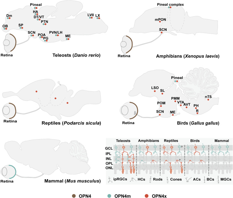
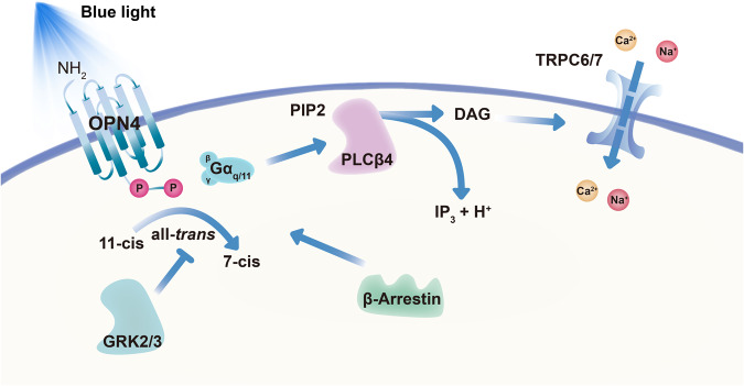
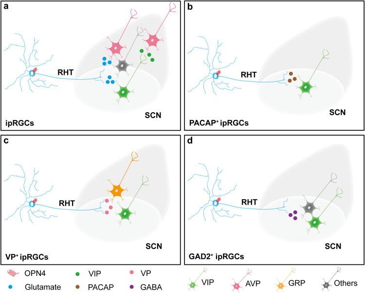
Melanopsin (OPN4) is a light-sensitive protein that plays a vital role in the regulation of circadian rhythms and other nonvisual functions. Current research on OPN4 has focused on mammals; more evidence is needed from non-mammalian vertebrates to fully assess the significance of the non-visual photosensitization of OPN4 for circadian rhythm regulation. There are species differences in the regulatory mechanisms of OPN4 for vertebrate circadian rhythms, which may be due to the differences in the cutting variants, tissue localization, and photosensitive activation pathway of OPN4. We here summarize the distribution of OPN4 in mammals, birds, and teleost fish, and the classical excitation mode for the non-visual photosensitive function of OPN4 in mammals is discussed. In addition, the role of OPN4-expressing cells in regulating circadian rhythm in different vertebrates is highlighted, and the potential rhythmic regulatory effects of various neuropeptides or neurotransmitters expressed in mammalian OPN4-expressing ganglion cells are summarized among them.


Conflict of interest statement

The authors declare no competing interests.

Figures

Fig. 1
Fig. 1. Distribution of OPN4 in teleost fish, amphibian, reptiles, birds, and mammals on generalized sagittal sections.
In teleost fish and birds, two orthologs of OPN4 are distributed in the retina, brain, and pineal gland,,,–. For teleosts, amphibians, and reptiles, splice variants of OPN4 are classified as OPN4m and OPN4x. In the reptile brain, OPN4x expression has been detected in the telencephalon, mesencephalon, and rhombencephalon, but the specific nuclei are still unclear. It is important to note that the available evidence does not determine the cell type of OPN4x in the inner nuclear layer or whether OPN4m is present in RGCs in reptiles. Mammalian OPN4 is mainly expressed in the retina, which integrates more complex photosensitive functions and widely projects to different brain regions through different subtypes of ipRGCs to regulate various physiological functions. Brackets indicate representative species. AC amacrine cell, AVT area ventralis of tsai, BC bipolar cell, Dm the medial zone of the dorsal telencephalic region, DT dorsal thalamus, HA habenula, HC horizontal cell, ipRGC intrinsically photosensitive retinal ganglion cell, LH lateral hypothalamic nucleus, LSO lateral septal organ, LVII facial lobe, LX vagal lobe, ME median eminence, MGC Muller glial cell, mPON magnocellular preoptic nucleus, nTS nucleus tractus solitarius, PG preglomerular area, PH plexus of horsley, PMM nucleus premammillaris, POA preoptic area, POM medial preoptic nucleus, PTN posterior tuberal nucleus, PVN periventricular nucleus, R raphe nucleus, SCN suprachiasmatic nucleus, SL nucleus septalis lateralis, SP subpallium, VT ventral thalamus, VTA ventral tegmental area.
Fig. 2
Fig. 2. Activation and termination of OPN4 in M1-subtype ipRGCs of mammals.
The OPN4-mediated light-sensitive pathways are predominantly triggered by the downstream Gq/11, PLCβ4, and TRPC6/7 cation channels in mammals. Retinaldehyde is covalently bonded to the transmembrane structure in OPN4, and light (especially near 480 nm) can change its conformation from an 11-cis to an all-trans state to a 7-cis state (silent state). It will trigger downstream Gq/11 coupling, causing PLCβ4 to break down PIP2 into DAG and IP3, where DAG activates the opening of the TRPC6/7 cation channels. The activated C-terminus of OPN4 is phosphorylated in response to GRK2/3, resulting in inactivation. This process may also involve β-Arrestin 2. In addition, β-Arrestin 1 leads to the isomer regeneration of OPN4, which serves subsequent light activation. DAG diacylglycerol, Gq/11 G protein subunit alpha q/11, GRK2/3 G protein-coupled receptor kinase 2/3, PLCβ4 phospholipase C-beta 4, PIP2 phosphatidylinositol bisphosphate, IP3 inositol triphosphate, TRPC6/7 transient receptor potential cation channel subfamily C member 6/7.
Fig. 3
Fig. 3. The light entrainment of OPN4 on circadian rhythms may involve multiple neural projection pathways, including neurotransmitters or neuropeptides.
ipRGCs are a class of retinal ganglion cells that express OPN4 (red) and can transmit OPN4-mediated photosensitive signals to the SCN via RHT projections. a Vglut2, but not Vglut1, packages glutamate (solid blue circles) into synaptic vesicles in these axons. These ipRGCs axons mainly make synaptic contacts with VIP neurons (green), AVP (pink), and other light-responsive neurons (gray) in the SCN,. The VIP neurons form part of the SCN core region and may communicate with AVP neurons via VIP receptor type 2. b Some ipRGC axons can also release PACAP (solid brown circles) to regulate VIP neurons via VPAC2 and PAC1 receptors. In addition, some OPN4-expressing ipRGCs also expressed VP (solid pink circles). c The axons of these ipRGCs are glutamatergic and VP-positive, and light stimulation can affect their secretion of VP. VP+ ipRGCs showed synaptic co-localization with GRP (yellow) and VIP neurons, but VP+ ipRGCs were not directly connected to AVP neurons. d Some ipRGCs expressed GAD2 and could transmit GABA (solid purple circles) to the SCN regions. These GABAergic signals can excite or inhibit some SCN neurons, including VIP neurons, and maintain the homeostasis of the central rhythms. AVP arginine vasopressin, GAD2 glutamic acid decarboxylase 2, GABA γ-aminobutyric acid, GRP gastrin releasing peptide, ipRGC intrinsically photosensitive retinal ganglion cell, RHT retinal hypothalamic tract, SCN supra-chiasmatic nucleus, VIP vasoactive intestinal peptide, VP vasopressin, PACAP pituitary adenylate cyclase-activating peptide.

References

    1. Yokoyama S. Evolution of dim-light and color vision pigments. Annu. Rev. Genomics Hum. Genet. 2008;9:259–282. - PubMed
    1. Musilova Z, Salzburger W, Cortesi F. The visual opsin gene repertoires of teleost fishes: evolution, ecology, and function. Annu. Rev. Cell Dev. Biol. 2021;37:441–468. - PubMed
    1. Tu DC, et al. Physiologic diversity and development of intrinsically photosensitive retinal ganglion cells. Neuron. 2005;48:987–999. - PubMed
    1. Douglas RH. The pupillary light responses of animals; a review of their distribution, dynamics, mechanisms and functions. Prog. Retin. Eye Res. 2018;66:17–48. - PubMed
    1. Lucas RJ, et al. Diminished pupillary light reflex at high irradiances in melanopsin-knockout mice. Science. 2003;299:245–247. - PubMed

Publication types