. 2023 Sep 28;12(9):1896-1911.
doi: 10.21037/tlcr-23-118. Epub 2023 Aug 23.

The differential prognostic implications of PD-L1 expression in the outcomes of Filipinos with EGFR-mutant NSCLC treated with tyrosine kinase inhibitors

Affiliations

The differential prognostic implications of PD-L1 expression in the outcomes of Filipinos with EGFR-mutant NSCLC treated with tyrosine kinase inhibitors

Herdee Gloriane C Luna et al. Transl Lung Cancer Res. .

Abstract

Background: The tumor immune microenvironment influences tumor evolution in non-small cell lung cancer (NSCLC). Yet, the prognostic value of programmed death-ligand 1 (PD-L1) in epidermal growth factor receptor (EGFR)-mutant NSCLC remains controversial. Additionally, prognostic studies in Filipinos with EGFR-mutant NSCLC remain unexplored to this day.

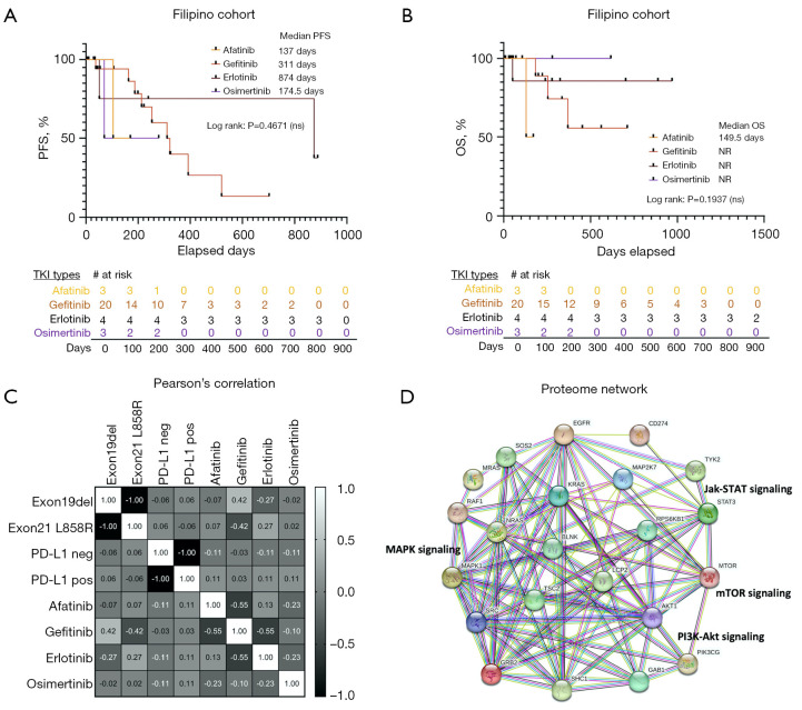
Methods: We prospectively studied the outcomes of EGFR-mutant NSCLC in Filipino cohort, and retrospectively verified the survival trend using The Cancer Genome Atlas (TCGA) cohort. Kaplan-Meier method and generalized linear regression were used to assess survival. Expression and DNA methylation of cluster of differentiation 274 (CD274, gene that codes for PD-L1) were examined from TCGA tumor profiles. Pearson's correlation was used to correlate PD-L1 expression with outcomes associated with occurrence of EGFR mutations, tyrosine kinase inhibitor (TKI) types, and programmed cell death protein 1 (PD-1) expression. Proteome network analysis was used to examine the correlation between drug resistance and PD-L1.

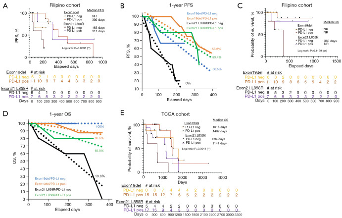
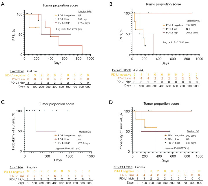
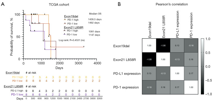
Results: PD-L1 positivity was associated with significantly longer progression-free survival (PFS; P=0.0096) but had a significantly contrasting influence in the overall survival (OS; P=0.0011). PD-L1 positivity (in both protein and RNA) was associated with longer median OS (mOS) in exon21 L858R, whereas, negativity was associated with longer mOS in exon19 deletion (exon19del). Stratification (high, low, negative) of PD-L1 expression lacked significant prognostic value (all P>0.05). PD-L1/CD274 expression (P<0.05) and DNA methylation (P<0.001) vary significantly among NSCLC subtypes and in different disease stages. Erlotinib treatment produced the longest median progression-free survival (mPFS; 874 days) relative to other EGFR-TKIs (137-311 days). PD-L1 lacked a significant correlation with EGFR-TKIs. Consistent with the immune-regulation activities of PD-1, higher expression leads to relatively shorter mOS. PD-1 correlated positively with PD-L1 expression and occurrence of exon21 L858R.

Conclusions: PD-L1 differentially influenced the outcomes of Filipinos with EGFR-mutant NSCLC. NSCLC subtypes, disease stage, and PD-1 expression may impact the collective outcomes associated with PD-L1 and EGFR-sensitizing mutations.

Keywords: EGFR tyrosine kinase inhibitor (EGFR-TKI); Filipinos NSCLC; Programmed death ligand-1 (PD-L1); epidermal growth factor receptor mutations (EGFR mutations); non-small cell lung cancer (NSCLC).


Conflict of interest statement

Conflicts of Interest: All authors have completed the ICMJE uniform disclosure form (available at https://tlcr.amegroups.com/article/view/10.21037/tlcr-23-118/coif). The authors have no conflicts of interest to declare.

Figures

Figure 1
Figure 1
The influence of PD-L1 positivity and negativity in the survival of EGFR-mutant NSCLC with or without PD-L1 expression. The Kaplan-Meier method was used to analyze the median PFS and median OS. P values of the curves were determined using the log-rank test. A GLR model was used to evaluate 1-year survival rates. (A) KM curves showing the probability of PFS, and (B) the predicted 1-year PFS rates among Filipinos with EGFR-mutant NSCLC. (C) KM curves showing the probability of survival, and (D) the predicted 1-year OS rates among Filipinos with EGFR-mutant NSCLC. (E) KM curve showing the probability of survival among TCGA cohort. **, P<0.01; ns, not significant. In GLR, the survival proportions were plotted in continuous line and the predicted survival rates were plotted in broke line. PD-L1, programmed death-ligand 1; neg, negative; pos, positive; EGFR, epidermal growth factor receptor; NSCLC, non-small cell lung cancer; PFS, progression-free survival; OS, overall survival; KM, Kaplan-Meier; TCGA, The Cancer Genome Atlas; GLR, generalized linear regression.
Figure 2
Figure 2
The influence of PD-L1 protein expression levels (negative, low, high) in the survival of Filipinos with EGFR-mutant NSCLC. Protein expression levels were stratified by TPS using IHC. The Kaplan-Meier method was used to analyze the median PFS of patients with (A) exon19del and (B) exon21 L858R and the median OS of patients with (C) exon19del and (D) exon21 L858R. The P values of the curves were determined using the log-rank test. ns, not significant. The median OS and median PFS of certain patient groups that had no 50% survival reduction throughout the observation period were considered NR. PD-L1, programmed death-ligand 1; EGFR, epidermal growth factor receptor; NSCLC, non-small cell lung cancer; TPS, tumor proportion scoring; IHC, immunohistochemistry; PFS, progression-free survival; OS, overall survival; NR, not reached.
Figure 3
Figure 3
The influence of PD-L1 RNA expression levels (negative, low, high) in the survival of TCGA cohort with EGFR-mutant NSCLC. RNA expression levels were stratified using the mean cut off value of PD-L1 RNA-seq abundance reads in FPKM. The Kaplan-Meier method was used to analyze the median OS of patients with (A) exon19del and (B) exon21 L858R. The P values of the curves were determined using the log-rank test. ns, not significant. (C) Violin plot of PD-L1 expression expressed as TPM in two major NSCLC histology types, LUAD and LUSC, in various disease stages (I–IV). The expression levels were analyzed from NSCLC patients in the TCGA cohort using GEPIA web tool. (D) Scatter plot comparing the RNA expression levels (in TPM) between tumors and normal tissue. Differences in the expression were evaluated using one-way ANOVA through the GEPIA web tool. *, P<0.05. (E) CD274 (also known as PD-L1) gene methylation profiles of NSCLC patients from TCGA cohort analyzed using the aggregated methylation mean Beta value of all five CpG probes within the promoter region of CD274. Comparison of methylation profiles were assessed using paired sample t-test. ***, P<0.001; ns, not significant. (F) Pearson’s correlation between CD274 DNA methylation and gene expression from TCGA cohort. The Pearson’s coefficient (R) and P values (p) were measured in each cytosine-guanine (cg) sites that corresponded to a specific CpG probe. Methylation analysis were performed using SMART web interface. PD-L1, programmed death-ligand 1; neg, negative; TCGA, The Cancer Genome Atlas; EGFR, epidermal growth factor receptor; NSCLC, non-small cell lung cancer; FPKM, fragments per kilobase of exon per million reads mapped; OS, overall survival; TPM, transcripts per million; LUAD, lung adenocarcinoma; LUSC, lung squamous cell carcinoma; GEPIA, Gene Expression Profiling Interactive Analysis; ANOVA, analysis of variance; CD274, cluster of differentiation 274; SMART, Shiny Methylation Analysis Resource Tool.
Figure 4
Figure 4
Survival of Filipinos with EGFR-mutant NSCLC treated with different TKIs and correlation with PD-L1 expression. The Kaplan-Meier method was used to analyze the (A) median PFS and (B) median OS of Filipinos with EGFR-mutant NSCLC. The P values of the curves were determined using the log-rank test. ns, not significant. (C) Pearson’s correlation of EGFR-TKIs with EGFR mutations and PD-L1 expression. The heatmap score (range, −1 to 1) represents the Pearson’s coefficient. (D) Proteome network of proteins associated with signaling pathways involved in EGFR-TKI resistance and PD-L1 expression. The protein interaction network was analyzed using STRING interaction network. EGFR, epidermal growth factor receptor; NSCLC, non-small cell lung cancer; TKIs, tyrosine kinase inhibitors; PD-L1, programmed death-ligand 1; PFS, progression-free survival; OS, overall survival.
Figure 5
Figure 5
The influence of PD-1 RNA expression levels (low and high) in the survival of TCGA cohort with PD-L1 positive EGFR-mutant NSCLC. PD-1 RNA expression levels were stratified using the mean cut off value of PD-1 RNA-seq abundance reads in FPKM. (A) The Kaplan-Meier method was used to analyze the median OS of patients with exon19del and exon21 L858R mutants positive for PD-L1. The P values of the curves were determined using the log-rank test. ns, not significant. (B) Pearson’s correlation of PD-1 with the occurrence of certain EGFR-sensitizing mutation, and PD-L1 expression. The heatmap score (range, −1 to 1) represents the Pearson’s coefficient. PD-1, programmed cell death protein 1; TCGA, The Cancer Genome Atlas; PD-L1, programmed death-ligand 1; EGFR, epidermal growth factor receptor; NSCLC, non-small cell lung cancer; FPKM, fragments per kilobase of exon per million reads mapped; OS, overall survival.

References

    1. Sung H, Ferlay J, Siegel RL, et al. Global Cancer Statistics 2020: GLOBOCAN Estimates of Incidence and Mortality Worldwide for 36 Cancers in 185 Countries. CA Cancer J Clin 2021;71:209-49. 10.3322/caac.21660 - DOI - PubMed
    1. WHO-GLOBOCAN. World Health Organization International Agency for Re-search on Cancer. 2020 Available online: https://gco.iarc.fr/today/data/factsheets/cancers/39-All-cancers-fact-sh... (accessed July 26, 2022).
    1. Mirhadi S, Tam S, Li Q, et al. Integrative analysis of non-small cell lung cancer patient-derived xenografts identifies distinct proteotypes associated with patient outcomes. Nat Commun 2022;13:1811. 10.1038/s41467-022-29444-9 - DOI - PMC - PubMed
    1. Shi Z, Zheng X, Shi R, et al. Radiological and Clinical Features associated with Epidermal Growth Factor Receptor Mutation Status of Exon 19 and 21 in Lung Adenocarcinoma. Sci Rep 2017;7:364. 10.1038/s41598-017-00511-2 - DOI - PMC - PubMed
    1. Blandin Knight S, Crosbie PA, Balata H, et al. Progress and prospects of early detection in lung cancer. Open Biol 2017;7:170070. 10.1098/rsob.170070 - DOI - PMC - PubMed