Case Reports
. 2023 Sep 27;19(5):91.
doi: 10.3892/mco.2023.2687. eCollection 2023 Nov.

A rare intrahepatic splenosis mimicking hepatocellular carcinoma: A case report

Affiliations
Case Reports

A rare intrahepatic splenosis mimicking hepatocellular carcinoma: A case report

Issei Umeda et al. Mol Clin Oncol. .

Abstract

Intrahepatic splenosis (IHS) is a rare disease that is considered to result from heterotopic autotransplantation or implantation of splenic tissue after splenic trauma or surgery. A 46-year-old man with a treatment history of a left lateral liver segmentectomy and splenectomy for a road traffic injury 30 years earlier presented to Sakai City Medical Center (Sakai, Japan) with acute abdominal pain in November 2019. Physical examination showed no significant signs, and serum data were normal. Computed tomography revealed a hypodense mass measuring 2.5x1.7 cm in segment 7 of the liver. Gadoxetic acid-enhanced magnetic resonance imaging showed early enhancement in the arterial phase and washout in the delayed phase. Therefore, laparoscopic surgery was performed with a preoperative diagnosis of hepatocellular carcinoma. Pathological examination of the tumor showed IHS. The postoperative course was uneventful, and the patient developed no new abnormal region in the liver during 2 years of follow-up. The present study presented a case of IHS assumed to be hepatocellular carcinoma. IHS should be considered as a differential diagnosis of a liver mass detected years after splenic trauma or surgery, even in cases with imaging patterns suggesting malignancy.

Keywords: intrahepatic splenosis; liver tumor; splenectomy; trauma.


Conflict of interest statement

The authors declare that they have no competing interests.

Figures

Figure 1
Figure 1
Abdominal ultrasonography and abdominal plain-computed tomography imaging of the IHS. (A) An abdominal ultrasonography revealed a 2.4x1.4 cm isoechoic lesion (arrows) with a hypoechoic zone at the margins in segment 7 of the liver. (B) An abdominal plane-computed tomography scan revealed a hypodense mass (arrows) measuring 2.5x1.7 cm in segment 7 of the liver.
Figure 2
Figure 2
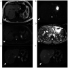
Magnetic resonance imaging of the IHS (arrows). The IHS was detected with (A) a homogeneous hypointensity in T1-weighted images, (B) slight hyperintensity in T2-weighted images. (C) Lesion showed a heterogeneous hyperintensity in diffusion-weighted images and (D) signal reduction in apparent diffusion coefficient. After the injection of gadoxetic acid, (E) the lesion appeared heterogeneously hyperintense during the early phase and (F) relatively hypointense during the delayed phase. HIS, intrahepatic splenosis.
Figure 3
Figure 3
During the surgery. (A) Tumor was buried in the liver and adherent to the diaphragm. It had a capsule and was soft texture. (B) Drainage vessels of the tumor were recognized from the inferior phrenic artery and vein.
Figure 4
Figure 4
Histopathologic findings. (A) Resected specimen was dark red and 2.7 cm in size. (B) Hematoxylin and eosin staining (magnification, x20). The resected specimen was composed of white and red pulp surrounded by a fibrous capsule (W: white pulp, R: red pulp). Scale bar, 200 µM.

References

    1. Gandhi D, Sharma P, Garg G, Songmen S, Solanki S, Singh T. Intrahepatic splenosis demonstrated by diffusion weighted MRI with histologic confirmation. Radiol Case Rep. 2020;15:602–606. doi: 10.1016/j.radcr.2020.02.022. - DOI - PMC - PubMed
    1. Menth M, Herrmann K, Haug A, Raziorrouh B, Zachoval R, Jung CM, Otto C. Intra-hepatic splenosis as an unexpected cause of a focal liver lesion in a patient with hepatitis C and liver cirrhosis: A case report. Cases J. 2009;2(8335) doi: 10.4076/1757-1626-2-8335. - DOI - PMC - PubMed
    1. Toh WS, Chan KS, Ding CSL, Tan CH, Shelat VG. Intrahepatic splenosis: A world review. Clin Exp Hepatol. 2020;6:185–198. doi: 10.5114/ceh.2020.99509. - DOI - PMC - PubMed
    1. Rickert CH, Maasjosthusmann U, Probst-Cousin S, August C, Gullotta F. A unique case of cerebral spleen. Am J Surg Pathol. 1998;22:894–896. doi: 10.1097/00000478-199807000-00011. - DOI - PubMed
    1. Baldari G, Sindoni A, Belletti A, Baldari S, Ruffini L. (99m)Tc disphosphonate uptake due to splenosis. Incidental finding 60 years after splenectomy. Clin Nucl Med. 2015;40:533–535. doi: 10.1097/RLU.0000000000000752. - DOI - PubMed

Publication types