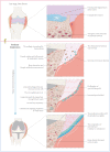
Nano wear particles and the periprosthetic microenvironment in aseptic loosening induced osteolysis following joint arthroplasty
- PMID: 37854857
- PMCID: PMC10579613
- DOI: 10.3389/fcimb.2023.1275086
Nano wear particles and the periprosthetic microenvironment in aseptic loosening induced osteolysis following joint arthroplasty
Abstract
Joint arthroplasty is an option for end-stage septic arthritis due to joint infection after effective control of infection. However, complications such as osteolysis and aseptic loosening can arise afterwards due to wear and tear caused by high joint activity after surgery, necessitating joint revision. Some studies on tissue pathology after prosthesis implantation have identified various cell populations involved in the process. However, these studies have often overlooked the complexity of the altered periprosthetic microenvironment, especially the role of nano wear particles in the etiology of osteolysis and aseptic loosening. To address this gap, we propose the concept of the "prosthetic microenvironment". In this perspective, we first summarize the histological changes in the periprosthetic tissue from prosthetic implantation to aseptic loosening, then analyze the cellular components in the periprosthetic microenvironment post prosthetic implantation. We further elucidate the interactions among cells within periprosthetic tissues, and display the impact of wear particles on the disturbed periprosthetic microenvironments. Moreover, we explore the origins of disease states arising from imbalances in the homeostasis of the periprosthetic microenvironment. The aim of this review is to summarize the role of relevant factors in the microenvironment of the periprosthetic tissues, in an attempt to contribute to the development of innovative treatments to manage this common complication of joint replacement surgery.
Keywords: aseptic loosening; homeostatic imbalance; joint arthroplasty; joint prothesis; microenvironment.
Copyright © 2023 Xie, Peng, Fu, Jin, Wang, Li, Zheng, Lyu, Deng and Ma.
Conflict of interest statement
The authors declare that the research was conducted in the absence of any commercial or financial relationships that could be construed as a potential conflict of interest.
Figures


Similar articles
-
[VASCULARITY STUDY ON PERIPROSTHETIC TISSUES AROUND ASEPTIC LOOSENING AFTER TOTAL HIP ARTHROPLASTY].Zhongguo Xiu Fu Chong Jian Wai Ke Za Zhi. 2016 Jan;30(1):39-43. Zhongguo Xiu Fu Chong Jian Wai Ke Za Zhi. 2016. PMID: 27062844 Chinese.
-
Value of multidetector computed tomography for the differentiation of delayed aseptic and septic complications after total hip arthroplasty.Skeletal Radiol. 2020 Jun;49(6):893-902. doi: 10.1007/s00256-019-03355-1. Epub 2020 Jan 3. Skeletal Radiol. 2020. PMID: 31900512
-
[Research progress on wear particles and periprosthetic osteolysis after artificial joint replacement].Zhongguo Gu Shang. 2016 Oct 25;29(10):968-972. doi: 10.3969/j.issn.1003-0034.2016.10.017. Zhongguo Gu Shang. 2016. PMID: 29285918 Review. Chinese.
-
Osteolysis around total knee arthroplasty: a review of pathogenetic mechanisms.Acta Biomater. 2013 Sep;9(9):8046-58. doi: 10.1016/j.actbio.2013.05.005. Epub 2013 May 10. Acta Biomater. 2013. PMID: 23669623 Free PMC article. Review.
-
Blockade of XCL1/Lymphotactin Ameliorates Severity of Periprosthetic Osteolysis Triggered by Polyethylene-Particles.Front Immunol. 2020 Aug 4;11:1720. doi: 10.3389/fimmu.2020.01720. eCollection 2020. Front Immunol. 2020. PMID: 32849609 Free PMC article.
Cited by
-
Fused exosomal targeted therapy in periprosthetic osteolysis through regulation of bone metabolic homeostasis.Bioact Mater. 2025 Apr 8;50:171-188. doi: 10.1016/j.bioactmat.2025.04.006. eCollection 2025 Aug. Bioact Mater. 2025. PMID: 40248188 Free PMC article.
-
Prosthesis survival situation and complications following total hip arthroplasty in hemophilic patients: a systematic review.BMC Musculoskelet Disord. 2025 Jul 9;26(1):672. doi: 10.1186/s12891-025-08919-y. BMC Musculoskelet Disord. 2025. PMID: 40634963 Free PMC article.
-
Impact of fixation method on femoral bone loss: a retrospective evaluation of stem loosening in first-time revision total hip arthroplasty among two hundred and fifty five patients.Int Orthop. 2024 Sep;48(9):2339-2350. doi: 10.1007/s00264-024-06230-4. Epub 2024 Jun 1. Int Orthop. 2024. PMID: 38822836 Free PMC article.
-
Leptin potentiates bone loss at skeletal sites distant from focal inflammation in female ob/ob mice.J Endocrinol. 2025 Feb 17;264(3):e240324. doi: 10.1530/JOE-24-0324. Print 2025 Mar 1. J Endocrinol. 2025. PMID: 39882726
-
Diagnosis of periprosthetic loosening of total hip and knee arthroplasty using 68Gallium-Zoledronate PET/CT.Arch Orthop Trauma Surg. 2024 Nov;144(11):4775-4781. doi: 10.1007/s00402-024-05562-5. Epub 2024 Oct 1. Arch Orthop Trauma Surg. 2024. PMID: 39352482 Free PMC article.
References
-
- Athanasou N. A. (2002). The pathology of joint replacement. Curr. Diagn. Pathol. 8, 26–32. doi: 10.1054/cdip.2001.0092 - DOI
Publication types
MeSH terms
LinkOut - more resources
Full Text Sources
Miscellaneous

