. 2023 Nov 20;260(1):e230157.
doi: 10.1530/JOE-23-0157. Print 2024 Jan 1.

Diffuse traumatic brain injury substantially alters plasma growth hormone in the juvenile rat

Affiliations

Diffuse traumatic brain injury substantially alters plasma growth hormone in the juvenile rat

J Bryce Ortiz et al. J Endocrinol. .

Abstract

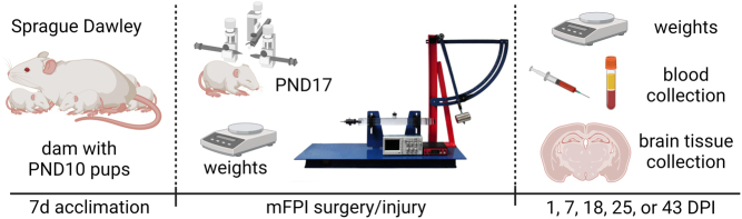
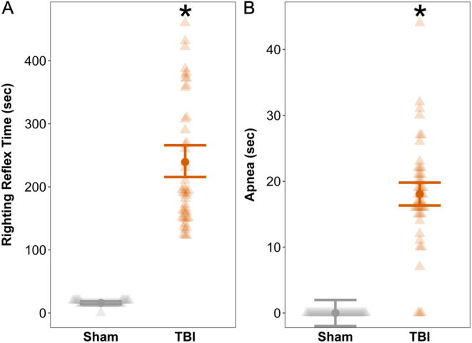
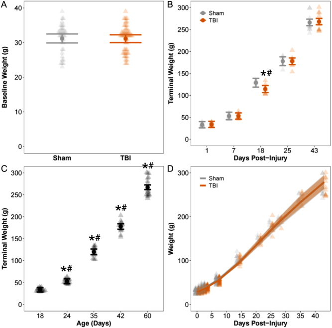
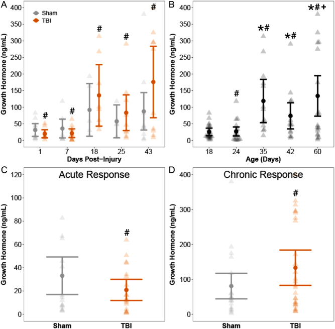
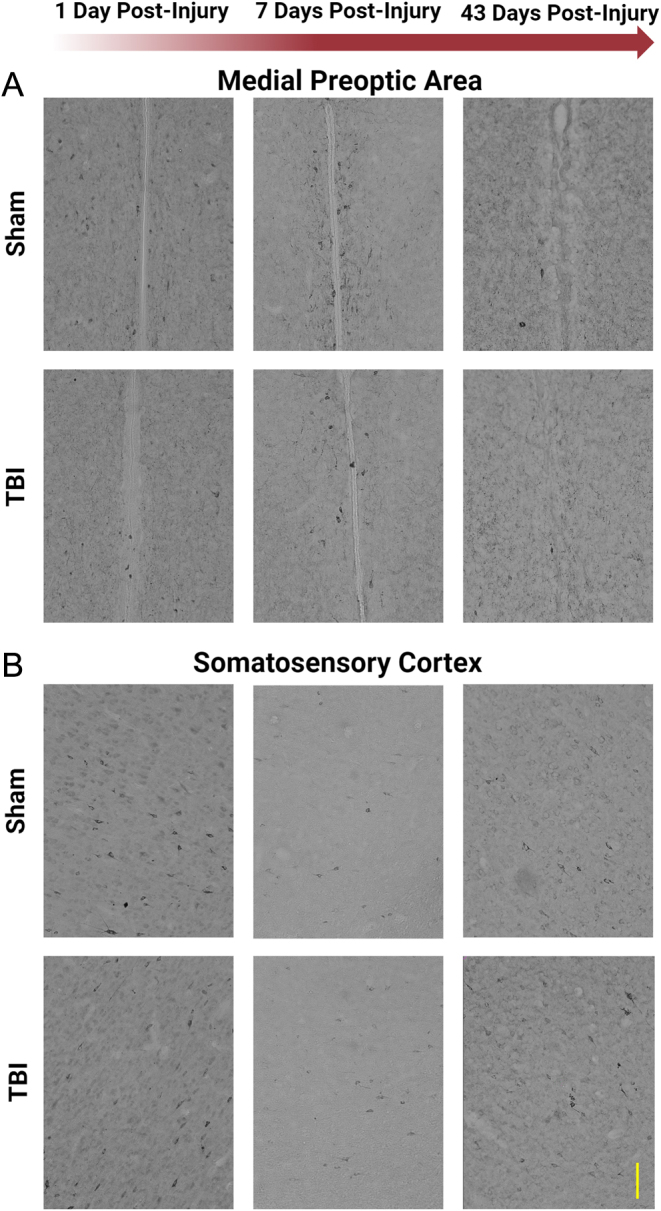
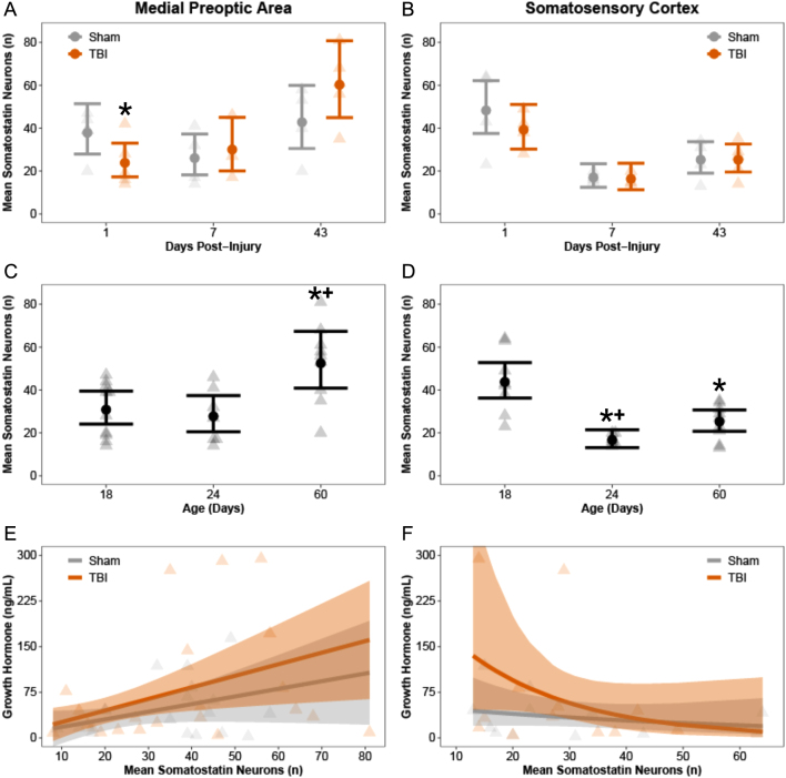
Traumatic brain injury (TBI) can damage the hypothalamus and cause improper activation of the growth hormone (GH) axis, leading to growth hormone deficiency (GHD). GHD is one of the most prevalent endocrinopathies following TBI in adults; however, the extent to which GHD affects juveniles remains understudied. We used postnatal day 17 rats (n = 83), which model the late infantile/toddler period, and assessed body weights, GH levels, and number of hypothalamic somatostatin neurons at acute (1, 7 days post injury (DPI)) and chronic (18, 25, 43 DPI) time points. We hypothesized that diffuse TBI would alter circulating GH levels because of damage to the hypothalamus, specifically somatostatin neurons. Data were analyzed with generalized linear and mixed effects models with fixed effects interactions between the injury and time. Despite similar growth rates over time with age, TBI rats weighed less than shams at 18 DPI (postnatal day 35; P = 0.03, standardized effect size [d] = 1.24), which is around the onset of puberty. Compared to shams, GH levels were lower in the TBI group during the acute period (P = 0.196; d = 12.3) but higher in the TBI group during the chronic period (P = 0.10; d = 52.1). Although not statistically significant, TBI-induced differences in GH had large standardized effect sizes, indicating biological significance. The mean number of hypothalamic somatostatin neurons (an inhibitor of GH) positively predicted GH levels in the hypothalamus but did not predict GH levels in the somatosensory cortex. Understanding TBI-induced alterations in the GH axis may identify therapeutic targets to improve the quality of life of pediatric survivors of TBI.

Keywords: concussion; development; growth hormone deficiency; pediatric; puberty; somatostatin.


Conflict of interest statement

The authors declare that they have no conflict of interest that could be perceived as prejudicing the impartiality of the research reported.

Figures

Figure 1
Figure 1
Study design. Postnatal day (PND) 10 rats were received with their dam and acclimated for 7 days. PND 17 rats were subjected to midline fluid percussion injury (mFPI) or control sham surgery. Body weights were taken at surgery, weekly post injury, and at the time of tissue collection. Blood and tissue samples were collected at 1, 7, 18, 25, or 43 days post injury (DPI).
Figure 2
Figure 2
Diffuse TBI suppressed acute neurological reflexes. Righting reflex time, time from the initial impact until the rat spontaneously righted itself from a supine position, and apnea time were measured as indicators of injury severity. Results shown are predicted marginal effects point estimates and 95% confidence intervals from generalized linear models; background triangles denote observed data points. Rats that were subjected to TBI had longer (A) righting reflex times and (B) apnea times compared to uninjured shams. * indicates statistically significant difference (P < 0.05).
Figure 3
Figure 3
Brain-injured rats had lower body weights at 18 days post injury (DPI) compared to sham. Results shown as predicted marginal effects point estimates and 95% confidence intervals; background triangles denote observed data points. (A) There were no statistically significant differences in baseline body weights taken prior to surgery/injury. (B) There were no brain injury-induced differences in terminal body weights of rats at 1, 7, 25, or 43 days post injury (DPI). * indicates P < 0.05 compared to sham. However, brain-injured rats weighed significantly less than uninjured shams at 18 DPI. (C) Terminal body weights increased over time with animal age. Compared to postnatal day 18, rats had higher body weights at postnatal days 24, 35, 42, and 60. * indicates P < 0.05 compared to postnatal day 18. (D) Terminal body weight increased nonlinearly over time with animal age at a mean rate of λ = 1.19, or 19% per day (95% CI = 17–22%). * indicates statistically significant difference (P < 0.05); # indicates biologically significant difference (d ≥ 1.0).
Figure 4
Figure 4
Growth hormone (GH) levels increased over time with animal age and were altered by TBI. Results shown as predicted marginal effects point estimates and 95% confidence intervals; background triangles denote observed data points. (A) There were no differences in plasma GH levels between sham and TBI rats at any terminal time point. (B) Compared to postnatal day 18, GH levels were comparable at postnatal day 24 but were significantly elevated at postnatal days 35, 42, and 60. GH levels at postnatal day 60 were also elevated compared to postnatal day 24. * indicates P < 0.05 compared to postnatal day 18; + indicates P < 0.05 compared to postnatal day 24. (C–D) We further assessed TBI-induced changes during the acute period (1 and 7 DPI) and the chronic period (18, 25, and 43 DPI). Compared to shams, GH levels were lower in the TBI group during the acute period, but higher in the TBI group during the chronic period. Those differences were not statistically significant but had very large effect sizes indicating biological significance. * indicates statistically significant difference (P < 0.05); # indicates biologically significant difference (d ≥ 1.0).
Figure 5
Figure 5
Representative images of somatostatin neurons in the medial preoptic area and somatosensory cortex. Somatostatin-stained neurons in the (A) medial preoptic area and (B) the somatosensory cortex from rats subjected to diffuse TBI or a control sham surgery. Representative images were taken at 1, 7, and 43 days post injury. Scale bar = 100 µm.
Figure 6
Figure 6
Mean number of somatostatin neurons predicted growth hormone (GH) levels. Changes in somatostatin neurons over time and whether neurons predicted GH levels were assessed in the (A, C, E) medial preoptic area and (B, D, F) somatosensory cortex. In both the (A) medial preoptic area and (B) somatosensory cortex, TBI rats had less somatostatin neurons at 1 day post injury (DPI) compared to shams, whereas no differences existed at 7 or 43 DPI. (C) The mean number of somatostatin neurons increased over time with animal age in the medial preoptic area, whereas (D) the mean number of somatostatin neurons decreased with age in the somatosensory cortex. (E) Irrespective of sham or TBI (i.e. treatment groups combined), the number of neurons was a positive predictor of GH in the medial preoptic area; for every additional neuron, an average of 1.27 ng/mL increase in GH was predicted. (F) In contrast, irrespective of sham or TBI, the number of neurons was not a significant predictor of GH in the somatosensory cortex. * indicates P < 0.05 compared to sham; + indicates P < 0.05 compared to postnatal day 18.

Similar articles

References

    1. Aberg ND Johansson I Aberg MA Lind J Johansson UE Cooper-Kuhn CM Kuhn HG & Isgaard J. 2009Peripheral administration of GH induces cell proliferation in the brain of adult hypophysectomized rats. Journal of Endocrinology 201141–150. (10.1677/JOE-08-0495) - DOI - PubMed
    1. Acerini CL Tasker RC Bellone S Bona G Thompson CJ & Savage MO. 2006Hypopituitarism in childhood and adolescence following traumatic brain injury: the case for prospective endocrine investigation. European Journal of Endocrinology 155663–669. (10.1530/eje.1.02284) - DOI - PubMed
    1. Agha A Phillips J & Thompson CJ. 2007Hypopituitarism following traumatic brain injury (TBI). British Journal of Neurosurgery 21210–216. (10.1080/02688690701253331) - DOI - PubMed
    1. Aimaretti G, Ambrosio MR, Benvenga S, Borretta G, De Marinis L, De Menis E, Di Somma C, Faustini-Fustini M, Grottoli S, Gasco V, et al.2004Hypopituitarism and growth hormone deficiency (GHD) after traumatic brain injury (TBI). Growth Hormone and IGF Research 14(Supplement A) S114–S117. (10.1016/j.ghir.2004.03.025) - DOI - PubMed
    1. Aimaretti G, Ambrosio MR, Di Somma C, Gasperi M, Cannavo S, Scaroni C, De Marinis L, Baldelli R, Bona G, Giordano G, et al.2005Hypopituitarism induced by traumatic brain injury in the transition phase. Journal of Endocrinological Investigation 28984–989. (10.1007/BF03345336) - DOI - PubMed

Publication types