. 2023 Nov 14;30(12):e230062.
doi: 10.1530/ERC-23-0062. Print 2023 Dec 1.

Mammary gland development and EDC-driven cancer susceptibility in mesenchymal ERα-knockout mice

Affiliations

Mammary gland development and EDC-driven cancer susceptibility in mesenchymal ERα-knockout mice

Clarissa Wormsbaecher et al. Endocr Relat Cancer. .

Abstract

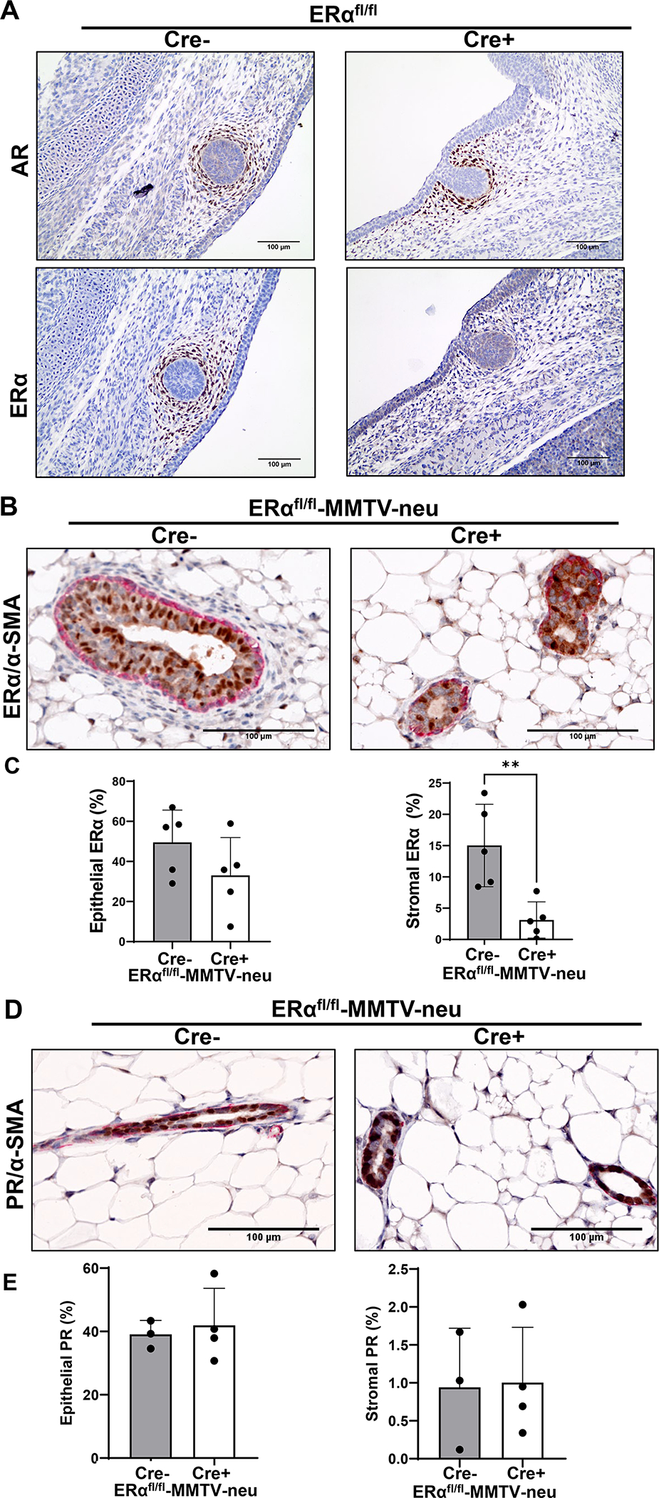
Development of the mammary gland requires both proper hormone signaling and cross talk between the stroma and epithelium. While estrogen receptor (ERα) expression in the epithelium is essential for normal gland development, the role of this receptor in the stroma is less clear. Moreover, several lines of evidence suggest that mouse phenotypes of in utero exposure to endocrine disruption act through mesenchymal ERα in the developing fetus. We utilized a Twist2-cre mouse line to knock out mesenchymal ERα. Herein, we assessed mammary gland development in the context of mesenchymal ERα deletion. We also tested the effect of in utero bisphenol A (BPA) exposure to alter the tumor susceptibility in the mouse mammary tumor virus-neu (MMTV-neu) breast cancer mouse model. Mesenchymal ERα deletion resulted in altered reproductive tract development and atypical cytology associated with estrous cycling. The mammary gland demonstrated mature epithelial extension unlike complete ERα-knockout mice, but ductal extension was delayed and reduced compared to ERα-competent mice. Using the MMTV-Neu cancer susceptibility model, ERα-intact mice exposed to BPA had reduced tumor-free survival and overall survival compared to BPA-exposed mice having mesenchymal ERα deletion. This difference is specific for BPA exposure as vehicle-treated animals had no difference in tumor development between mice expressing and not expressing mesenchymal ERα. These data demonstrate that mesenchymal ERα expression is not required for ductal extension, nor does it influence cancer risk in this mouse model but does influence the cancer incidence associated with in utero BPA exposure.

Keywords: MMTV-Neu; endocrine disruptors; estrogen receptor; mammary gland.


Conflict of interest statement

Declaration of Interest

There are no conflicts of interest that would affect the impartiality of this research.

Figures

Figure 1
Figure 1. Efficient knockout of mesenchymal ERα in ERαΔM mice.
A To validate mesenchymal ERα knockout in ERαΔM mice, mammary buds were first identified at embryonic day 13.5 (E13.5) by IHC for the androgen receptor (AR) (top). Sequential sections were stained for estrogen receptor alpha (ERα) (bottom). Images taken at 20X magnification. B Representative images of IHC of mammary glands from control and mesenchymal ERα knockout ERαΔM-MMTV-neu mice from 12–14 week old mice, dual stained with ERα (brown)/α-SMA (red). Images are at 20X magnification. C Graphs show quantification of percentage of ERα positivity in the epithelial compartment (left) and the stromal compartment (right). Graphed as mean ± standard deviation, each dot represents one mouse, five mice from four litters were assessed in the control group and five mice from five litters were assessed in the ERαΔM-MMTV-neu group. Significance determined by a t-test, p=0.0061. D Representative images of IHC of mammary glands from control and mesenchymal ERα knockout ERαΔM-MMTV-neu mice from 12–14 week old mice, dual stained with PR/α-SMA, focused on ductal areas. Images are at 20X magnification. E Graphs show quantification of percentage of PR positivity in the epithelial compartment (left) and the stromal compartment (right). Graphed as mean ± standard deviation, each dot represents one mouse, three mice from three litters were assessed in the control group and four mice from three litters were assessed in the ERαΔM-MMTV-neu group.
Figure 2
Figure 2. Mesenchymal ERα knockout mice have altered response to estrous cycling.
A Vaginal cytology samples in each stage of estrous cycling from control and ERαΔM-MMTV-neu mice. Images taken at 10X magnification. B Reoccurring vaginal cytology sample in ERαΔM-MMTV-neu mice. Image taken at 10X magnification. Filled-in arrow heads indicating squamous epithelial cells and open arrow heads indicating leukocytes. C Images of reproductive tract from control and ERαΔM-MMTV-neu mice collected in the estrus stage (left) and in the diestrus stage (right) as measured by vaginal cytology. D H&E staining of vaginal sections collected from mice in the estrus stage as measured by vaginal cytology. Images taken at 20X magnification. E Estradiol (left) and progesterone (right) serum levels of control mice collected in both estrus (filled in circles) and diestrus stages (open circles), compared to serum levels from ERαΔM-MMTV-neu mice. ERαΔM-MMTV-neu serum collected from mice in stage closest to estrus per vaginal cytology in closed circles and collected in stage closest identified to diestrus per vaginal cytology in open circles. Graphed as mean ± standard deviation, each dot represents one mouse.
Figure 3
Figure 3. ERαΔM-MMTV-neu have delayed epithelial elongation at 4.7 weeks and reduced epithelial area in adulthood.
A Mammary glands were collected in control and ERαΔM-MMTV-neu mice at 4.7 weeks, whole mounted, and carmine-aluminum stained to look at epithelial elongation. Images taken at 1X magnification. B Epithelial elongation was measured in 4.7 week whole mounts by measuring from the lymph node, plus or minus to the most distal point of epithelial elongation in stained mammary whole mounts with quantification graphed to the right. Graphed as mean ± standard deviation, each dot represents one mouse, and mice from six separate litters were analyzed. Significance determined by a t-test, p=0.0017. C The number of terminal end buds was quantified in whole mount images of 4.7 week old mice. Graphed as mean ± standard deviation, each dot represents one mouse, and mice from six separate litters were analyzed. Significance determined by a t-test, p<0.0001. D Mammary glands were collected in control and ERαΔM-MMTV-neu mice at about 14 weeks, whole mounted, and stained to look at epithelial area. Images taken at 0.6X magnification. E Epithelial area was measured in 14 week whole mounts by measuring the pixels in the epithelium as a percentage of total pixels in the whole mammary fat pad. Graphed as mean ± standard deviation, each dot represents one mouse, and mice from at least five separate litters were analyzed. Significance determined by a t-test, p=0.0045. E Whole mounted mammary glands collected at 28–30 weeks of age from control and ERαΔM-MMTV-neu mice. Images taken at 0.6X magnification. F Representative images of IHC of mammary glands from control and mesenchymal ERα knockout (ERαΔM-MMTV-neu) mice from adult 14 week old mice, dual stained with Ki67 (brown)/α-SMA (red), focused on ductal areas. Images are at 20X magnification. G Graphs show quantification of percentage of Ki67 positivity in the epithelial compartment (left) and the stromal compartment (right). Graphed as mean ± standard deviation, each dot represents one mouse, five mice from five litters were assessed in both groups. H Representative images of IHC of mammary glands from control and mesenchymal ER α knockout ERαΔM-MMTV-neu mice from adult 14week old mice, dual stained with k14(brown)/ck8 (red), focused on ductal areas. Images are at 20X magnification.
Figure 4
Figure 4. Mesenchymal ERα knockout mice exposed in utero to BPA have better outcomes compared to mice with mesenchymal ERα. A
Tumors from control mice with mesenchymal ERα and mice without mesenchymal ERα (ERαΔM-MMTV-neu) which were exposed in utero to oil or BPA were harvested once reached exclusion criteria and stained with H&E after fixation. Representative images from H&E on tumors, taken at 10X magnification and 20X magnification (B). C Representative images of Ki67 IHC of tumors from control and ERαΔM-MMTV-neu mice focused on tumor edges. Images are at 20X magnification. D Graph shows quantification of percentage of Ki67 positivity at the tumor edges. Graphed as mean ± standard deviation, each dot represents one mouse, four mice from at least three separate litters were assessed for each group. E Kaplan-meier curves graphing tumor free survival (time until observation of first tumor) and overall survival (time until mice reached exclusion) (F) Significance determined by a log rank test, each cohort contains mice from at least eight separate litters.

References

    1. 2011. National Toxicology Program. Specifications for the Conduct of Studies to Evaluate the Reproductive and Developmental Toxicity of Chemical, Biological and Physical Agents in Laboratory Animals for the National Toxicology Program (NTP).
    1. BANKHEAD P, LOUGHREY MB, FERNÁNDEZ JA, DOMBROWSKI Y, MCART DG, DUNNE PD, MCQUAID S, GRAY RT, MURRAY LJ, COLEMAN HG, JAMES JA, SALTO-TELLEZ M & HAMILTON PW 2017. QuPath: Open source software for digital pathology image analysis. Sci Rep, 7, 16878. - PMC - PubMed
    1. BOCCHINFUSO WP & KORACH KS 1997. Mammary gland development and tumorigenesis in estrogen receptor knockout mice. J Mammary Gland Biol Neoplasia, 2, 323–34. - PubMed
    1. BUCHANAN DL, KURITA T, TAYLOR JA, LUBAHN DB, CUNHA GR & COOKE PS 1998. Role of stromal and epithelial estrogen receptors in vaginal epithelial proliferation, stratification, and cornification. Endocrinology, 139, 4345–52. - PubMed
    1. BUCHANAN DL, SETIAWAN T, LUBAHN DB, TAYLOR JA, KURITA T, CUNHA GR & COOKE PS 1999. Tissue compartment-specific estrogen receptor-alpha participation in the mouse uterine epithelial secretory response. Endocrinology, 140, 484–91. - PubMed

Publication types

Substances