. 2023 Dec 12;11(6):e0135923.
doi: 10.1128/spectrum.01359-23. Epub 2023 Oct 19.

In silico virtual screening for the identification of novel inhibitors against dihydrodipicolinate reductase (DapB) of Mycobacterium tuberculosis, a key enzyme of diaminopimelate pathway

Affiliations

In silico virtual screening for the identification of novel inhibitors against dihydrodipicolinate reductase (DapB) of Mycobacterium tuberculosis, a key enzyme of diaminopimelate pathway

Nupur Angrish et al. Microbiol Spectr. .

Abstract

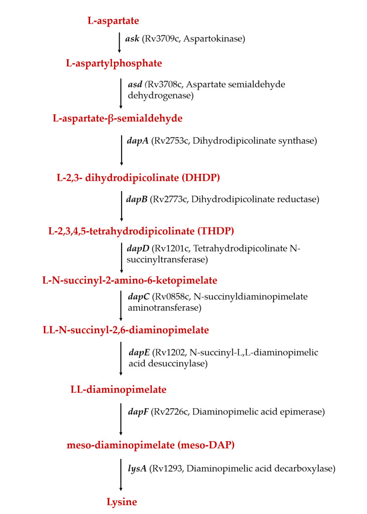
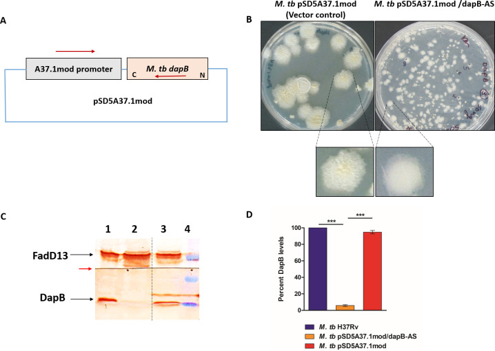
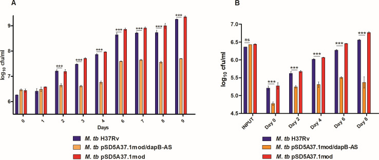
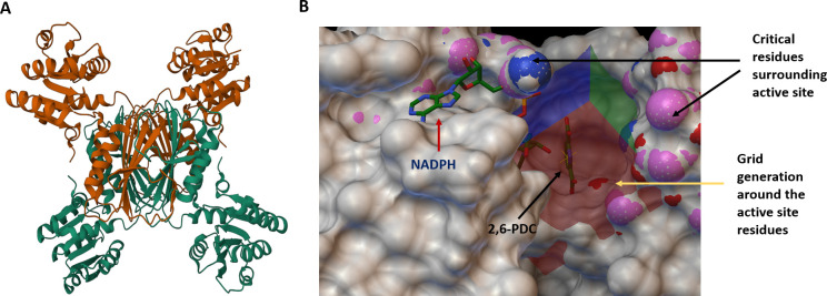
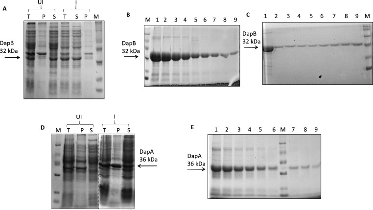
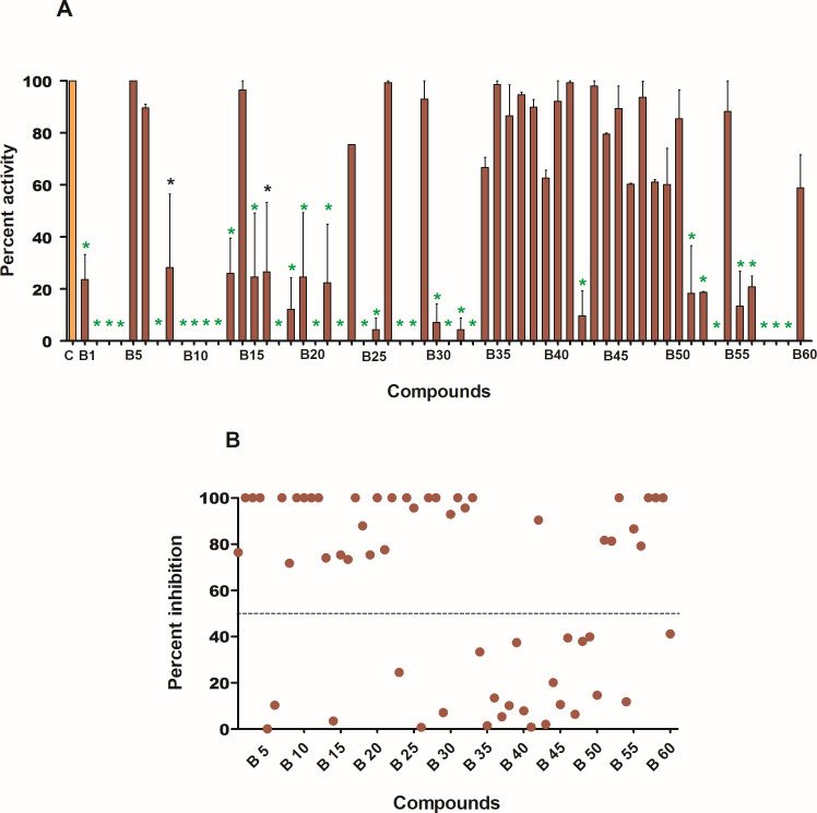
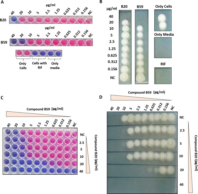
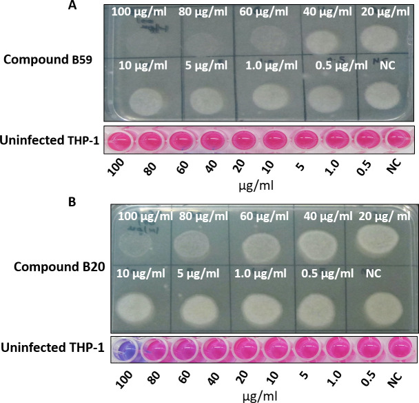
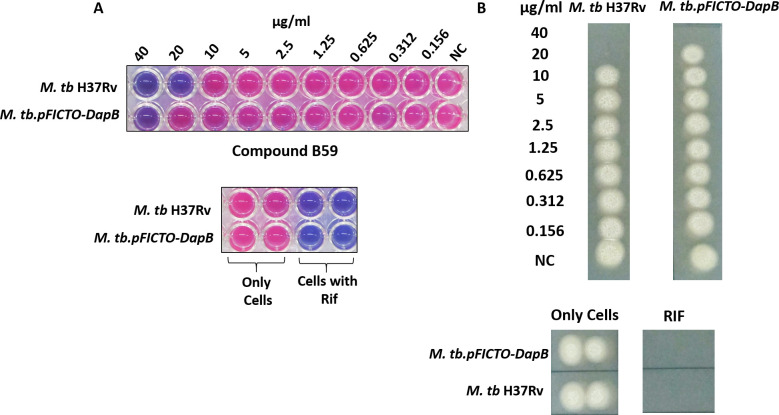
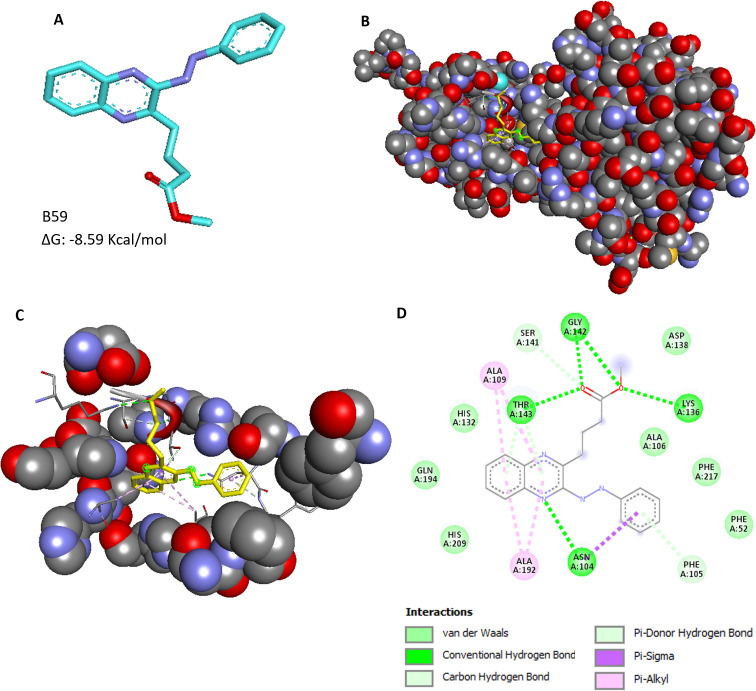
Non-compliance to lengthy antituberculosis (TB) treatment regimen, associated side effects, and emergence of drug-resistant strains of Mycobacterium tuberculosis (M. tb) emphasize the need to develop more effective anti-TB drugs. Here, we have evaluated the role of M. tb dihydrodipicolinate reductase (DapB), a component of the diaminopimelate pathway, which is involved in the biosynthesis of both lysine and mycobacterial cell wall. We showed that DapB is essential for the in vitro as well as intracellular growth of M. tb. We further utilized M. tb DapB, as a target for identification of inhibitors by employing in silico virtual screening, and conducted various in vitro screening assays to identify inhibitors with potential to inhibit DapB activity and in vitro and intracellular growth of M. tb with no significant cytotoxicity against various mammalian cell lines. Altogether, M. tb DapB serves as an important drug target and a hit molecule, namely, 4-(3-Phenylazoquinoxalin-2-yl) butanoic acid methyl ester has been identified as an antimycobacterial molecule in our study.

Keywords: Mycobacterium tuberculosis; tuberculosis; virtual screening.


Conflict of interest statement

The authors declare no conflict of interest.

Figures

Fig 1
Fig 1
Schematic diagram showing various steps of the diaminopimelate pathway in Mycobacterium tuberculosis.
Fig 2
Fig 2
Construction of M. tb dapB knockdown mutant strain. (A) Schematic representation showing dapB gene cloned in an antisense orientation downstream to an A37.1mod promoter in a pSD5A37.1mod promoter. (B) Colony morphology of M. tb vector control strain (M. tb pSD5A37.1mod) and antisense knockdown mutant strain (M. tb pSD5A37.1mod/dapB-AS). dapB antisense mutant strain showed smooth and small-sized colonies as compared with rough and large-sized colonies in vector control. (C) Confirmation of dapB antisense knockdown in M. tb by immunoblot analysis. Lysates containing 70 µg protein each of M. tb (lane 1), M. tb pSD5A37.1mod/dapB-AS (lane 2), and M. tb pSD5A37.1mod (lane 3) were electrophoresed on 12.5% polyacrylamide gel. The proteins were transferred on PVDF (polyvinylidene difluoride) membrane, and the blot was cut into two at the red arrow. Immunoblotting was carried out by using anti-DapB antibody for the lower part of the membrane and with anti-fadD13 antibody for the upper part. Mycobacterium tuberculosis FadD13, a mycobacterial cytosolic protein unrelated to the DAP pathway, was employed as a loading control. Dashed line shows that different lanes (non-adjacent) from the same immunoblot have been brought together. Lane 4—pre-stained protein molecular weight markers (from the top: 50, 37, and 25 kDa). (D) The bar diagram shows percent inhibition of DapB protein levels in M. tb vector control and antisense knockdown mutant strain as compared with M. tb H37Rv after normalizing the data with FadD13 levels (loading control). Quantification of the protein levels in the immunoblot was performed by using ImageJ software. The data are represented as the mean ± SEM (error bars) of two independent experiments, and representative immunoblot is shown.
Fig 3
Fig 3
Involvement of DapB in in vitro and intracellular growth of M. tb. (A) In vitro growth of M. tb H37Rv (blue), M. tb pSD5A37.1mod/dapB-AS (orange), and M. tb pSD5A37.1mod (red) strains in 7H9 medium at 37°C, 200 rpm by CFU analysis. Graph showed significant difference in the growth of antisense knockdown mutant strain as compared with the wild-type M. tb and vector control strains. (B) Intracellular growth of M. tb H37Rv (blue), M. tb pSD5A37.1mod/dapB-AS (orange), and M. tb pSD5A37.1mod (red) strains inside THP-1 macrophages at different time points. A significant difference in the intracellular growth of M. tb H37Rv (blue) and M. tb pSD5A37.1mod/dapB-AS (orange) strains was observed. The data are represented as the mean ± SEM (error bars) of at least two independent experiments. (*P < 0.05, **P < 0.01, and ***P < 0.001, two-way ANOVA, Bonferroni post-tests).
Fig 4
Fig 4
Crystal structure of DapB and docking sites employed for virtual screening. (A) The figure shows the three-dimensional structure of DapB (PDB ID: 1YL5) in its apo form. (B) Virtual screening was carried out against the active site of DapB. The critical residues around the active site such as Glu-130, His-132, His-133, Lys-136, Asp-138, Ala-139, Pro-140, Ser-141, Gly-142, Thr-143, and Ala-144 are highlighted as colored surface and were used for grid preparation for performing docking. NADPH (cofactor of DapB) and 2, 6-PDC (substrate analog) are shown by red and black colored arrows, respectively. Figure is prepared by using Autodock4.2.
Fig 5
Fig 5
Expression and purification of recombinant proteins (DapB and DapA). (A) Dihydrodipicolinate reductase expression was induced with 0.5  mM IPTG (isopropyl-β-d-thiogalactopyranoside) at 37°C for 3 hours. (B) 6X-His tagged recombinant DapB was purified by Ni-NTA affinity chromatography. Eluted protein fractions were loaded on 12.5% polyacrylamide gel (lanes 1–9). (C) Ni-NTA purified DapB (lane 1) was loaded on a Sephadex G-200 gel filtration column, and eluted fractions (lanes 2–9) were electrophoresed on 12.5% polyacrylamide gel. (D) Expression of DapA was induced with 0.5 mM IPTG at 18°C, overnight. Dashed line represents that sections from different gel pictures have been brought together. (E) Figure shows eluted fractions (lanes 1–9) of 6X-His tagged recombinant DapA purified by Ni-NTA affinity chromatography. M denotes protein molecular weight marker (from the top: 97, 66, 43, 29, 20, and 14 kDa). For Fig. 5A and D, UI, uninduced sample; I, induced sample; T, total cell lysate; P, pellet; S, supernatant.
Fig 6
Fig 6
Evaluation of inhibitory potential of the compounds against enzymatic activity of DapB. (A) Compounds were screened against DapB activity by using a standardized coupled biochemical assay at a concentration of 100 µg/mL. Bar graph represents percent activity in the presence of inhibitors (brown-colored bars). Percent activity in the control (without inhibitor) is considered as 100% and is represented by orange-colored bar. The data are represented as the mean ± SEM (error bars) of at least two independent experiments. Black-colored stars represent P < 0.01, and green-colored stars represent P < 0.001 by one-way ANOVA, Tukey’s multiple comparison test. (B) Scatter plot of all the compounds showing percent inhibition of DapB enzymatic activity at a concentration of 100 µg/mL (derived from the percent activity data given in 6A). Each dot represents mean percent inhibition of two independent experiments. Thirty-six compounds showed more than 50% inhibition (denoted by dashed line) of DapB activity.
Fig 7
Fig 7
Evaluation of inhibitory potential of the compounds against in vitro growth of M. tb by resazurin microtiter assay. (A) Mycobacterium tuberculosis cells were incubated with varying concentrations of compounds (0.156 µg/mL–40 µg/mL) for 7 days at 37°C in a 96-well plate, followed by the addition of resazurin dye and visualization of change in the color of dye. Blue-colored wells indicate non-viable M. tb cells whereas pink-colored wells indicate viable mycobacterial cells. Wells containing rifampicin (0.25 µg/mL) and only MB7H9 medium were used as positive and negative controls, respectively. (B) MIC99 value was determined by spotting an aliquot from each well onto a 7H11 agar plate to check the growth of bacteria. Wells with blue-color dye in Fig. 7A showed no growth on agar plates. NC, well without compound. (C) Evaluation of interaction between compounds B59 and B20 by employing checkerboard assay. Mycobacterium tuberculosis cells were incubated with varying concentrations of compounds B59 and B20 diluted along the x-axis and y-axis in a 96-well format for 7 days at 37°C, and the growth of the bacteria was analyzed by resazurin dye-based method. (D) MIC99 value of each compound in combination with the other compound was determined by spotting an aliquot from each well on to 7H11 agar plates. NC, well without compound. MIC99 is considered as the concentration of the well that showed no visible growth on the agar plate.
Fig 8
Fig 8
Evaluation of the ability of compounds (B59 and B20) to inhibit the intracellular growth of the pathogen. Phorbol 12-myristate 13-acetate (PMA)-activated THP-1 macrophages were infected with M. tb H37Rv, and the growth of pathogen in the presence of varying concentrations of compounds was visualized on agar plates after 5 days of infection. (A) Compound B59 displayed inhibition of M. tb growth at 60 µg/mL and above. The bottom panel depicts cytotoxicity exhibited by compound B59 toward macrophages by resazurin microtiter dye. Compound B59 did not display any cytotoxicity toward the macrophages. (B) Compound B20 displayed inhibition of M. tb growth at a concentration of 80 µg/mL and above. It exhibited toxicity toward THP-1 macrophages at a concentration of 100 µg/mL. NC, well without compound.
Fig 9
Fig 9
Evaluation of the target specificity of compound B59 by employing resazurin dye reduction assay. (A) Varying concentrations of compound B59 (0.156 µg/mL to 40 µg/mL), along with appropriate controls, were incubated with various mycobacterial strains (M. tb H37Rv and M. tb.pFICTO-dapB) for 7 days at 37°C followed by the addition of resazurin dye and visualization of change in the color of dye. Blue-colored wells indicate non-viable M. tb cells whereas pink-colored wells indicate viable mycobacterial cells. (B) MIC99 value was evaluated by spotting an aliquot from each well onto agar plates to assess the growth of bacteria.
Fig 10
Fig 10
Binding mode analysis of the hit compound, B59. (A) Chemical structure of the compound B59, 4-(3-Phenylazoquinoxalin-2-yl) butanoic acid methyl ester (cyan: carbon, purple: nitrogen, and red: oxygen). (B) Figure displays binding of compound B59 at the active site of DapB. (C) Pocket residues of receptor interacting with the ligand, B59. (D) Key interactions of compound B59 with the residues of receptor. The receptor-ligand interaction diagrams were generated by using BIOVIA Discovery Studio 2021.

References

    1. Bagcchi S. 2023. WHO’s global tuberculosis report 2022. Lancet Microbe 4:e20. doi:10.1016/S2666-5247(22)00359-7 - DOI - PubMed
    1. Pai M, Behr MA, Dowdy D, Dheda K, Divangahi M, Boehme CC, Ginsberg A, Swaminathan S, Spigelman M, Getahun H, Menzies D. 2016. Tuberculosis (primer). Nat Rev Dis Primers 2. doi:10.1038/nrdp.2016.76 - DOI - PubMed
    1. Yelamanchi SD, Surolia A. 2021. Targeting amino acid metabolism of Mycobacterium tuberculosis for developing inhibitors to curtail its survival. IUBMB Life 73:643–658. doi:10.1002/iub.2455 - DOI - PubMed
    1. Shaku M, Ealand C, Kana BD. 2020. Cell surface biosynthesis and remodeling pathways in mycobacteria reveal new drug targets. Front Cell Infect Microbiol 10:603382. doi:10.3389/fcimb.2020.603382 - DOI - PMC - PubMed
    1. Vilchèze C. 2020. Mycobacterial cell wall: a source of successful targets for old and new drugs. Applied Sciences 10:2278. doi:10.3390/app10072278 - DOI

Publication types

LinkOut - more resources