. 2023 Dec 12;11(6):e0253823.
doi: 10.1128/spectrum.02538-23. Epub 2023 Oct 19.

Anaphylatoxin signaling activates macrophages to control intracellular Rickettsia proliferation

Affiliations

Anaphylatoxin signaling activates macrophages to control intracellular Rickettsia proliferation

Mustapha Dahmani et al. Microbiol Spectr. .

Abstract

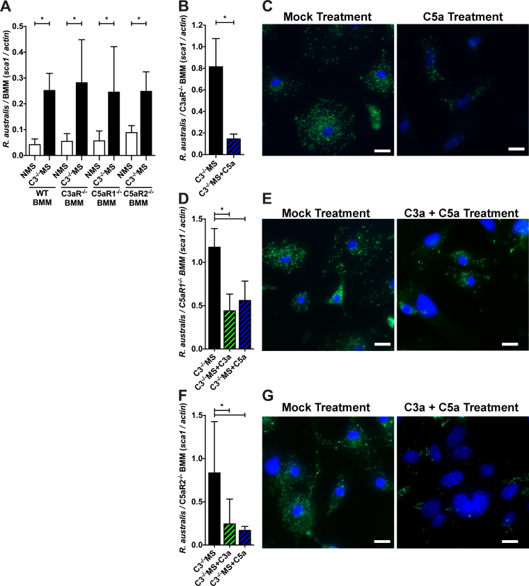
Pathogenic Rickettsia species are extremely dangerous bacteria that grow within the cytoplasm of host mammalian cells. In most cases, these bacteria are able to overpower the host cell and grow within the protected environment of the cytoplasm. However, a dramatic conflict occurs when Rickettsia encounter innate immune cells; the bacteria can "win" by taking over the host, or the bacteria can "lose" if the host cell efficiently fights the infection. This manuscript examines how the immune complement system is able to detect the presence of Rickettsia and alert nearby cells. Byproducts of complement activation called anaphylatoxins are signals that "activate" innate immune cells to mount an aggressive defensive strategy. This study enhances our collective understanding of the innate immune reaction to intracellular bacteria and will contribute to future efforts at controlling these dangerous infections.

Keywords: Rickettsia; anaphylatoxin; complement system; intracellular bacteria; macrophage.


Conflict of interest statement

The authors declare no conflict of interest.

Figures

FIG 1
FIG 1
Murine anaphylatoxins reduce Rickettsia proliferation in RAW264.7 cells. (A and B) Presence of the anaphylatoxins C3a (A) and C5a (B) in the media after 3 days of culture of R. parkeri-infected RAW264.7 cells in the presence of NMS and C3−/− MS, as determined by an enzyme-linked immunosorbent assay. (C and D) Proliferation of R. parkeri (C) and R. rickettsii (D) in RAW264.7 cells cultivated with complement-active serum (NMS) or complement-deficient serum (C3−/− MS), as determined by the quantitative PCR ratio of Rickettsia sca1 to murine actin DNA. (E and F) Quantity of R. parkeri (E) and R. rickettsii (F) after 3 days of culture in RAW264.7 cells with complement-deficient serum (C3−/− MS) supplemented with phosphate-buffered saline, mouse C3a peptide, mouse C5a peptide, or both C3a and C5a peptides. (G) Presence of R. parkeri after 3 days of culture in RAW264.7 cells with complement-active serum (NMS), complement-deficient serum (C3−/− MS), or complement-active serum with anaphylatoxin receptor antagonists PMX53 and SB290170 (NMS + inhibitors). *P < 0.05 by (A,B) Student’s t-test; (C,D) one-way ANOVA with Sidák’s multiple comparison of matched days; (E,F,G) one-way ANOVA with Dunnett’s multiple comparison test to the NMS/mock-treated control. All columns represent at least five data points, and experiments were repeated to ensure reproducibility.
FIG 2
FIG 2
Human anaphylatoxins restrict Rickettsia proliferation in THP-1 cells. (A) R. parkeri proliferation in human THP-1 monocytes cultivated with either complement-active NHS or complement-deficient hiHS, as determined by the qPCR ratio of Rickettsia sca1 to human gapdh DNA. (B) Growth of R. parkeri cultivated for 3 days in THP-1 cells with either NHS or complement-deficient EDTA-inactivated human serum. (C) Quantity of R. parkeri in THP-1 cells cultivated in complement hiHS supplemented with PBS, human C3a peptide, human C5a peptide, or both C3a and C5a peptides. (D) Quantity of R. parkeri after 3 days of growth in THP-1 cells cultivated in complement-active serum (NHS) or NMS plus the anaphylatoxin receptor antagonists PMX53 and SB290170 (NHS + inhibitors). (E) R. parkeri proliferation in EA.hy926 human endothelial cells cultivated complement-active NHS or complement-inactivated NHS + EDTA. (F) R. parkeri growth in endothelial (EA.hy926) and macrophage (THP-1) co-culture. R. parkeri proliferates in ea.hy926 or co-culture in complement-inactive serum, but growth is restricted in EA.hy926/THP-1 co-culture containing complement-active serum. *P < 0.05 by (A) one-way ANOVA with Sidák’s multiple comparison of matched days; (B,D,E) Student’s t-test; (C) one-way ANOVA with Dunnett’s multiple comparison test to mock-treated control; (F) one-way ANOVA with Tukey’s multiple comparison test of all columns. All columns represent at least five data points, and experiments were repeated to ensure reproducibility.
FIG 3
FIG 3
Anaphylatoxins restrict Rickettsia australis proliferation in C57BL/6J bone marrow macrophages. (A, B, C) R. australis proliferation in C57BL/6J BMMs cultured in complement-active NMS or complement-deficient C3−/− MS as determined by (A) qPCR ratio of R. australis sca1 to murine actin, (B) fluorescent microscopic imaging, and (C) visual quantification of R. australis and BMM nuclei in over 600 BMMs when cultured for 3 days in the presence of NMS or C3−/− MS. (D, E, F) Quantity of R. australis after culture in BMM grown with complement-deficient C3−/− MS supplemented with PBS, mouse C3a peptide, mouse C5a peptide, or both C3a and C5a peptides as determined by (D) qPCR, (E) fluorescence microscopy, and (F) visual quantification of R. australis in 600 BMMs. *P < 0.05 by (A) one-way ANOVA with Sidák’s multiple comparison of matched days; (C,F) Student’s t-test; (D) one-way ANOVA with Dunnett’s multiple comparison test to C3−/− MS. Scale bar = 50 µm. All columns represent at least five data points, and experiments were repeated to ensure reproducibility. The scatter plot with mean and SEM represents at least 600 different individual cells.
FIG 4
FIG 4
The anaphylatoxin receptors C3aR1, C5aR1, and C5aR2 are essential to the restriction of Rickettsia australis growth in murine BMM. (A) Quantity R. australis after 3 days of growth in WT, C3aR−/−, C5aR1−/−, and C5aR2−/− BMMs cultivated in the presence of complement-active NMS or complement-deficient C3−/− MS as determined by the qPCR ratio of R. australis sca1 to murine actin DNA. (B, C) R. australis growth in C3aR−/− BMM cultured with complement-deficient C3−/− mouse serum supplemented with PBS or mouse C5a peptide as determined by qPCR (B) and Rickettsia (green) and DAPI (blue) microscopy (C). (D,E) Quantity of R. australis after 3 days of growth in C5aR1−/− BMM cultured with complement-deficient C3−/− mouse serum supplemented with PBS, mouse C3a peptide, or mouse C5a peptide as determined by qPCR (D) and Rickettsia (green) and DAPI (blue) microscopy (E). (F, G) Quantity of R. australis after 3 days of growth in C5aR2−/− BMM cultured with complement-deficient C3−/− mouse serum supplemented with PBS, mouse C3a peptide, or mouse C5a peptide as determined by qPCR (F) and Rickettsia (green) and DAPI (blue) microscopy (G). *P < 0.05 by (A) one-way ANOVA with Sidák’s multiple comparison of matched receptor mutants; (B,D,F) Student’s t-test. Scale bar = 20 µm. All columns represent at least five data points, and experiments were repeated to ensure reproducibility.
FIG 5
FIG 5
Anaphylatoxins modulate the phenotype of murine BMM, leading to decreased Rickettsia proliferation. (A) ERK1/2 phosphorylation as compared to total ERK1/2 and actin from R. australis-infected BMMs treated with PBS or anaphylatoxins C3a and C5a. (B) Representative images of R. australis cultured for 1 h in BMM pretreated with complement-deficient C3−/− MS or C3−/− MS supplemented with mouse C3a and C5a peptides as determined by confocal microscopy using the endosomal/lysosomal marker lysosomal-associated membrane protein 1 (LAMP-1), anti-Rickettsia, and DAPI. (C) Subsequent quantification of R. australis-LAMP1 co-localization in over 100 single BMMs. (D) Quantification of cDNA from Rickettsia-infected BMMs cultured in the presence of complement-active NMS or complement-deficient C3−/− MS. Macrophage activation markers, pro-inflammatory cytokines, and anaphylatoxin receptor mRNA were quantified by qPCR as compared to actin cDNA. *P < 0.05 by Students t-test. Scale bar = 5 µm. All columns represent at least five data points, and experiments were repeated to ensure reproducibility. LAMP-1 colocalization was observed for 100 individual cells in each column and was repeated to ensure reproducibility.

References

    1. Laukaitis HJ, Macaluso KR. 2021. Unpacking the intricacies of Rickettsia–vector interactions. Trends Parasitol 37:734–746. doi:10.1016/j.pt.2021.05.008 - DOI - PMC - PubMed
    1. Eremeeva ME. 2012. Molecular epidemiology of rickettsial diseases in North America. Ticks Tick Borne Dis 3:332–337. doi:10.1016/j.ttbdis.2012.10.022 - DOI - PubMed
    1. Biggs HM, Behravesh CB, Bradley KK, Dahlgren FS, Drexler NA, Dumler JS, Folk SM, Kato CY, Lash RR, Levin ML, Massung RF, Nadelman RB, Nicholson WL, Paddock CD, Pritt BS, Traeger MS. 2016. Diagnosis and management of tickborne rickettsial diseases: rocky mountain spotted fever and other spotted fever group rickettsioses, ehrlichioses, and anaplasmosis — United States. a practical guide for health care and public health professionals. MMWR Recomm Rep 65:1–44. doi:10.15585/mmwr.rr6502a1 - DOI - PubMed
    1. Robinson MT, Satjanadumrong J, Hughes T, Stenos J, Blacksell SD. 2019. Diagnosis of spotted fever group Rickettsia infections: the Asian perspective. Epidemiol Infect 147:e286. doi:10.1017/S0950268819001390 - DOI - PMC - PubMed
    1. Salje J. 2021. Cells within cells: rickettsiales and the obligate intracellular bacterial lifestyle. Nat Rev Microbiol 19:375–390. doi:10.1038/s41579-020-00507-2 - DOI - PubMed

LinkOut - more resources