

Revisiting the History of Odontoma, with Special Reference to Its Original Illustration
- PMID: 37856052
- PMCID: PMC10739675
- DOI: 10.1007/s12105-023-01593-3
Revisiting the History of Odontoma, with Special Reference to Its Original Illustration
Abstract
Background: Practically every facet of the most common odontogenic tumor, odontoma, has been covered by an extensive volume of literature. However, uncertainty about its precise history has persisted.
Materials and methods: The historical evolution of odontoma was traced with reference to the original illustrations that accompanied European and American reports published at the beginning of the 19th century and also at the turn of the century.
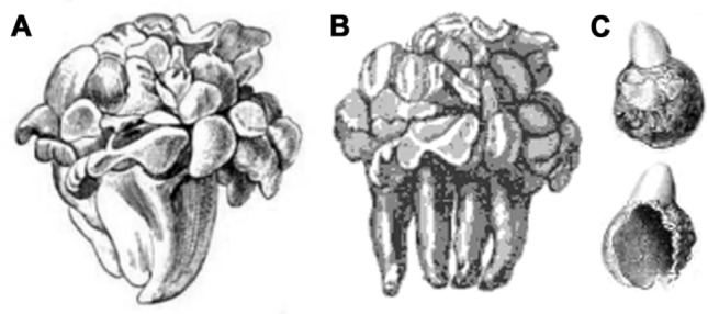
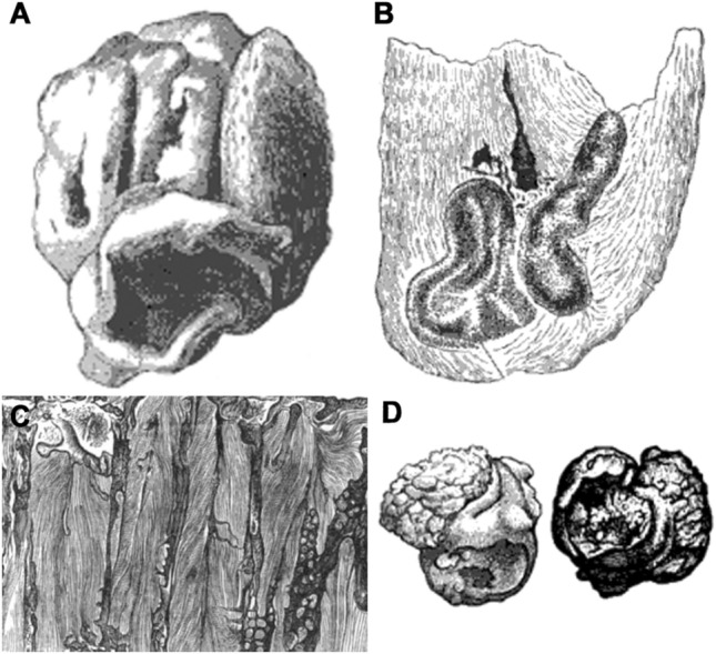
Results: The prevailing views regarding the first description of odontoma by Oudet of Paris in 1809 and the original designation "odontome" by Broca of Paris in 1867 are not entirely accurate. Before Broca's suggested term, "exostose dentaire" (dental exostosis) and "tumeur dentaire" (dental tumor) proposed by Oudet and Forget of Paris, respectively, were popular terms adopted in France, while in Briatin the terms "warty tooth" and "supernumerary teeth" proposed by Salter and Tomes of London, respectively, were widely coined. The original illustrations of complex odontoma were published by Wedl of Vienna in 1851, and in 1862 Tomes published the first drawing of compound odontoma denticles. Before the advent of diagnostic radiography in the early 1900s, spontaneous exposure or eruption of odontoma followed by secondary infection was very common. In 1887-1888, Bland Sutton of London criticized Broca's monumental research and formulated the first modern classification which, in essence, remains valid today. At that time, large osteomas of the maxilla were inappropriately classified as odontomas by many pathologists because of Bland Sutton's influential view. Interestingly, the first radiographic evidence of odontoma was published by the American oral surgeon Gilmer in 1899.
Conclusion: In view of their fundamental achievements, the names of Wedl, Salter, Broca and Bland Sutton have been closely associated with the true history of odontoma.
Keywords: Conceptual controversy; Historical review; Odontoma; Original illustration.
© 2023. The Author(s), under exclusive licence to Springer Science+Business Media, LLC, part of Springer Nature.
Conflict of interest statement
The authors declare no competing interests.
Figures


References
-
- Rognetta (1835) Dexième mémoire sur les exostoses; de leurs causes, de leur mode de développement, et des accidens qu’elles peuvent occasioner. Gaz Méd Paris 2nd ser. 3(45):705–710
-
- Virchow R, editor. Die Krankhaften Geschwülste. Berlin: Verlag; 1864. pp. 53–59.
-
- Broca P. Recherches sur un nouveau groupe de tumeurs désigné sous le nom d’odontômes. Compt Rend Acad Sci. 1867;65(December 30):1117–1121.
-
- Broca P (ed) (1867) Recherches sur un nouveau groupe de tumeurs désignés sous le nom odontomes. P Asselin, Paris
-
- Broca P. Recherches sur un nouveau groupe de tumeurs désigné sous le nom d’odontômes. Gaz Hebd Méd Chir 2nd Ser. 1868;5(2):19–21.
Publication types
MeSH terms
LinkOut - more resources
Full Text Sources
