. 2023 Nov 1;145(43):23702-23714.
doi: 10.1021/jacs.3c08135. Epub 2023 Oct 19.

DNA-Histone Cross-Link Formation via Hole Trapping in Nucleosome Core Particles

Affiliations

DNA-Histone Cross-Link Formation via Hole Trapping in Nucleosome Core Particles

Tingyu Wen et al. J Am Chem Soc. .

Abstract

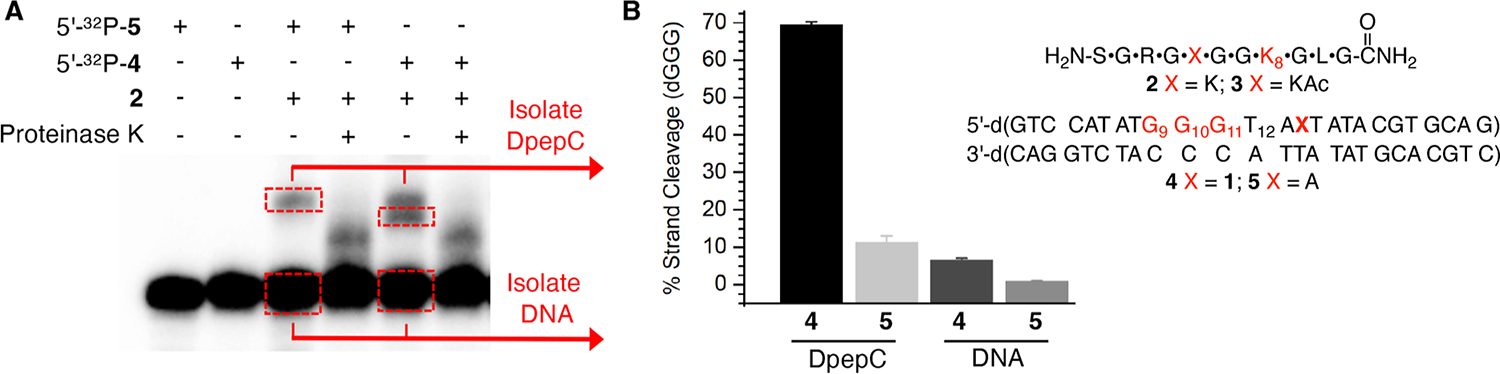
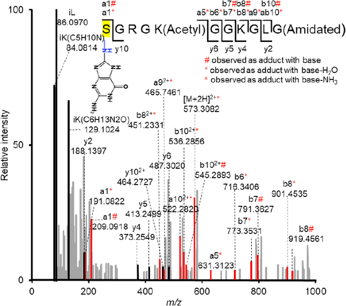
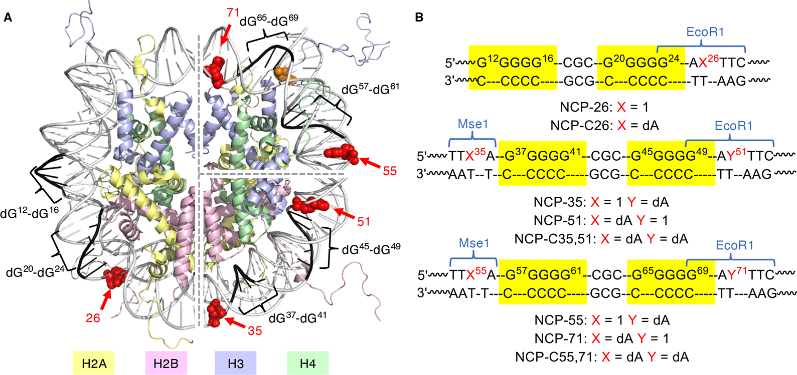
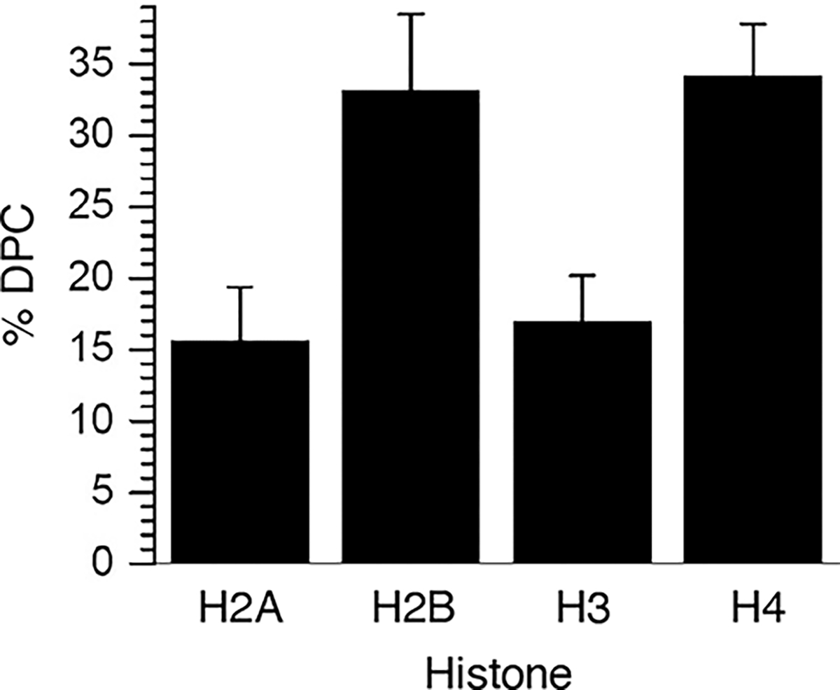
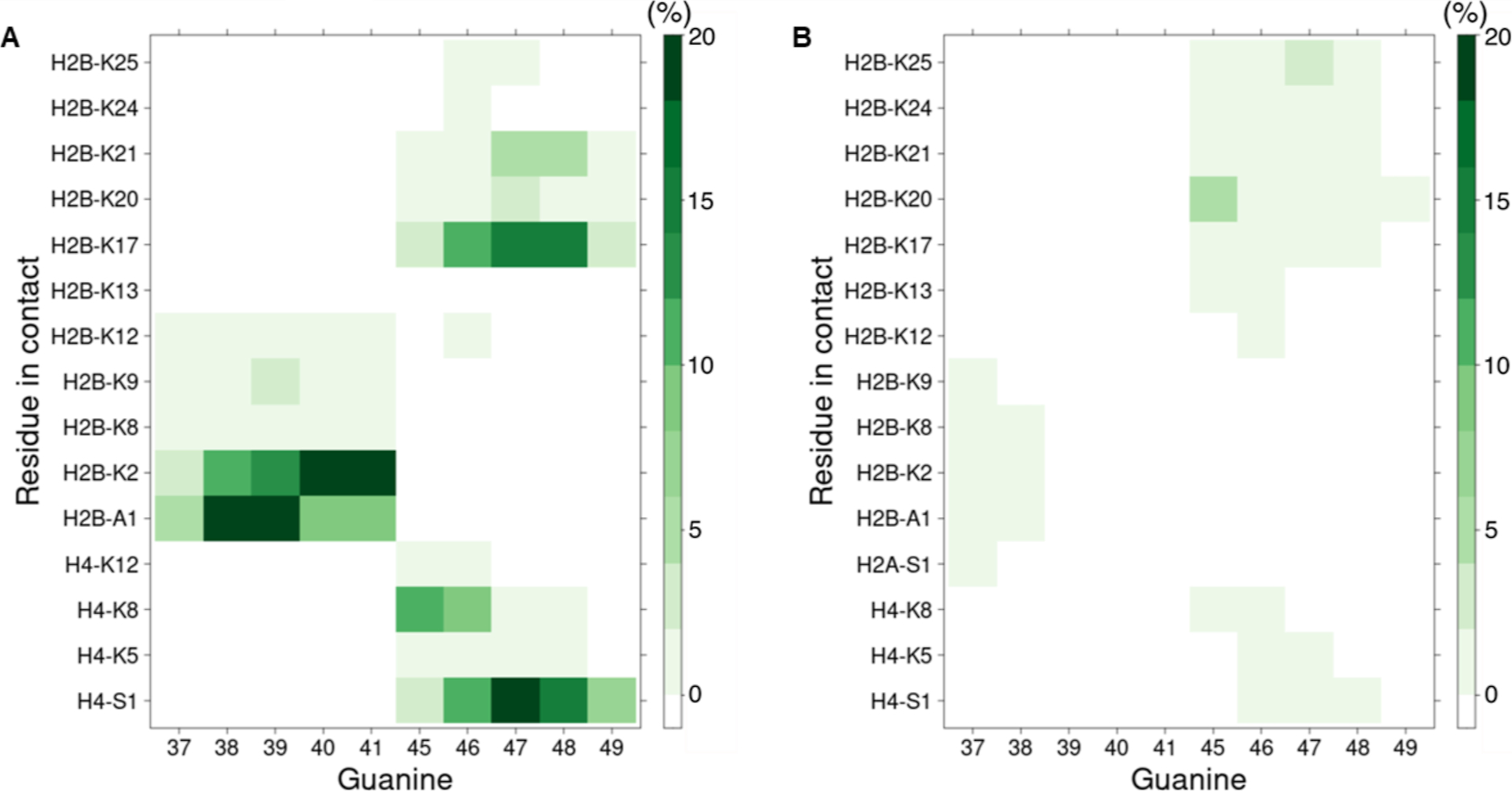
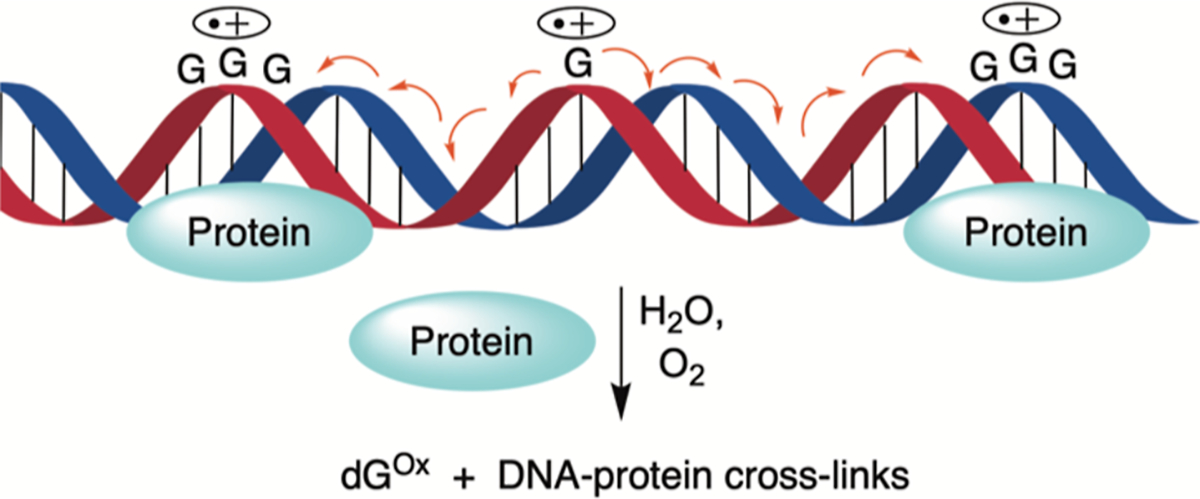
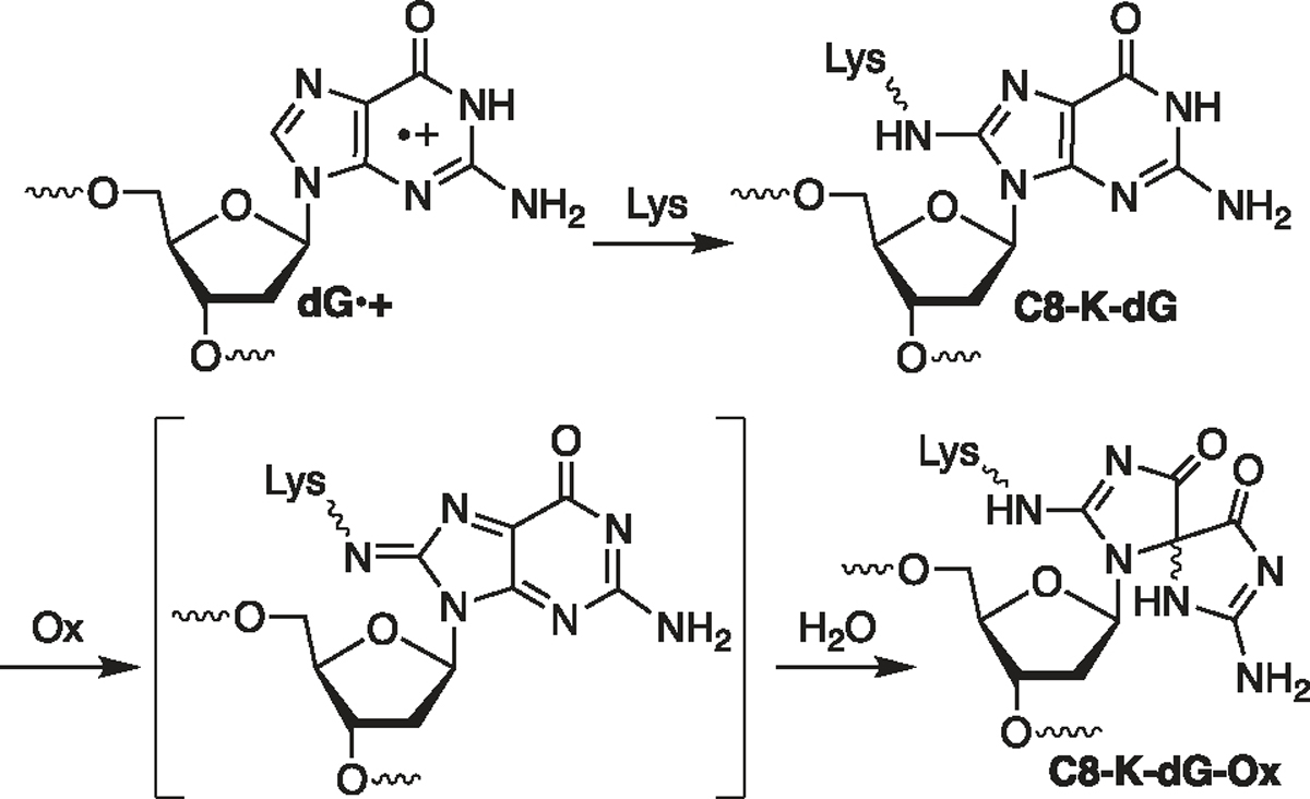
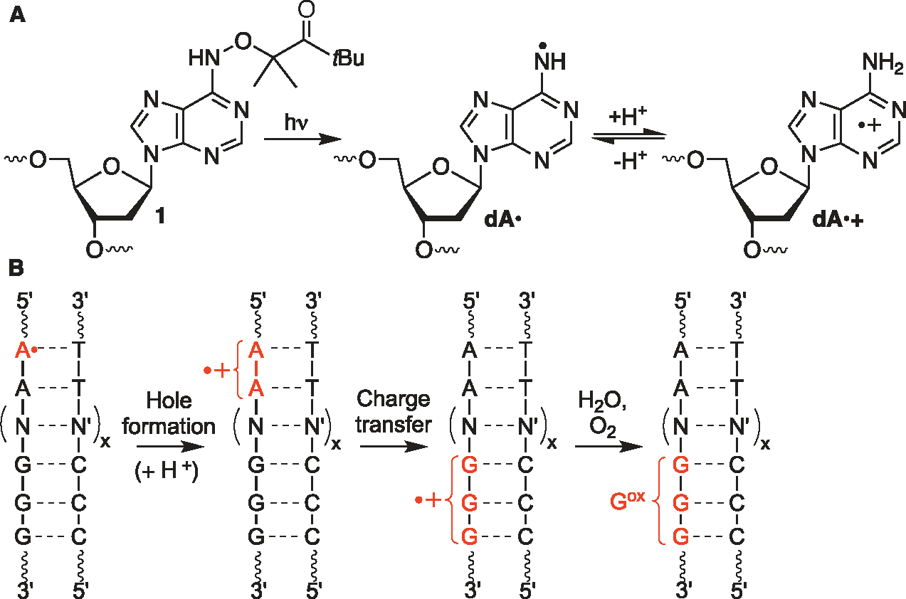
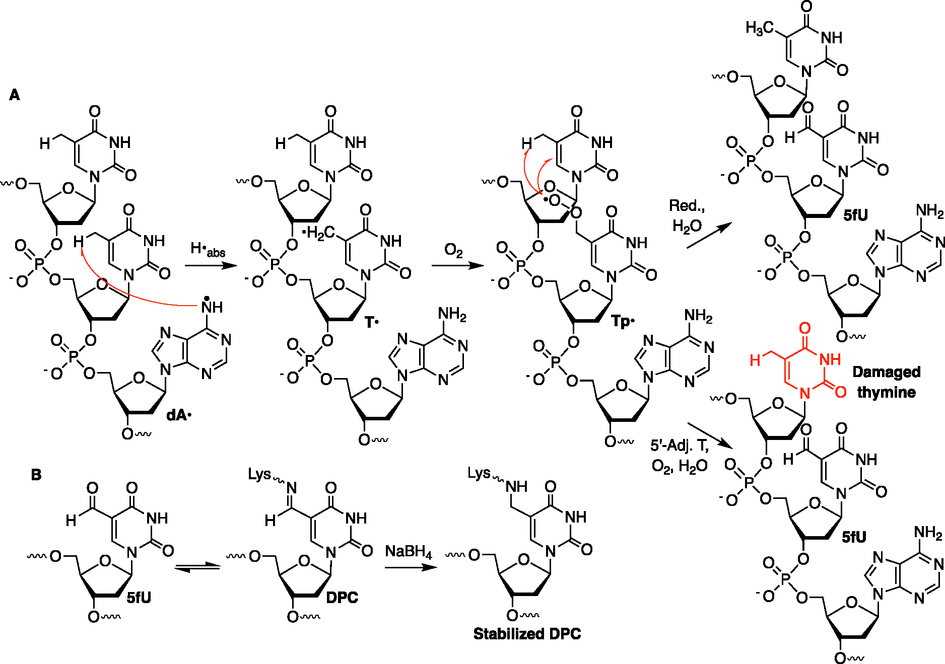
Radical cations (holes) produced in DNA by ionizing radiation and other oxidants yield DNA-protein cross-links (DPCs). Detailed studies of DPC formation in chromatin via this process are lacking. We describe here a comprehensive examination of DPC formation within nucleosome core particles (NCPs), which are the monomeric component of chromatin. DNA holes are introduced at defined sites within NCPs that are constructed from the bottom-up. DPCs form at DNA holes in yields comparable to those of alkali-labile DNA lesions that result from water trapping. DPC-forming efficiency and site preference within the NCP are dependent on translational and rotational positioning. Mass spectrometry and the use of mutant histones reveal that lysine residues in histone N-terminal tails and amino termini are responsible for the DPC formation. These studies are corroborated by computational simulation at the microsecond time scale, showing a wide range of interactions that can precede DPC formation. Three consecutive dGs, which are pervasive in the human genome, including G-quadruplex-forming sequences, are sufficient to produce DPCs that could impact gene expression.


Conflict of interest statement

The authors declare no competing financial interest.

Figures

Figure 1.
Figure 1.
DNA–peptide cross-link formation. (A) Representative 20% denaturing PAGE showing DpepC formation between 5′-32P-4 or 5′-32P-5 and H4 1–11 peptide (2) at pH 7.5 upon photolysis. (B) Percent of piperidine cleavage at the dGGG sequence in isolated DpepC or isolated uncross-linked DNA plotted as the ave. ± std. dev of 3 independent reactions.
Figure 2.
Figure 2.
MS/MS spectrum of H4 1–11-K5-Ac (M = 1013.5720) containing C8-Lys-dG (M of base = 149.0338) obtained from photolysis of 3 and 4 detected by LC–MS/MS (exp. m/z = 582.3145, calcd m/z = 582.3107). Color code: Ions containing modification, red; unmodified ion, black; ion from other peptide(s), gray.
Figure 3.
Figure 3.
Nucleosome core particles. (A) Core particle structure highlighting dG5 tracts, positions at which 1 is incorporated and H3-Y41. The structure was generated by superimposing two NCP structures (pdb: 1kx5 and 3lz0). (B) Nucleosomal DNA sequences in the region of dG5 tracts. See Figure S4A for the entire nucleosomal DNA sequences.
Figure 4.
Figure 4.
DPC formation in NCPs. (A) DPC formation in NCP-35. Denaturing PAGE analysis of piperidine-treated isolated DPCs from (B) NCP-35 and NCP-51, (C) NCP-55, NCP-55, H3-Y41F NCP-71 and H3-Y41F NCP-71, and (D) NCP-26.
Figure 5.
Figure 5.
Solvent accessibilities and histone tail interactions at dG5 tracts in NCP-C35,51 and NCP-C55,71. (A,B) SASA of dG5 tracts in (A) NCP-C35,51 and (B) NCP-C55,71. The guanines are colored according to their SASA from white (25 Å) to bright blue (50 Å). (C,D) SASA of guanines within dG5 tracts of (C) NCP-C35,51 and (D) NCP-C55,71 with (blue bar) or without (blue and striped bar) histone tails. (E) dG5 tracts in NCP-C35,51 with 40 conformations of H4 (light green) and H2B (pink) tails from MD simulations. (F) dG tracts in NCP-C51,71 with 40 conformations of H3 (light blue) and H4 (light green) tails from MD simulations. dG5 tracts are highlighted in black. Major cross-linked dG sites within those tracts are highlighted in red. The structures were generated from MD simulations that utilized pdb: 3lz0 as a starting point.
Figure 6.
Figure 6.
Individual histone protein contribution to DPCs formed by hole trapping upon the photolysis of WT NCP-51.
Figure 7.
Figure 7.
Contact map between dG5 tracts and histone tails in NCP-C35,51. (A) Contact map between ammonium group of lysines or of N-terminal residues and major groove exposed heavy atoms of guanines. (B) Contact map between the ammonium group of lysines or N-terminal residues and phosphate groups. The heat map extends from white (no contact) to dark green (at least one contact is observed for more than 20% of the trajectory frames).
Figure 8.
Figure 8.
Correspondence between modified residues detected by LC–MS/MS of NCP-51 and residues interacting with dG5 tracts in MD simulations of NCP-C35,51. (A) Two perspectives of the NCP. Modified residues detected only by LC–MS/MS (red), interacting residues detected only computationally (cyan), or residues detected both computationally and by LC–MS/MS (dark violet). Asterisks (*) indicate that MS2 spectra do not contain enough fragment ions to distinguish modification at this lysine from that at the adjacent lysine. The structures were generated from an MD simulation snapshot based upon pdb: 3lz0. (B) Histone N-terminal tail sequences [same color assignment as in (A)].
Figure 9.
Figure 9.
Nucleosome core particles containing dG3 tracts. The structure was generated by superimposing two NCP structures (pdb: 1kx5 and 3lz0). The local DNA sequences can be found in the Supporting Information.
Scheme 1.
Scheme 1.
Charge Migration through DNA and the Formation of Hole Trapping Products on dG
Scheme 2.
Scheme 2.
Lysine Trapping and Further Oxidation of dG•+
Scheme 3.
Scheme 3.
Hole Generation from 1 and Subsequent Charge Transfer in DNA
Scheme 4.
Scheme 4.
DPC and Tandem Lesion Formation dA• Generation in 5′-dTT1 Sequences

References

    1. Wei X; Peng Y; Bryan C; Yang K Mechanisms of DNA-Protein Cross-Link Formation and Repair. Biochim. Biophys. Acta 2021, 1869, 140669. - PubMed
    1. Nakano T; Mitsusada Y; Salem AMH; Shoulkamy MI; Sugimoto T; Hirayama R; Uzawa A; Furusawa Y; Ide H Induction of DNA-Protein Cross-Links by Ionizing Radiation and Their Elimination from the Genome. Mut. Res. 2015, 771, 45–50. - PubMed
    1. Vaz B; Popovic M; Newman JA; Fielden J; Aitkenhead H; Halder S; Singh AN; Vendrell I; Fischer R; Torrecilla I; Drobnitzky N; Freire R; Amor DJ; Lockhart PJ; Kessler BM; McKenna GW; Gileadi O; Ramadan K Metalloprotease Sprtn/Dvc1 Orchestrates Replication-Coupled DNA-Protein Crosslink Repair. Mol. Cell 2016, 64, 704–719. - PMC - PubMed
    1. Stingele J; Bellelli R; Alte F; Hewitt G; Sarek G; Maslen SL; Tsutakawa SE; Borg A; Kjær S; Tainer JA; Skehel JM; Groll M; Boulton SJ Mechanism and Regulation of DNA-Protein Crosslink Repair by the DNA-Dependent Metalloprotease Sprtn. Mol. Cell 2016, 64, 688–703. - PMC - PubMed
    1. Weickert P; Li H-Y; Götz MJ; Dürauer S.; Yaneva D.; Zhao S.; Cordes J.; Acampora AC.; Forne I.; Imhof A.; Stingele J. Sprtn Patient Variants Cause Global-Genome DNA-Protein Crosslink Repair Defects. Nat. Commun. 2023, 14, 352. - PMC - PubMed

Publication types

LinkOut - more resources