. 2023 Oct 20;382(6668):eadi8237.
doi: 10.1126/science.adi8237. Epub 2023 Oct 20.

Associative and predictive hippocampal codes support memory-guided behaviors

Affiliations

Associative and predictive hippocampal codes support memory-guided behaviors

Can Liu et al. Science. .

Abstract

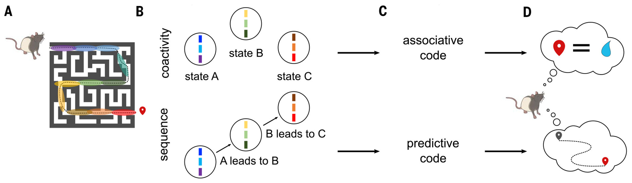
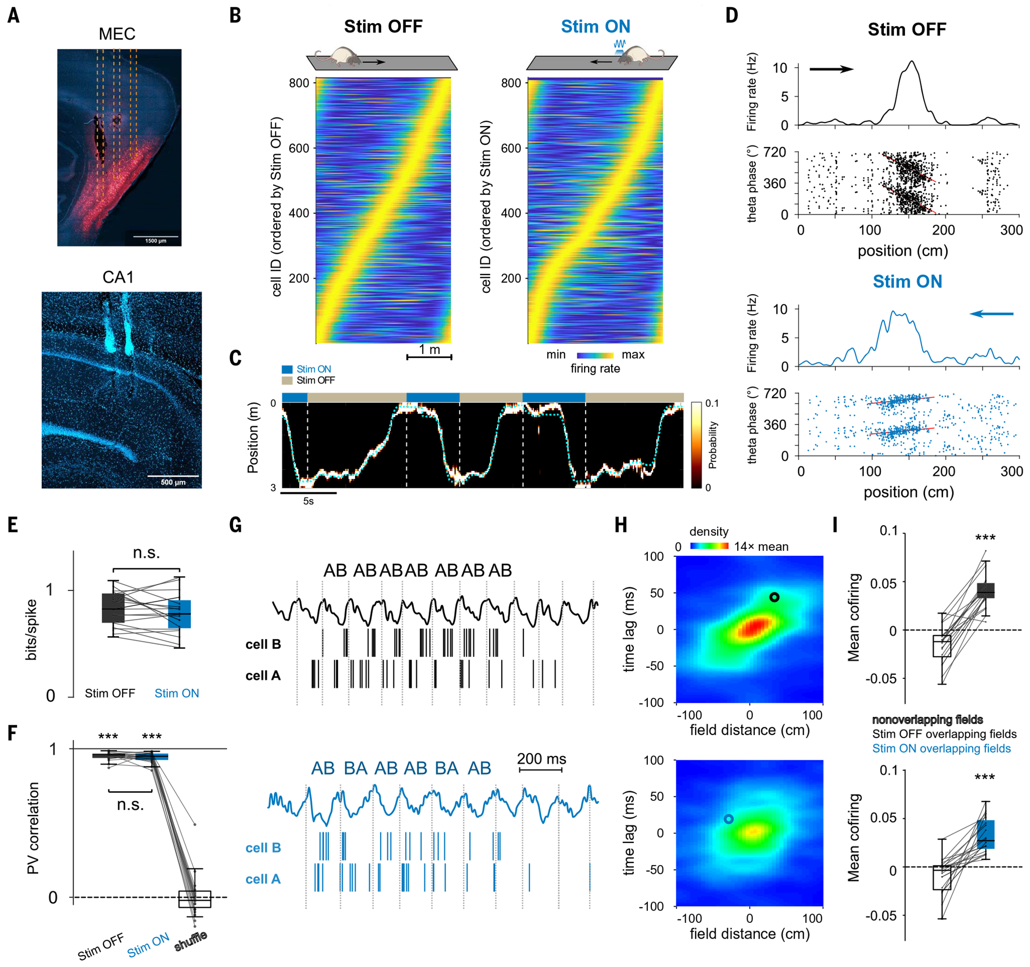
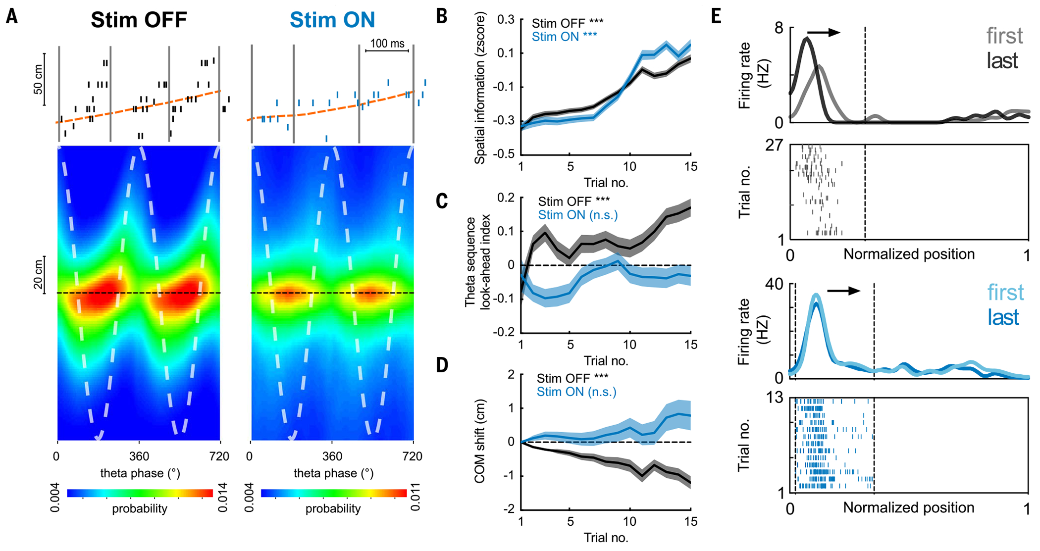
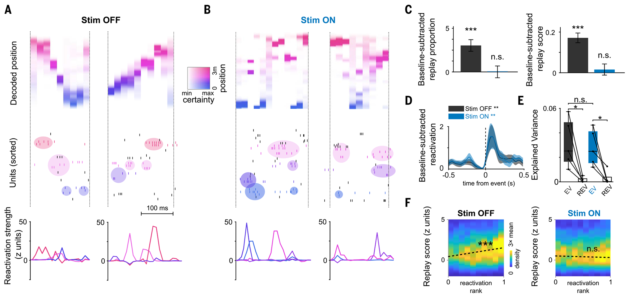
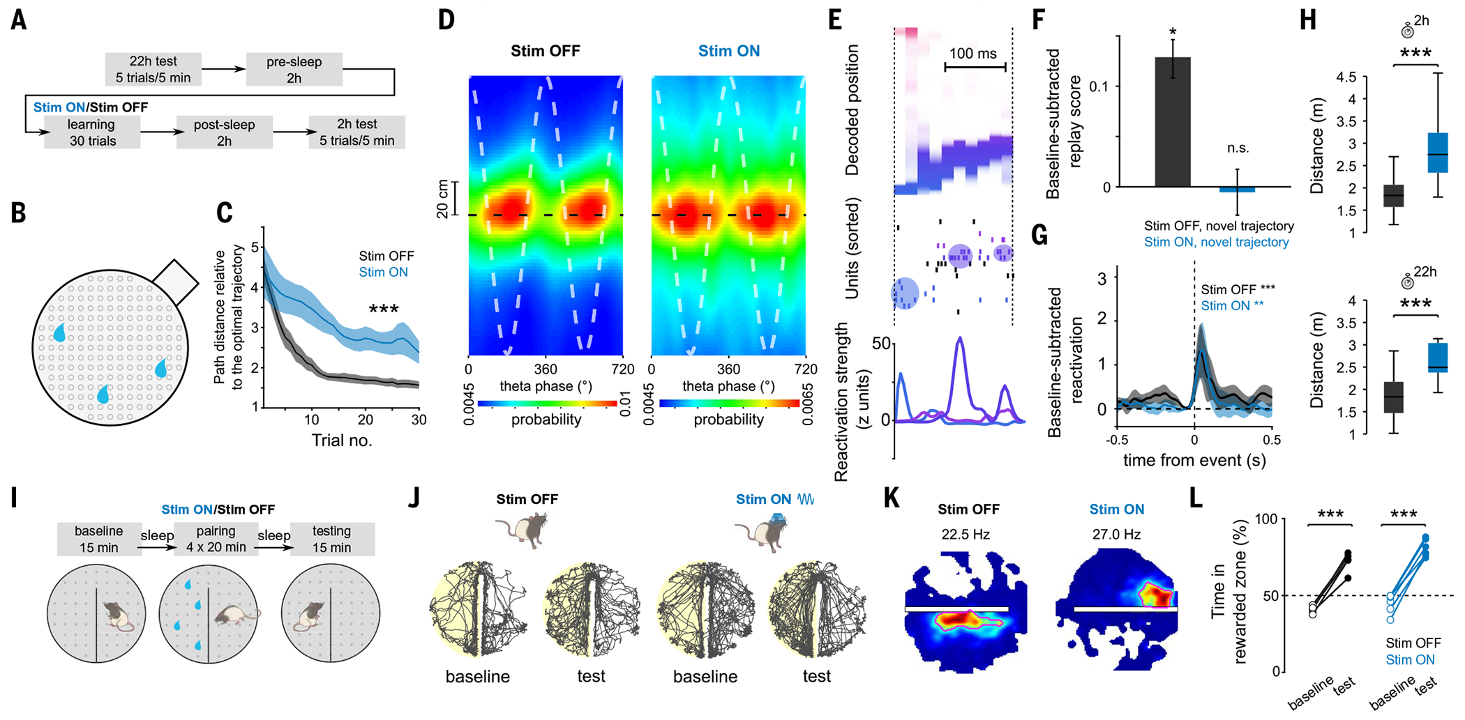
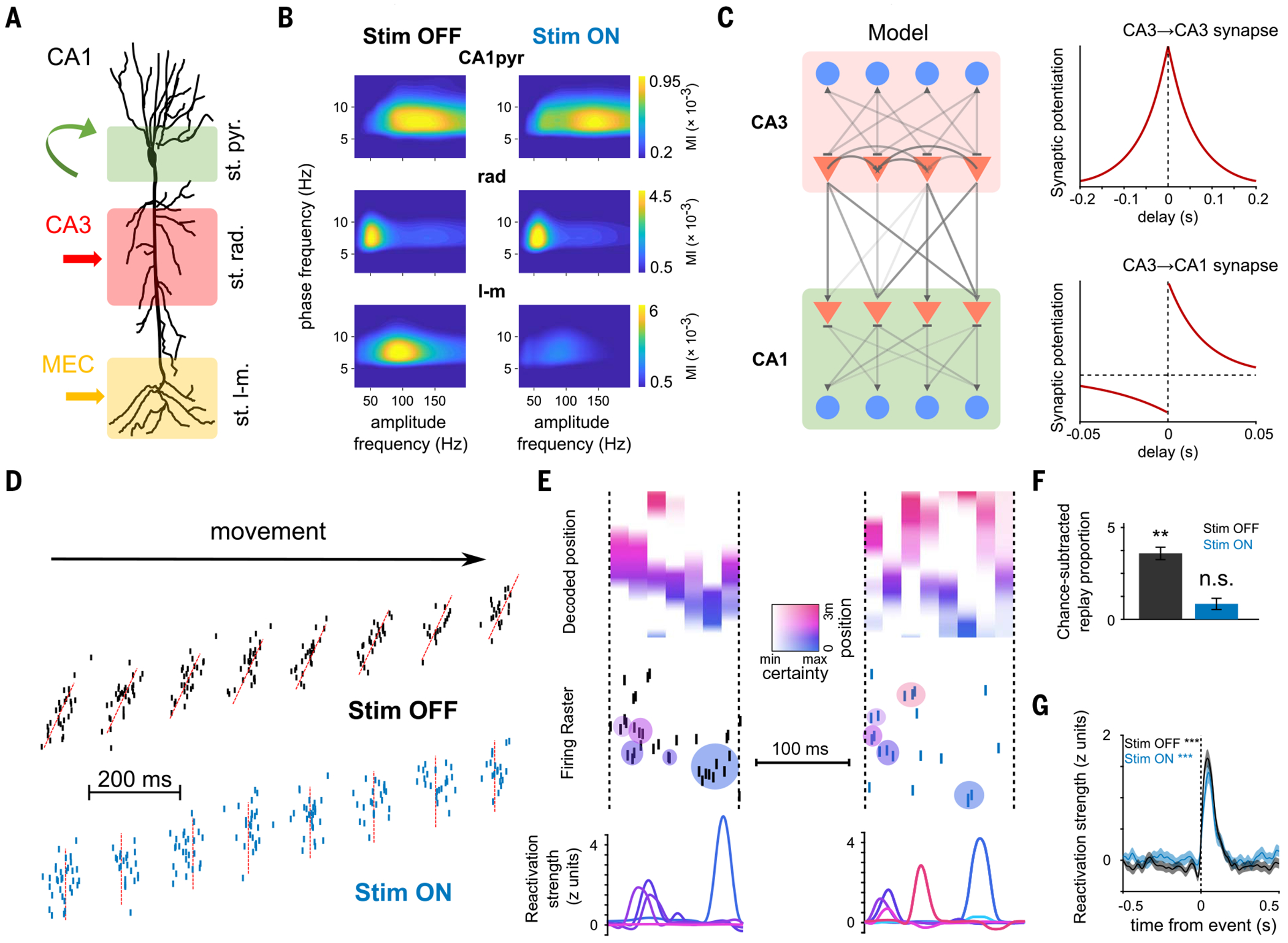
Episodic memory involves learning and recalling associations between items and their spatiotemporal context. Those memories can be further used to generate internal models of the world that enable predictions to be made. The mechanisms that support these associative and predictive aspects of memory are not yet understood. In this study, we used an optogenetic manipulation to perturb the sequential structure, but not global network dynamics, of place cells as rats traversed specific spatial trajectories. This perturbation abolished replay of those trajectories and the development of predictive representations, leading to impaired learning of new optimal trajectories during memory-guided navigation. However, place cell assembly reactivation and reward-context associative learning were unaffected. Our results show a mechanistic dissociation between two complementary hippocampal codes: an associative code (through coactivity) and a predictive code (through sequences).


Conflict of interest statement

Competing interests: The authors declare that they have no competing interests.

Figures

Fig. 1.
Fig. 1.. Two hypothesized memory codes in the hippocampus.
(A) As a rat learns to navigate a maze to find a reward, place cells activate along its spatial trajectory (color areas represent place fields). (B) During navigation, but also during offline periods, place cells with overlapping fields are active together, forming functional assemblies (top), and these assemblies form a sequence that reproduces an animal's learned trajectory (bottom). (C) We propose that the coactivity of cell assemblies represents an associative code to learn discrete states in the world, while their sequential activation forms a predictive code to learn behaviorally relevant state transitions. (D) An associative memory code may be sufficient for some types of memory, such as associating a place with a reward. Other types of memory-guided behavior, such as flexibly navigating a complex environment, would require a predictive model.
Fig. 2.
Fig. 2.. Optogenetic perturbation of MEC gamma timing impairs temporal but not rate coding features of CA1 place cells.
(A) Histological sections showing (top) expression of mDlx-ChR2-mCherry in MEC GABAergic cells (red) and optic fibers’ tracks (dashed lines) and (bottom) probe locations in CA1. (B) Rate maps for CA1 pyramidal cells (n = 815) in the Stim OFF and Stim ON directions. (C) Example Bayesian decoding of CA1 population spike trains during running behavior. Dashed cyan line represents animal’s actual position. The color map indicates probability of a decoded location. (D) Representative place cells for the Stim OFF (top) and Stim ON (bottom) directions. (Top) Firing rate as a function of spatial positions. (Bottom) Theta phase–position raster of place cell spikes. Red line represents the linear circular regression. (E) Spatial information was not significantly different between cells in the Stim OFF and Stim ON directions (P = 0.39; Wilcoxon signed-rank test). (F) Representation stability as measured by the population vector (PV) correlation between even and odd trials in both directions was higher than shuffled data (OFF/ON versus shuffle: P = 6 × 10⁻⁸/P = 6 × 10⁻⁸, Wilcoxon signed-rank test) and not different between the ON and the OFF directions (P = 0.35, Wilcoxon signed-rank test). (G) Examples of theta firing relationships for cell pairs with overlapping place fields in OFF (top) and ON (bottom) directions. The cells cofired in the same theta cycles in both cases, but the order of firing (AB) is only consistent in the OFF direction. (H) Average normalized density plot between place field distance and theta timescale firing lag for all overlapping place cell pairs in OFF (top) and ON (bottom) running directions. Theta compression was disrupted in Stim ON only (P = 9 × 10⁻⁵, Wilcoxon signed-rank test between theta compression slopes; see methods). Circles indicate the cell pairs shown in (G). (I) Cofiring of overlapping place cells in the same theta cycles was preserved in both directions (overlapping versus nonoverlapping place cells in OFF/ON: P = 1 × 10⁻⁴/P = 9 × 10⁻⁵, Wilcoxon signed-rank test; OFF versus ON: P = 0.067). ***P < 0.001.
Fig. 3.
Fig. 3.. Impaired development of place cell theta sequences and predictive properties.
(A) (Top) Example theta sequences. (Bottom) Average (n = 20 sessions in five rats) decoded position estimated across different theta phases, relative to the actual animal position (dashed black line). Stim ON theta sequences were impaired (Stim OFF versus Stim ON quadrant scores: P = 7 × 10−141; weighted correlation: P = 4 × 10⁻²², Wilcoxon rank sum test, n = 17,992/13,265 theta cycles). (B) Increase in spatial information of place cells across laps in both Stim ON (blue) and Stim OFF (black) conditions (***P = 1.8 × 10⁻¹¹/1.2 × 10−5 for Stim OFF/ON, Wilcoxon rank sum tests comparing the 1st versus the 15th lap). (C) Increase in theta sequence look-ahead index in the Stim OFF (***P = 4.0 × 10⁻⁶), but not the Stim ON, direction (P = 0.71, Wilcoxon signed-rank tests). (D) Backward shifting of PF center-of-mass (COM) overlaps on the Stim OFF trajectory (***P = 1.3 × 10−69) but not the Stim ON trajectory (P = 0.16, Wilcoxon signed-rank tests). COM shift relative to the first lap is shown (>0, forward shifting to future; < 0, backward shifting to past). (E) Example place cells showing backward shifting place fields on the Stim OFF trajectory (top) and stable fields on the Stim ON trajectory (bottom). Arrows depict the animal’s running direction. Curves on top show smoothed firing rate for the first and last laps, and raster plots below show spikes across all laps. For all these analyses, n = 20 sessions from five rats were used.
Fig. 4.
Fig. 4.. Impaired replay with preserved reactivation of a novel experience.
(A and B) Examples of replay and reactivation for (A) the Stim OFF and (B) the Stim ON experience, respectively. (Top) Decoded position during replay events (color coded by decoded position). (Middle) Raster plots of neuronal firing; colored circles indicate activations of assemblies composed of neurons with nearby place fields (colored ticks; colors of assemblies reflect their peak activation position). (Bottom) Assembly reactivation strength curves. (C) Increase in proportion significant replay (left) and sequence scores (right) of SWRs in post-task sleep as compared with pre-task sleep. Unlike the OFF direction, there was no significant replay in the ON direction (n = 4437 events; Stim OFF: studenťs t test, ***P = 7 × 10⁻¹¹; Stim ON: P = 0.29 for proportions; and Wilcoxon signed-rank test: OFF ***P = 3 × 10⁻¹⁷ and ON P = 0.29 for scores). Only the first session in the maze was included (n = 5 sessions). (D) Reactivation strength of task-related assemblies centered on post-task sleep SWR, increased relative to baseline (Stim OFF post-task sleep versus baseline sleep during SWRs: Wilcoxon signed-rank test, n = 31 components, **P = 0.0096; Stim ON post-task sleep versus baseline sleep during SWRs: Wilcoxon signed-rank test, n = 27 components, **P = 0.0026). (E) Reactivation of pairwise neuronal correlation as measured by explained variance (EV) in post-task sleep was significant for both directions (Stim OFF: *P = 0.031; Stim ON: Wilcoxon signed-rank test, *P = 0.031; Stim OFF versus Stim ON, Wilcoxon signed-rank test, P = 0.81; n = 5 sessions). (F) Correlation between replay scores and assembly reactivation strength for post-task SWRs computed independently for Stim OFF and Stim ON replay events. Reactivation and replay were correlated in Stim OFF (Pearson’s correlation coefficient r = 0.11, ***P = 3 × 10⁻¹²) but not in Stim ON (Pearson’s r = −0.028, P = 0.06).
Fig. 5.
Fig. 5.. Internally generated sequences are needed for memory-guided behavior.
(A) Task structure of the cheeseboard task: Each day, animals learned a novel trajectory to get three hidden rewards in a circular arena. (B) Schematic of the cheeseboard setup. (C) Learning performance measured as distance traveled relative to optimal trajectory. ANOVA with repeated measures showed a significant main effect of group (F1,43 = 133.4, P < 10−10; n = 33 and 11 sessions for Stim OFF and Stim ON, respectively). (D) Average decoded position versus theta phase, relative to actual animal position (dashed black line). Stim ON theta sequences were degraded (quadrant scores were lower in Stim ON than in Stim OFF: n = 5958/2695 cycles for Stim OFF and Stim ON, respectively, P = 2 × 10⁻³; weighted correlations were lower in Stim ON than in Stim OFF: P = 3 × 10⁻⁴, rank sum test). Two cycles are shown for visibility. (E) Example of a decoded replay event. Decoded linearized position (top), spike raster (middle), and assembly reactivation strength (bottom) were shown for the same replay event. (F) Increase in replay score in post-task sleep as compared with pre-task sleep. Unlike the Stim OFF condition, there was no significant replay in the Stim ON condition (Stim OFF: P = 3 × 10⁻²⁰; signed-rank test, n = 11,460 events; Stim ON: P = 0.41, n = 8689 events). (G) Reactivation strength of task-related assemblies centered on post-task sleep SWR (Stim OFF post-task sleep versus baseline sleep during SWRs: n = 36 components, P = 4.71 × 10⁻⁴; Stim ON post-task sleep versus baseline sleep during SWRs: n = 22 components, P = 3.19 × 10⁻³, signed-rank test). (H) Memory performance during 2 and 22 hour post-learning recall tests (P = 6.8 × 10⁻⁴/7.9 × 10−4 for 2 and 22 hour tests, rank sum test; n = 25/11 for Stim OFF/ON sessions from five rats). (I) Task structure of CPP task. (J) Example rat paths for Stim OFF (left) and Stim ON (right) baseline and testing sessions on CPP task. The side rewarded during pairing is highlighted in yellow. (K) Example place fields near the boundary for Stim OFF (left) and Stim ON (right) conditions, illustrating place field elongation along the boundary, which was disrupted in the Stim ON condition. Red circles emphasize the shape of the place field. (L) CPP memory performance: both Stim ON and Stim OFF training resulted in animals spending more active time in rewarded versus unrewarded side (Stim OFF: paired t test, n = 5 sessions, P = 2.85 × 10⁻⁴; Stim ON: paired t test, n = 6 sessions, P = 2.10 × 10⁻⁵).
Fig. 6.
Fig. 6.. Circuit mechanisms for coactivity and sequence hippocampal dynamics.
(A) Schema depicting the inputs to CA1 pyramidal neurons stratified along their somatodendritic axis. Local inhibitory inputs are dominant in the pyramidal layer (st. pyr.), CA3 inputs target proximal apical dendrites in the stratum radiatum (st. rad.), and entorhinal inputs target distal dendrites in the stratum lacunosum-moleculare (st. l-m.). (B) Gamma amplitude-theta phase comodulograms for each layer-specific LFP component (see methods) displayed modulation in a specific gamma sub-band (averaged data from n = 10 sessions from four rats): CA1pyr in gammaF (100 to 160 Hz), rad in gammaS (30 to 60 Hz), and LM in gammaM (60 to 110 Hz). (Right) MEC perturbation selectively impaired LM gammaM but not CA1pyr or rad oscillations (**P = 0.002, Wilcoxon signed-rank test; n = 13 sessions from five rats). (C) (Left) Model schematic depicting a subnetwork of CA3 cells projecting to a subnetwork of CA1 cells. Triangles represent pyramidal neurons, and circles represent inhibitory interneurons. (Right) STDP rules used to train different synapses within the network during learning trials. (D) In Stim OFF simulations (top), place cells displayed phase precession and prominent theta sequences. In Stim ON simulations (bottom), phase precession was disrupted, and theta sequences were abolished. (E) Example replay events simulated by the model after Stim OFF (left) and Stim ON (right) learning. Decoded position, spike raster, and assembly reactivation strength as in Fig. 4. (F) Increase in proportion of “SWR” events with significant replay in post-task “sleep” epochs in Stim OFF and Stim ON protocols. Replay improvement was above chance levels in Stim OFF but not Stim ON simulations (Stim OFF: P = 0.0039, Wilcoxon signed-rank test, n = 6585 events; Stim ON: P = 0.19, n = 6031 events). (G) Reactivation strength of task-related assemblies centered on post-task sleep SWR-like events increase relative to baseline (Stim OFF post-task sleep versus baseline sleep during SWR-like events: Wilcoxon signed-rank test, n = 362 components, ***P = 5 × 10−44; Stim ON post-task sleep versus baseline sleep during SWR-like events: Wilcoxon signed-rank test, n = 312 components, ***P = 1 × 10⁻²⁷).

References

    1. Nakazawa K. et al., Requirement for hippocampal CA3 NMDA receptors in associative memory recall. Science 297, 211–218 (2002). doi: 10.1126/science.1071795; - DOI - PMC - PubMed
    1. Tovote P, Fadok JP, Lüthi A, Neuronal circuits for fear and anxiety. Nat. Rev. Neurosci 16, 317–331 (2015). doi: 10.1038/nrn3945; - DOI - PubMed
    1. Barker GRI, Warburton EC, When is the hippocampus involved in recognition memory? J. Neurosci 31, 10721–10731 (2011). doi: 10.1523/JNEUR0SCI.6413-10.2011; - DOI - PMC - PubMed
    1. Beylin AV et al., The role of the hippocampus in trace conditioning: Temporal discontinuity or task difficulty? Neurobiol. Learn. Mem 76, 447–461 (2001). doi: 10.1006/nlme.2001.4039; - DOI - PubMed
    1. MacDonald CJ, Lepage KQ, Eden UT, Eichenbaum H, Hippocampal “time cells” bridge the gap in memory for discontiguous events. Neuron 71, 737–749 (2011). doi: 10.1016/j.neuron.2011.07.012; - DOI - PMC - PubMed