. 2023 Nov:67:102929.
doi: 10.1016/j.redox.2023.102929. Epub 2023 Oct 10.

Arginase2 mediates contrast-induced acute kidney injury via facilitating nitrosative stress in tubular cells

Affiliations

Arginase2 mediates contrast-induced acute kidney injury via facilitating nitrosative stress in tubular cells

Ling-Yun Zhou et al. Redox Biol. 2023 Nov.

Abstract

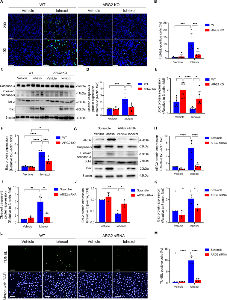
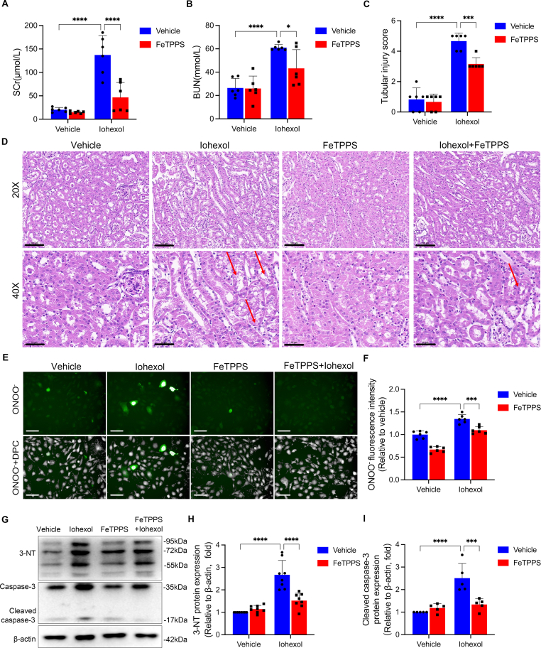
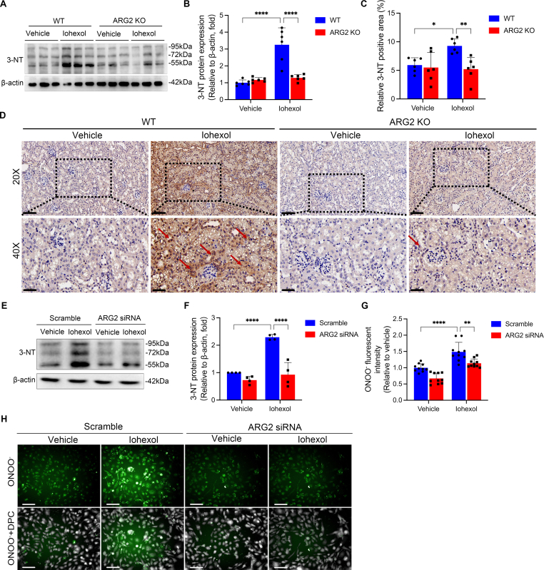
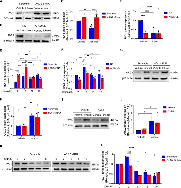
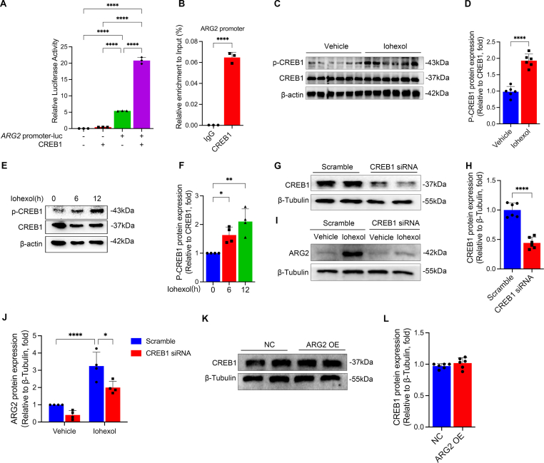
Contrast-induced acute kidney injury(CI-AKI) is the third cause of AKI. Although tubular injury has been regarded as an important pathophysiology of CI-AKI, the underlying mechanism remains elusive. Here, we found arginase2(ARG2) accumulated in the tubules of CI-AKI mice, and was upregulated in iohexol treated kidney tubular cells and in blood samples of CI-AKI mice and patients, accompanied by increased nitrosative stress and apoptosis. However, all of the above were reversed in ARG2 knockout mice, as evidenced by the ameliorated kidney dysfunction and the tubular injury, and decreased nitrosative stress and apoptosis. Mechanistically, HO-1 upregulation could alleviate iohexol or ARG2 overexpression mediated nitrosative stress. Silencing and overexpressing ARG2 was able to upregulate and downregulate HO-1 expression, respectively, while HO-1 siRNA had no effect on ARG2 expression, indicating that ARG2 might inhibit HO-1 expression at the transcriptional level, which facilitated nitrosative stress during CI-AKI. Additionally, CREB1, a transcription factor, bound to the promoter region of ARG2 and stimulated its transcription. Similar findings were yielded in cisplatin- or vancomycin-induced AKI models. Taken together, ARG2 is a crucial target of CI-AKI, and activating CREB1/ARG2/HO-1 axis can mediate tubular injury by promoting nitrosative stress, highlighting potential therapeutic strategy for treating CI-AKI.

Keywords: Apoptosis; Arginase2; Contrast-induced AKI; Key target; Kidney tubular cells; Nitrosative stress.


Conflict of interest statement

Declaration of competing interest Authors declare that there are no conflicts of interest.

Figures

Image 1
Graphical abstract
Fig. 1
Fig. 1
ARG2 is upregulated in the CI-AKI mice kidneys and primary tubular epithelial cells, and increased in blood samples of CI-AKI mice and patients. (A) Heat map of proteomics in iohexol-induced AKI mice. P-Value <0.05 and log2 fold change (FC) > 2. (B) Heat map of transcriptomics in iohexol-induced AKI mice. P-Value <0.0001 and log2 fold change (FC) > 3. (C–E) The ARG2 protein and mRNA expressions were evaluated in the kidneys of mice treated with iohexol. (F) Representative images of immunohistochemistry staining of ARG2 in the kidneys of iohexol-induced AKI mice. The red arrows indicate deeper ARG2-positive staining of renal tubules. Scale, 20×, 100 μm, 40×, 50 μm. (G) Representative images of the immunofluorescence colocalization of CD31, F4/80, LTL, PNA, and DBA with ARG2 in the kidney of iohexol-induced AKI mice. The following specific markers were used: endothelial, CD31; macrophage, F4/80; proximal tubule, lotus tetragonolobus lectin (LTL); distal tubule, peanut agglutinin (PNA); and collecting duct, dolichos biflorus agglutinin (DBA). The white arrows indicate positive tubules with colocalization of ARG2 and specific tubular markers. Scale, 50 μm. (H–J) ARG2 protein and mRNA expressions in primary tubular epithelial cells treated with iohexol. (K) Representative image of ARG2 and mito-tracker immunofluorescence colocalization in HK-2 cells. The orange arrows indicate that iohexol causes a decrease in mitochondrial fluorescence. The white arrows indicate the normal colocalization of ARG2 with mitochondria. The yellow arrows indicate that iohexol leads to reduced colocalization of ARG2 with mitochondria. Scale, 20 μm. (L and M) Serum levels of ARG2 in CI-AKI mice (L) and patients (M). *P < 0.05, **P < 0.01, ***P < 0.001, ****P < 0.0001. mean ± SD, n = 5-6 in mice, n = 3–6 in HK-2 cells.
Fig. 2
Fig. 2
ARG2 inhibition and deficiency significantly ameliorate iohexol-induced AKI. (A–D) Renal function and renal tissue injury of iohexol-induced AKI mice treated with nor-NOHA were assessed by SCr, BUN, and tubular injury score. Scale, 20×, 100 μm, 40×, 50 μm. (E–H) Renal function and renal tissue injury of ARG2 KO and WT mice injected by iohexol via tail vein were assessed by SCr, BUN, and tubular injury score. The red arrows indicate the vacuolar degeneration of renal tubules. Scale, 20×, 100 μm, 40×, 50 μm *P < 0.05, **P < 0.01, ***P < 0.001, ****P < 0.0001. mean ± SD, n = 6.
Fig. 3
Fig. 3
ARG2 deficiency significantly reduces apoptosis in iohexol-induced AKI mice and HK-2 cells. (A and B) Representative images of TUNEL staining in ARG2 KO and WT mice treated with iohexol. TUNEL-positive cells and nuclei were indicated by green and blue fluorescence, respectively. (C–F) The protein expressions of caspase-3, cleaved caspase-3, Bcl-2, and Bax were evaluated with western blotting in ARG2 KO and WT mice administrated with iohexol. β-actin was used as the loading control. (G–K) Western blotting was used to evaluate the protein expressions of ARG2, caspase-3, cleaved caspase-3, Bcl-2, and Bax in HK-2 cells transfected with ARG2 siRNA and incubated with iohexol. (L and M) TUNEL staining was performed to examine apoptotic HK-2 cells transfected with ARG2 siRNA and incubated with iohexol. TUNEL-positive cells and nuclei were indicated by green and blue fluorescence, respectively. *P < 0.05, **P < 0.01, ***P < 0.001, ****P < 0.0001. mean ± SD, n = 6 in mice, n = 3-6 in HK-2 cells.
Fig. 4
Fig. 4
FeTPPS rescues nitrosative stress caused by iohexol in mice and HK-2 cells. (A–D) Renal function and renal tissue injury of AKI mice induced by iohexol treated with or without FeTPPS were assessed by SCr, BUN, and tubular injury score. The red arrows indicate the vacuolar degeneration of renal tubules. (E and F) The level of ONOO was evaluated with immunofluorescence in HK-2 cells cultured with FeTPPS and iohexol. Green fluorescence indicates ONOO. The number of cells was counted by DPC. DPC, Digital phase contrast. Scale, 100 μm. (G–I) The protein expressions of 3-NT, caspase-3 and cleaved caspase-3 were evaluated by western blotting in HK-2 cells cultured with FeTPPS and iohexol. *P < 0.05, **P < 0.01, ***P < 0.001, ****P < 0.0001. mean ± SD, n = 6 in mice, n =5-8 in HK-2 cells.
Fig. 5
Fig. 5
ARG2 mediates nitrosative stress in iohexol-induced AKI and HK-2 cells. (A and B) The protein expression of 3-NT was evaluated by western blotting in ARG2 KO and WT mice treated with iohexol. (C and D) Representative images of immunohistochemistry staining of 3-NT in the kidneys of ARG2 KO and WT mice treated with iohexol. The red arrows indicate deeper 3-NT-positive staining of renal tubules. Scale, 20×, 100 μm, 40×, 50 μm. (E and F) The expression of 3-NT was evaluated with western blotting in HK-2 cells transfected with ARG2 siRNA and incubated with iohexol. (G and H) The level of ONOO was evaluated with immunofluorescence in HK-2 cells transfected with ARG2 siRNA and incubated with iohexol. Green fluorescence indicates ONOO. The number of cells was counted by DPC. DPC, Digital phase contrast. Scale, 100 μm *P < 0.05, **P < 0.01, ****P < 0.0001. mean ± SD, n = 6 in mice, n = 3-10 in HK-2 cells.
Fig. 6
Fig. 6
HO-1 is involved in ARG2 mediated nitrosative stress. (A) Correlation between ARG2 protein expression and significantly different proteins of proteomics. X-axis, R with ARG2; Y-axis, P-value. (B) The HO-1 mRNA expression was evaluated in mice treated with iohexol. (C and D) The HO-1 protein expression was evaluated by western blotting in ARG2 KO and WT mice administrated with iohexol. (E–G) The HO-1 mRNA and protein expressions were evaluated in primary tubular epithelial cells incubated with iohexol. (H and I) The level of ONOO was evaluated by immunofluorescence in HK-2 cells transfected with HO-1 overexpression plasmid or cultured with iohexol. Green fluorescence indicates ONOO. The number of cells was counted by DPC. DPC, Digital phase contrast. Scale, 100 μm. (J and K) The 3-NT protein expression was evaluated by western blotting in HK-2 cells incubated with iohexol and transfected with ARG2 overexpression plasmid and HO-1 overexpression plasmid. *P < 0.05, **P < 0.01, ***P < 0.001, ****P < 0.0001. mean ± SD, n = 6 in mice, n = 3-6 in cells.
Fig. 7
Fig. 7
ARG2 inhibits the expression of HO-1 at the transcriptional level in HK-2 cell cultured with iohexol. (A–D) The HO-1 protein expression was examined by western blotting in HK-2 cells transfected with ARG2 siRNA or ARG2 overexpression plasmid, and incubated with iohexol. (E and F) The HO-1 mRNA expression was evaluated in HK-2 cells transfected with ARG2 siRNA or ARG2 overexpression, and incubated with iohexol. (G and H) The ARG2 protein expression was evaluated with western blotting in HK-2 cells transfected with HO-1 siRNA or cultured with iohexol. (I and J) The ARG2 protein expression was evaluated with western blotting in HK-2 cells cultured with CoPP and iohexol. (K and L) The HO-1 protein expression was evaluated with western blotting in HK-2 cells transfected with ARG2 siRNA or cultured with CHX. *P < 0.05, **P < 0.01, ***P < 0.001, ****P < 0.0001. mean ± SD, n = 3-6 in HK-2 cells.
Fig. 8
Fig. 8
CREB1 regulates ARG2 transcription. (A) Luciferase reporter gene results demonstrated that CREB1 combined with ARG2 promoter. (B) ChIP-PCR showed a direct combination and enrichment of CREB1 to the promoter region of ARG2. (C and D) The P-CREB1 and CREB1 protein expressions were evaluated in mice treated with iohexol. (E and F) The P-CREB1 and CREB1 protein expressions were evaluated in HK-2 cells cultured with iohexol at the indicated time points. (G–J) The CREB1 and ARG2 protein expression were evaluated in HK-2 cells transfected with CREB1 siRNA. (K and L) The CREB1 protein expression was evaluated in HK-2 cells transfected with ARG2 overexpression plasmid. *P < 0.05, **P < 0.01, ****P < 0.0001. mean ± SD, n = 6 in mice, n = 3-6 in HK-2 cells.
Fig. 9
Fig. 9
ARG2 may be a potential target of cisplatin-induced AKI. (A–C) ARG2 protein and mRNA expressions in the kidneys of cisplatin-induced AKI mice. (D) Representative images of immunohistochemistry staining of ARG2 in cisplatin-induced AKI mice kidneys. The red arrows indicate deeper ARG2-positive staining of renal tubules. Scale, 20×, 100 μm, 40×, 50 μm. (E) Representative images of the immunofluorescence colocalization of CD31, F4/80, LTL, PNA, and DBA with ARG2 in the kidney of cisplatin-induced AKI mice. The following specific markers were used: endothelial, CD31; macrophage, F4/80; proximal tubule, lotus tetragonolobus lectin (LTL); distal tubule, peanut agglutinin (PNA); and collecting duct, dolichos biflorus agglutinin (DBA). The white arrows indicate positive tubules with colocalization of ARG2 and specific tubular markers. Scale, 50 μm. (F and G) ARG2 protein expression in primary tubular epithelial cells treated by cisplatin. (H–K) Renal function and renal tissue injury of ARG2 KO and WT mice administrated with cisplatin were assessed by SCr, BUN, and tubular injury score. The red arrows showed the vacuolar degeneration, tubular necrosis, cast formation, and tubular dilation of renal tubules. (L and M) The HO-1 protein expression was evaluated by western blotting in ARG2 KO and WT mice administrated with cisplatin. *P < 0.05, **P < 0.01, ***P < 0.001, ****P < 0.0001. mean ± SD, n = 6.
Fig. 10
Fig. 10
ARG2 may be a potential target of vancomycin-induced AKI. (A–C) ARG2 protein and mRNA expressions in the kidneys of vancomycin-induced AKI mice. (D) Representative images of immunohistochemistry staining of ARG2 in vancomycin-induced AKI mice kidneys. The red arrows indicate deeper ARG2-positive staining of renal tubules. Scale, 20×, 100 μm, 40×, 50 μm. (E) Representative images of the immunofluorescence colocalization of CD31, F4/80, LTL, PNA, and DBA with ARG2 in the kidney of vancomycin-induced AKI mice. The following specific markers were used: endothelial, CD31; macrophage, F4/80; proximal tubule, lotus tetragonolobus lectin (LTL); distal tubule, peanut agglutinin (PNA); and collecting duct, dolichos biflorus agglutinin (DBA). The white arrows indicate positive tubules with colocalization of ARG2 and specific tubular markers. Scale, 50 μm. (F and G) ARG2 protein expression in primary tubular epithelial cells treated by vancomycin. (H–K) Renal function and renal tissue injury of ARG2 KO and WT mice administrated with vancomycin were assessed by SCr, BUN, and tubular injury score. The red arrows showed the vacuolar degeneration, tubular necrosis, cast formation, and tubular dilation of renal tubules. (L and M) The HO-1 protein expression was evaluated by western blotting in ARG2 KO and WT mice administrated with vancomycin. *P < 0.05, **P < 0.01, ***P < 0.001, ****P < 0.0001. mean ± SD, n = 6.

References

    1. Yang L., Xing G., Wang L., Wu Y., Li S., Xu G., He Q., Chen J., Chen M., Liu X., Zhu Z., Yang L., Lian X., Ding F., Li Y., Wang H., Wang J., Wang R., Mei C., Xu J., Li R., Cao J., Zhang L., Wang Y., Xu J., Bao B., Liu B., Chen H., Li S., Zha Y., Luo Q., Chen D., Shen Y., Liao Y., Zhang Z., Wang X., Zhang K., Liu L., Mao P., Guo C., Li J., Wang Z., Bai S., Shi S., Wang Y., Wang J., Liu Z., Wang F., Huang D., Wang S., Ge S., Shen Q., Zhang P., Wu L., Pan M., Zou X., Zhu P., Zhao J., Zhou M., Yang L., Hu W., Wang J., Liu B., Zhang T., Han J., Wen T., Zhao M., Wang H. Acute kidney injury in China: a cross-sectional survey. Lancet. 2015;386(10002):1465–1471. - PubMed
    1. Bejoy J., Qian E.S., Woodard L.E. Tissue culture models of AKI: from tubule cells to human kidney organoids. J. Am. Soc. Nephrol. 2022;33(3):487–501. - PMC - PubMed
    1. James M.T., Bhatt M., Pannu N., Tonelli M. Long-term outcomes of acute kidney injury and strategies for improved care. Nat. Rev. Nephrol. 2020;16(4):193–205. - PubMed
    1. Liu C., Yan S., Wang Y., Wang J., Fu X., Song H., Tong R., Dong M., Ge W., Wang J., Yang H., Wang C., Xia P., Zhao L., Shen S., Xie J., Xu Y., Ma P., Li H., Lu S., Ding Y., Jiang L., Lin Y., Wang M., Qiu F., Feng W., Yang L. Drug-induced hospital-acquired acute kidney injury in China: a multicenter cross-sectional survey. Kidney Dis. 2021;7(2):143–155. - PMC - PubMed
    1. Wang H., Gao T., Zhang R., Hu J., Wang Y., Wei J., Zhou Y., Dong H. The intellectual base and global trends in contrast-induced acute kidney injury: a bibliometric analysis. Ren. Fail. 2023;45(1) - PMC - PubMed

Publication types

LinkOut - more resources