. 2023 Dec 1;64(12):1956-1964.
doi: 10.2967/jnumed.123.265418.

Gadolinium-Based Nanoparticles Sensitize Ovarian Peritoneal Carcinomatosis to Targeted Radionuclide Therapy

Affiliations

Gadolinium-Based Nanoparticles Sensitize Ovarian Peritoneal Carcinomatosis to Targeted Radionuclide Therapy

Clara Diaz Garcia-Prada et al. J Nucl Med. .

Abstract

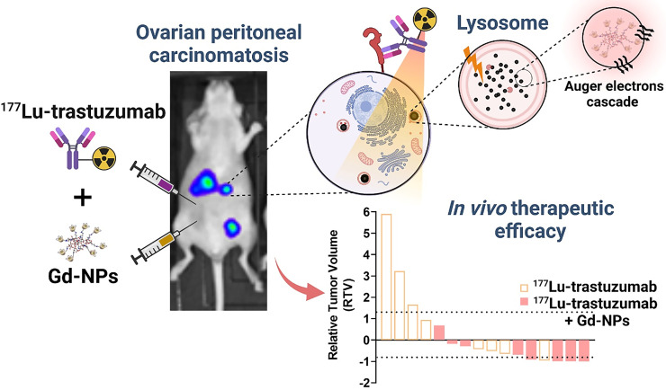
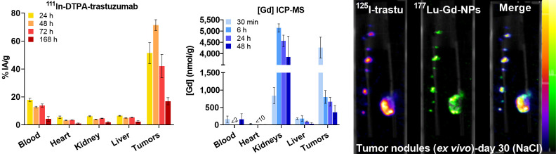
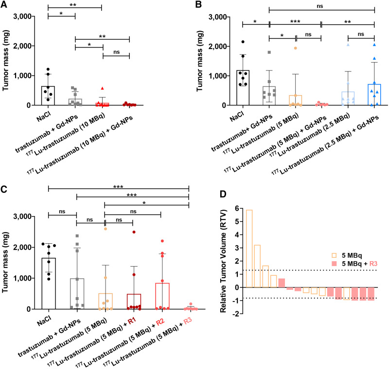
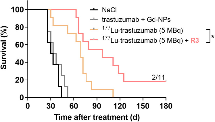
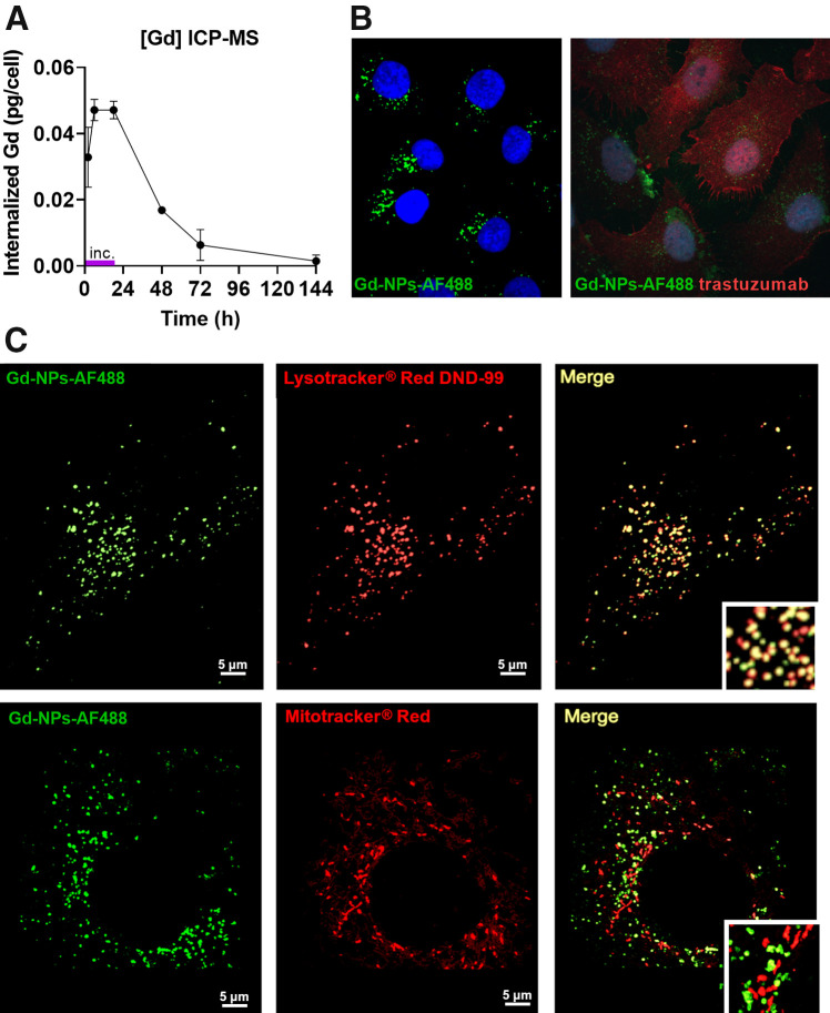
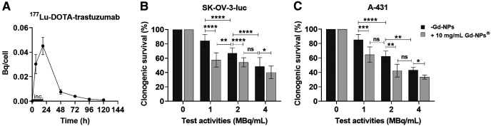
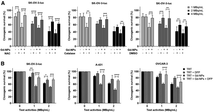
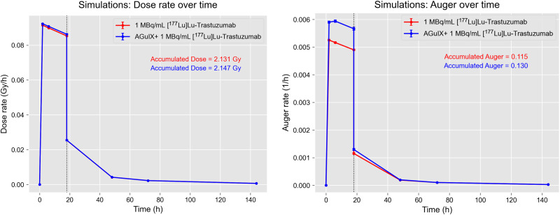
Ovarian cancer (OC) is the most lethal gynecologic malignancy (5-y overall survival rate, 46%). OC is generally detected when it has already spread to the peritoneal cavity (peritoneal carcinomatosis). This study investigated whether gadolinium-based nanoparticles (Gd-NPs) increase the efficacy of targeted radionuclide therapy using [177Lu]Lu-DOTA-trastuzumab (an antibody against human epidermal growth factor receptor 2). Gd-NPs have radiosensitizing effects in conventional external-beam radiotherapy and have been tested in clinical phase II trials. Methods: First, the optimal activity of [177Lu]Lu-DOTA-trastuzumab (10, 5, or 2.5 MBq) combined or not with 10 mg of Gd-NPs (single injection) was investigated in athymic mice bearing intraperitoneal OC cell (human epidermal growth factor receptor 2-positive) tumor xenografts. Next, the therapeutic efficacy and toxicity of 5 MBq of [177Lu]Lu-DOTA-trastuzumab with Gd-NPs (3 administration regimens) were evaluated. NaCl, trastuzumab plus Gd-NPs, and [177Lu]Lu-DOTA-trastuzumab alone were used as controls. Biodistribution and dosimetry were determined, and Monte Carlo simulation of energy deposits was performed. Lastly, Gd-NPs' subcellular localization and uptake, and the cytotoxic effects of the combination, were investigated in 3 cancer cell lines to obtain insights into the involved mechanisms. Results: The optimal [177Lu]Lu-DOTA-trastuzumab activity when combined with Gd-NPs was 5 MBq. Moreover, compared with [177Lu]Lu-DOTA-trastuzumab alone, the strongest therapeutic efficacy (tumor mass reduction) was obtained with 2 injections of 5 mg of Gd-NPs/d (separated by 6 h) at 24 and 72 h after injection of 5 MBq of [177Lu]Lu-DOTA-trastuzumab. In vitro experiments showed that Gd-NPs colocalized with lysosomes and that their radiosensitizing effect was mediated by oxidative stress and inhibited by deferiprone, an iron chelator. Exposure of Gd-NPs to 177Lu increased the Auger electron yield but not the absorbed dose. Conclusion: Targeted radionuclide therapy can be combined with Gd-NPs to increase the therapeutic effect and reduce the injected activities. As Gd-NPs are already used in the clinic, this combination could be a new therapeutic approach for patients with ovarian peritoneal carcinomatosis.

Keywords: Auger electrons; TRT; ovarian cancer; radioimmunotherapy; radiopharmaceutical; radiosensitization.


Figures

None
Graphical abstract
FIGURE 1.
FIGURE 1.
(Left) Biodistribution of [111In]In-DTPA-trastuzumab determined by ex vivo γ-counting of tumor nodules and organs collected at various times (4 per time point) after intraperitoneal injection. (Middle) Biodistribution of Gd-NPs by inductively coupled plasma mass spectrometry in tumor nodules and organs collected at various times after intraperitoneal injection (3 per time point). Results are mean ± SEM. (Right) Ex vivo SPECT/CT dual-isotope imaging of SK-OV-3-luc cell tumors collected from mice after intraperitoneal injection of [125I]I-trastuzumab and [177Lu]Lu-CuPRiX (NH TherAguix S.A. and Institut Lumière Matière) Gd-NPs. Merged images confirmed colocalization of trastuzumab and CuPRiX NPs. % IA = percentage injected activity; DTPA = diethylenetriaminepentaacetic acid; ICP-MS = inductively coupled plasma mass spectrometry; trastu = trastuzumab.
FIGURE 2.
FIGURE 2.
Determination of therapeutic efficacy of [177Lu]Lu-DOTA-trastuzumab combined with Gd-NPs. (A and B) Tumor mass was determined in mice treated with maximum tolerated activity (10 MBq) of [177Lu]Lu-DOTA-trastuzumab followed or not (48 h later) by 10 mg of Gd-NPs (A) and low (2.5 MBq) or intermediate (5 MBq) activity of [177Lu]Lu-DOTA-trastuzumab followed or not (48 h later) by 10 mg of Gd-NPs (B). (C) Tumor mass in mice treated with 5 MBq of [177Lu]Lu-DOTA-trastuzumab regimen 1 (1 injection of 4 mg of Gd-NPs/d for 5 d starting 48 h after [177Lu]Lu-DOTA-trastuzumab injection), regimen 2 (2 injections of 2 mg of Gd-NPs/d for 5 d starting 48 h after [177Lu]Lu-DOTA-trastuzumab), or regimen 3 (2 injections of 5 mg of Gd-NPs per day at 24 and 72 h after [177Lu]Lu-DOTA-trastuzumab). (D) Relative tumor volume at treatment end. Results are mean ± SD. *P < 0.05. **P < 0.01. ***P < 0.001. ns = not significant (Mann–Whitney t test).
FIGURE 3.
FIGURE 3.
Kaplan–Meier survival analysis of mice bearing intraperitoneal SK-OV-3-luc tumor cell xenografts. Mice received single intraperitoneal injection of NaCl, 25 μg of trastuzumab plus Gd-NPs (2 × 5 mg per day, 24 and 72 h after trastuzumab), 5 MBq of [177Lu]Lu-DOTA-trastuzumab, or 5 MBq of [177Lu]Lu-DOTA-trastuzumab plus Gd-NPs (2 × 5 mg per day, 24 and 72 h after TRT). R3 = regimen 3.
FIGURE 4.
FIGURE 4.
Gd-NP uptake and subcellular localization. (A) Cellular uptake was measured in SK-OV-3-luc cells by inductively coupled plasma mass spectrometry after 18 h of incubation with Gd-NPs, 10 mg/mL. Results are mean ± SD of 3 independent experiments performed in triplicate. (B) Cytoplasmic localization of Gd-NP Alexa Fluor 488 (Life Technologies Corp.) (left) and trastuzumab (right) by immunofluorescence analysis. (C) Colocalization of Gd-NP Alexa Fluor 488 with lysosomes (Lysotracker red DND-99; Molecular Probes, Inc.) but not with mitochondria (Mitotracker red CMXRos; Molecular Probes, Inc.). ICP-MS = inductively coupled plasma mass spectrometry.
FIGURE 5.
FIGURE 5.
[177Lu]Lu-DOTA-trastuzumab uptake and cytotoxic effects when combined with Gd-NPs. (A) [177Lu]Lu-DOTA-trastuzumab uptake in SK-OV-3-luc cells incubated with radiolabeled antibody, 1 MBq/mL, for 18 h. (B and C) Clonogenic survival of SK-OV-3-luc (B) and A-431 (C) cells incubated with [177Lu]Lu-DOTA-trastuzumab (0–4 MBq/mL) with or without Gd-NPs, 10 mg/mL, for 18 h. Results are mean ± SD of 3 independent experiments performed in triplicate. Role of oxidative stress in Gd-NP radiosensitizing effect was demonstrated using N-acetyl-l-cysteine, catalase, and dimethyl sulfoxide (Fig. 6A). Indeed, when SK-OV-3-luc cells were coincubated with Gd-NPs and each of these antioxidants, clonogenic survival increased. These results were confirmed using deferiprone, an iron chelator that prevents reactive oxygen species formation (Fig. 6B). *P < 0.05. **P < 0.01. ***P < 0.001. ****P < 0.0001. inc = incubation time (18 h); ns = not significant (Mann–Whitney t test).
FIGURE 6.
FIGURE 6.
Oxidative stress is involved in radiosensitizing effects of Gd-NPs. (A) SK-OV-3-luc cells were incubated with [177Lu]Lu-DOTA-trastuzumab, 1 MBq/mL, for 18 h with or without Gd-NPs, 10 mg/mL, with or without N-acetyl-l-cysteine (NAC), catalase, or dimethyl sulfoxide. Then, clonogenic survival was measured. (B) Clonogenic survival of SK-OV-3-luc, A-431, and OVCAR-3 cells incubated with [177Lu]Lu-DOTA-trastuzumab (0–4 MBq/mL) with or without Gd-NPs, 10 mg/mL, for 18 h in presence or not of DFP. Results are mean ± SD of 3 independent experiments performed in triplicate. *P < 0.05. **P < 0.01. ***P < 0.001. ****P < 0.0001. DFP = deferiprone; DMSO = dimethyl sulfoxide; NAC = N-acetyl-l-cysteine; ns = not significant (Mann–Whitney t test) compared with cells treated with [177Lu]Lu-DOTA-trastuzumab.
FIGURE 7.
FIGURE 7.
Physical aspects of TRT combined with Gd-NPs. Dose rate and accumulated dose in lysosomes at various time points for incubation and postincubation scenarios (left). Auger electron production rate and cumulative number in lysosomes under same conditions as described for left panel (right).

References

    1. Key Statistics for ovarian cancer. American Cancer Society website. https://www.cancer.org/cancer/ovarian-cancer/about/key-statistics.html. Revised January 12, 2023. Accessed September 11, 2023.
    1. Ledermann JA, Raja FA, Fotopoulou C, Gonzalez-Martin A, Colombo N, Sessa C. Newly diagnosed and relapsed epithelial ovarian carcinoma: ESMO clinical practice guidelines for diagnosis, treatment and follow-up. Ann Oncol. 2013;24:vi24–vi32. - PubMed
    1. Armstrong DK, Alvarez RD, Bakkum-Gamez JN, et al. . Ovarian cancer, version 2.2020, NCCN clinical practice guidelines in oncology. J Natl Compr Canc Netw. 2021;19:191–226. - PubMed
    1. Moore K, Colombo N, Scambia G, et al. . Maintenance olaparib in patients with newly diagnosed advanced ovarian cancer. N Engl J Med. 2018;379:2495–2505. - PubMed
    1. Oza AM, Cook AD, Pfisterer J, et al. . Standard chemotherapy with or without bevacizumab for women with newly diagnosed ovarian cancer (ICON7): overall survival results of a phase 3 randomised trial. Lancet Oncol. 2015;16:928–936. - PMC - PubMed

Publication types