. 2023 Oct 19;8(1):414.
doi: 10.1038/s41392-023-01660-9.

Astragaloside IV derivative HHQ16 ameliorates infarction-induced hypertrophy and heart failure through degradation of lncRNA4012/9456

Affiliations

Astragaloside IV derivative HHQ16 ameliorates infarction-induced hypertrophy and heart failure through degradation of lncRNA4012/9456

Jingjing Wan et al. Signal Transduct Target Ther. .

Abstract

Reversing ventricular remodeling represents a promising treatment for the post-myocardial infarction (MI) heart failure (HF). Here, we report a novel small molecule HHQ16, an optimized derivative of astragaloside IV, which effectively reversed infarction-induced myocardial remodeling and improved cardiac function by directly acting on the cardiomyocyte to reverse hypertrophy. The effect of HHQ16 was associated with a strong inhibition of a newly discovered Egr2-affiliated transcript lnc9456 in the heart. While minimally expressed in normal mouse heart, lnc9456 was dramatically upregulated in the heart subjected to left anterior descending coronary artery ligation (LADL) and in cardiomyocytes subjected to hypertrophic stimulation. The critical role of lnc9456 in cardiomyocyte hypertrophy was confirmed by specific overexpression and knockout in vitro. A physical interaction between lnc9456 and G3BP2 increased NF-κB nuclear translocation, triggering hypertrophy-related cascades. HHQ16 physically bound to lnc9456 with a high-affinity and induced its degradation. Cardiomyocyte-specific lnc9456 overexpression induced, but knockout prevented LADL-induced, cardiac hypertrophy and dysfunction. HHQ16 reversed the effect of lnc9456 overexpression while lost its protective role when lnc9456 was deleted, further confirming lnc9456 as the bona fide target of HHQ16. We further identified the human ortholog of lnc9456, also an Egr2-affiliated transcript, lnc4012. Similarly, lnc4012 was significantly upregulated in hypertrophied failing hearts of patients with dilated cardiomyopathy. HHQ16 also specifically bound to lnc4012 and caused its degradation and antagonized its hypertrophic effects. Targeted degradation of pathological increased lnc4012/lnc9456 by small molecules might serve as a novel promising strategy to regress infarction-induced cardiac hypertrophy and HF.


Conflict of interest statement

The authors declare no competing interests.

Figures
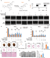
Fig. 1
Fig. 1
New molecule HHQ16 significantly improves cardiac function and reverses myocardial remodeling in HF mice. a Chemical structure of astragaloside IV and its derivative HHQ16. b Schematic overview of the experimental design in the mice model. Representative M-mode images (c) and statistical analysis of EF (%) and FS (%) (d) from mice underwent sham or LADL surgery for 4 weeks, then treated by daily intragastric administration of vehicle, enalapril, or HHQ16 at the doses indicated for 4 weeks. The cardiac function was detected by echocardiography at 4 (before admin.) and 8 (after admin.) weeks post-LADL (n = 7–10). e Representative photographs of mouse heart (left) and statistical analysis of heart weight to body surface area ratio (HW/BSA, right). Mice were treated as mentioned in b with the dose of HHQ16 at 10 mg/kg (n = 12–18). f Representative H&E of the myocardial tissues derived from mice treated as mentioned in e (n = 3). g Representative Masson’s trichrome staining (left) and quantification (right) of myocardial tissues in mice treated as mentioned in e (n = 3). h Representative electron microscopic images (left) and their quantification (right) of mitochondrial number in the myocardial tissues derived from mice treated as mentioned in e (n = 6). Data are presented as the means ± SEM. *P < 0.05, **P < 0.01, ***P < 0.001 by two-way ANOVA with Sidak’s multiple comparisons test (d) and one-way ANOVA with Tukey’s multiple comparisons test (e, g, h)
Fig. 2
Fig. 2
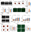
HHQ16 restrains cardiac hypertrophy by directly acts on cardiomyocytes. a The top 10 of KEGG analysis from the RNA-sequencing (mRNA) of myocardial tissues derived from mice treated as mentioned in Fig. 1e. b Red-labeled KEGG items in a were further enriched. Blue square: downregulated genes; Red square: upregulated genes. c Representative WGA staining (left) and its quantification (right) in myocardial tissue derived from mice treated as mentioned in Fig. 1e (n = 3). d qRT-PCR detection for hypertrophy biomarkers Anp, Bnp, and β-Mhc in myocardial tissues of mice treated as mentioned in Fig. 1e (n = 6–8). e Western blotting (upper) and its quantification (lower) for ANP, BNP, and β-MHC in myocardial tissues of mice treated as mentioned in Fig. 1e (n = 3). f Representative phalloidin staining (left) and its quantification (right) of HL-1 mouse cardiomyocytes treated with vehicle or ISO + PE (100 μM each) in the absence or presence of HHQ16 (100 nM) for 24 h (n = 3). g Western blotting (left) and its quantification (right) for ANP and BNP of neonatal mouse primary cardiomyocytes treated with vehicle or ISO (100 μM) in the absence or presence of HHQ16 (100 nM) for 24 h (n = 3). h qRT-PCR detection for Anp and Bnp in neonatal mouse primary cardiomyocytes treated as mentioned in g (n = 4). Data are presented as the means ± SEM. *P < 0.05, **P < 0.01, ***P < 0.001 by one-way ANOVA with Tukey’s multiple comparisons test (ch)
Fig. 3
Fig. 3
A new transcript lnc9456 is screened out to be strongly inhibited by HHQ16. a The discovery procedure of lnc9456 from the RNA-sequencing (lncRNAs) of myocardial tissues derived from mice treated as mentioned in Fig. 1e. Volcano plot shows the differentially expressed transcripts (DETs) discovery by using the P < 0.05 and foldchange (FC) ≥ 5 or ≤0.2 as the threshold of significant difference. Then, DETs analysis generates Venn diagram to identify 35 transcripts of interest. Finally, 35 transcripts were validated by qRT-PCR and lnc9456 was screened out. b qRT-PCR detection for lnc9456 in neonatal mouse primary cardiomyocytes treated with vehicle or ISO (100 μM) in the absence or presence of HHQ16 at the dose indicated for 12 h (left, n = 6) or at the dose of 100 nM for the time indicated (right, n = 4). c qRT-PCR detection for lnc9456 in HL-1 mouse cardiomyocytes treated with vehicle or Ang II (50 μM, left) or ISO + PE (100 μM each, right) in the absence or presence of HHQ16 (100 nM) for 24 h. (n = 4). Data are presented as the means ± SEM. *P < 0.05, **P < 0.01, ***P < 0.001 by one-way ANOVA with Tukey’s multiple comparisons test (ac)
Fig. 4
Fig. 4
Lnc9456 is critical for cardiomyocyte hypertrophy. a Synchronous expression of lnc9456 and hypertrophic biomarkers Anp or Bnp of myocardial tissues from mice after LADL surgery were detected by qRT-PCR at the indicated time points. (n = 6–10). b qRT-PCR detection for lnc9456, Anp and Bnp in neonatal mouse primary cardiomyocytes treated by vehicle or ISO (50, 100, 200 μM) for 12 h (n = 6). c Representative phalloidin staining (left) and its quantification (right) of HL-1 mouse cardiomyocytes transfected with control adenovirus (AdV-Vector) or lnc9456 overexpression adenovirus (AdV-lnc9456) for 48 h (n = 3). d qRT-PCR detection for hypertrophic biomarkers Anp, Bnp and β-Mhc of HL-1 mouse cardiomyocytes treated as mentioned in c (n = 6). e Western blotting (left) and its quantification (right) for ANP, BNP and β-MHC of HL-1 mouse cardiomyocytes treated as mentioned in c (n = 3). qRT-PCR detection for lnc9456 (f) and hypertrophic biomarkers Anp, Bnp, and β-Mhc (g) in HL-1 mouse cardiomyocytes. The cells were transfected with smart silencer RNA of lnc9456 (ssRNA-lnc9456) or its negative control (ssRNA-NC) for 48 h and then treated with vehicle or ISO + PE (100 μM each) for another 24 h (n = 4). Data are presented as the means ± SEM. *P < 0.05, **P < 0.01, ***P < 0.001 by one-way ANOVA with Dunnett’s multiple comparisons test (a, b), Student’s t test (ce) and two-way ANOVA with Sidak’s multiple comparisons test (f, g)
Fig. 5
Fig. 5
Lnc9456 interacts with G3BP2 and promotes the nuclear translocation of p65 subunit of NF-κB. a The top 7 proteins that may bind to lnc9456 were predicted by catRAPID omics module. b The top 20 of Gene Ontology (GO) analysis of interacting proteins pulled down by biotinylated lnc9456 probe and identified by mass spectrometry. c Representative western blotting (upper) and its quantification (lower) of G3BP2 in myocardial tissues derived from mice at the indicated time post-LADL (n = 4). d Representative western blotting (upper) and its quantification (lower) of G3BP2 in HL-1 mouse cardiomyocytes transfected with smart silencer RNA of lnc9456 (ssRNA-lnc9456) or its negative control (ssRNA-NC) for 72 h (n = 4). e Representative western blotting (left) and its quantification (right) of G3BP2 in HL-1 mouse cardiomyocytes transfected with control plasmid (OE-NC) or lnc9456 overexpression plasmid (OE-lnc9456) for 48 h (n = 4). f RNA pull-down and western blotting detection for the binding of G3BP2 to lnc9456 in HL-1 mouse cardiomyocytes treated as mentioned in e. g Co-IP detection for the binding of G3BP2 to IκBα in HL-1 mouse cardiomyocytes treated as mentioned in e. h Immunofluorescence staining for cellular localization of NF-κB p65 (green) in HL-1 mouse cardiomyocytes treated as mentioned in e (n = 3). Data are presented as the means ± SEM. *P < 0.05, **P < 0.01, ***P < 0.001 by one-way ANOVA with Tukey’s multiple comparisons test (c) and Student’s t test (d, e)
Fig. 6
Fig. 6
HHQ16 directly binds to lnc9456 with high-affinity and induces its degradation. a qRT-PCR detection for lnc9456 in HL-1 mouse cardiomyocytes. The cells were treated with vehicle or 100 nM of HHQ16 for 0, 1, 3 and 6 h in the presence of actinomycin D (0.5 μg/mL) (n = 4). b qRT-PCR detection for lnc9456 in HL-1 cardiomyocytes. The cells were transfected with AdV-Vector or AdV- lnc9456 for 24 h, then treated with vehicle or 100 nM of HHQ16 for another 24 h (n = 5–6). c Representative agarose gel electrophoresis image (upper) and its quantification (lower) of in vitro-transcribed lnc9456 incubated with vehicle or HHQ16 (100 nM) in the absence or presence of indicated volume of HL-1 mouse cardiomyocytes lysates (n = 3). d Representative agarose gel electrophoresis image (upper) and its quantification (lower) of in vitro-transcribed lnc9456 incubated with vehicle or HHQ16 (100 nM) for the time indicated in the presence of 1 μL of HL-1 mouse cardiomyocyte lysates (n = 3). e Representative agarose gel electrophoresis image (upper) and its quantification (lower) of in vitro-transcribed lnc9456 incubated with vehicle or HHQ16 (100 nM) at the indicated doses for 10 min in the presence of 1 μL of HL-1 mouse cardiomyocyte lysates (n = 3). f MST detection for the binding affinity of HHQ16 to lnc9456 (left) and negative control (lncRNA Mhrt, right). Kd value was automatically by the curve fitting. Data are presented as the means ± SEM. *P < 0.05, **P < 0.01, ***P < 0.001 by two-way ANOVA followed by Bonferroni’s post-hoc t-test (a) and one-way ANOVA with Tukey’s multiple comparisons test (be)
Fig. 7
Fig. 7
Lnc9456 is essential for HHQ16 to reverse cardiac hypertrophy and dysfunction in mice. Representative echocardiography images (a) and statistical analysis of EF (%) and FS (%) (b) in mice. These mice were heart orthotopically injected with cardiomyocyte-specific lnc94546 overexpression adeno-associated virus (AAV-lnc9456) driven by the cTnT promoter or its negative control (AAV-Vector). At 4th week post injection, AAV-lnc9456 mice were treated with 10 mg/kg of HHQ16 for 2 weeks, and the cardiac function was detected by echocardiography (n = 10–15). Representative photographs of mouse heart (c) and statistical analysis of heart weight (d). Mice were treated as mentioned in a (n = 10). e Representative WGA staining (left) and its quantification (right) of mouse myocardial tissue derived from mice treated as mentioned in a (n = 3). Representative echocardiography images (f) and statistical analysis of EF (%) and FS (%) (g) from male lnc9456f/f or lnc9456CKO mice underwent LADL surgery for 4 weeks, then were treated with vehicle or 10 mg/kg of HHQ16 for another 4 weeks. The cardiac function was detected by echocardiography at 4 (before admin.) and 8 (after admin.) weeks post-LADL (n = 3–4). Representative photographs of mouse hearts (h) and statistical analysis of heart weight to body surface area ratio (HW/BSA, i) in lnc9456f/f and lnc9456CKO mice treated as mentioned in f (n = 3–4). j Representative WGA staining (left) and its quantification (right) of myocardial tissue in lnc9456f/f and lnc9456CKO mice treated as mentioned in f (n = 3). Data are presented as the means ± SEM. *P < 0.05, **P < 0.01, ***P < 0.001 by one-way ANOVA with Tukey’s multiple comparisons test (b, d, e, i and j) and two-way ANOVA with Sidak’s multiple comparisons test (g)
Fig. 8
Fig. 8
Human ortholog lnc4012 is a bona fide target for HHQ16. a qRT-PCR detection for lnc4012 of myocardial tissues derived from human normal or failing heart (n = 4–7). b qRT-PCR detection for lnc4012 of AC16 human cardiomyocytes treated with vehicle or ISO (20, 50, 100 μM) for 24 h (n = 6). c Schematic diagram of full length lnc4012 (lnc4012-WT), lnc4012 truncated mutant1 (lnc4012-MUT1, Δ251-478 bp), and lnc4012 truncated mutant2 (lnc4012-MUT2, Δ351-378 bp) (upper). RNA pull-down and western blotting detection for the binding of lnc4012 to G3BP2 in AC16 human cardiomyocytes (lower) transfected with lnc4012-WT, lnc4012-MUT1 or lnc4012-MUT2. d RNA pull-down and western blotting detection for the binding of lnc4012 to IκBα in AC16 human cardiomyocytes transfected with control plasmid (OE-NC) or lnc4012 overexpression plasmid (OE-lnc4012) for 48 h. e Representative western blotting (left) and its quantification (right) for the expression of NF-κB p65 in nucleus or cytoplasm of AC16 human cardiomyocytes treated as mentioned in d (n = 4). f MST detection for the binding affinity of HHQ16 to lnc4012. Kd value was automatically by the curve fitting. g Representative agarose gel electrophoresis image (upper) and its quantification (lower) of in vitro-transcribed lnc4012 incubated with vehicle or HHQ16 (100 nM) for the time indicated in the absence or presence of AC16 human cardiomyocyte lysates (n = 3). h qRT-PCR detection for lnc4012 in Human Stem Cell Induced Differentiated Cardiomyocytes (HiPSC-CMs) treated with vehicle or ISO (50 μM) in the absence or presence of HHQ16 (100 nM) for 24 h (n = 4–5). Data are presented as the means ± SEM. *P < 0.05, **P < 0.01, ***P < 0.001 by Student’s t test (a), one-way ANOVA with Dunnett’s multiple comparisons test (b), two-way ANOVA with Sidak’s multiple comparisons test (e) and one-way ANOVA with Tukey’s multiple comparisons test (g, h)

References

    1. Frantz S, et al. Left ventricular remodelling post-myocardial infarction: pathophysiology, imaging, and novel therapies. Eur. Heart J. 2022;43:2549–2561. doi: 10.1093/eurheartj/ehac223. - DOI - PMC - PubMed
    1. Nakamura M, Sadoshima J. Mechanisms of physiological and pathological cardiac hypertrophy. Nat. Rev. Cardiol. 2018;15:387–407. doi: 10.1038/s41569-018-0007-y. - DOI - PubMed
    1. Kramer CM, et al. Regional myocyte hypertrophy parallels regional myocardial dysfunction during post-infarct remodeling. J. Mol. Cell Cardiol. 1998;30:1773–1778. doi: 10.1006/jmcc.1998.0741. - DOI - PubMed
    1. Zurek M, et al. Neuregulin-1 induces cardiac hypertrophy and impairs cardiac performance in post-myocardial infarction rats. Circulation. 2020;142:1308–1311. doi: 10.1161/CIRCULATIONAHA.119.044313. - DOI - PubMed
    1. Jilaihawi H, et al. Left ventricular hypertrophy and the risk of subsequent left ventricular remodeling following myocardial infarction. Am. J. Cardiol. 2003;91:723–726. doi: 10.1016/S0002-9149(02)03414-8. - DOI - PubMed

Publication types

Substances