. 2023 Oct 19;13(1):17874.
doi: 10.1038/s41598-023-44426-7.

Neuroprotective potential of intranasally delivered L-myc immortalized human neural stem cells in female rats after a controlled cortical impact injury

Affiliations

Neuroprotective potential of intranasally delivered L-myc immortalized human neural stem cells in female rats after a controlled cortical impact injury

Mari Amirbekyan et al. Sci Rep. .

Abstract

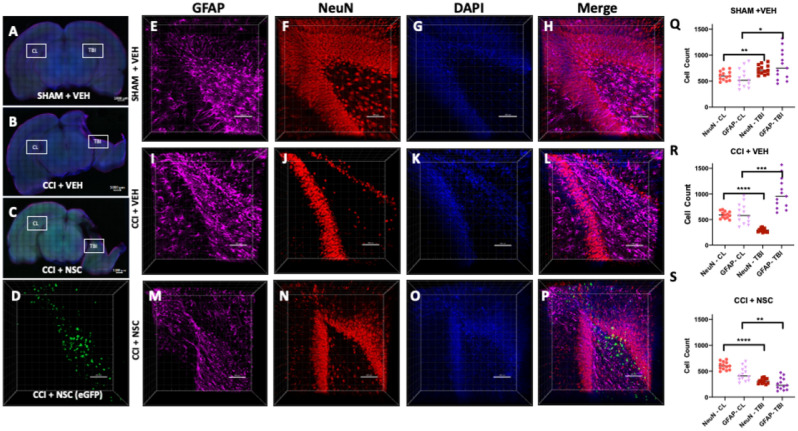
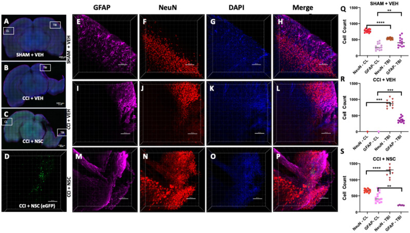
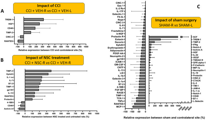
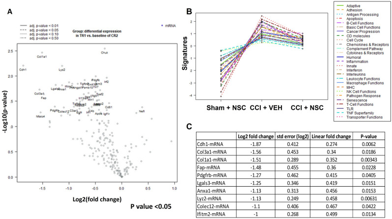
Efficacious stem cell-based therapies for traumatic brain injury (TBI) depend on successful delivery, migration, and engraftment of stem cells to induce neuroprotection. L-myc expressing human neural stem cells (LMNSC008) demonstrate an inherent tropism to injury sites after intranasal (IN) administration. We hypothesize that IN delivered LMNSC008 cells migrate to primary and secondary injury sites and modulate biomarkers associated with neuroprotection and tissue regeneration. To test this hypothesis, immunocompetent adult female rats received either controlled cortical impact injury or sham surgery. LMNSC008 cells or a vehicle were administered IN on postoperative days 7, 9, 11, 13, 15, and 17. The distribution and migration of eGFP-expressing LMNSC008 cells were quantified over 1 mm-thick optically cleared (CLARITY) coronal brain sections from TBI and SHAM controls. NSC migration was observed along white matter tracts projecting toward the hippocampus and regions of TBI. ELISA and Nanostring assays revealed a shift in tissue gene expression in LMNSC008 treated rats relative to controls. LMNSC008 treatment reduced expression of genes and pathways involved in inflammatory response, microglial function, and various cytokines and receptors. Our proof-of-concept studies, although preliminary, support the rationale of using intranasal delivery of LMNSC008 cells for functional studies in preclinical models of TBI and provide support for potential translatability in TBI patients.


Conflict of interest statement

The authors declare no competing interests.

Figures

Figure 1
Figure 1
Visualization of intranasally administered NSCs in the injured neurogenic zone. (AC) 1 mm-thick coronal brain sections were stained for astrocytes (far-red), maturing neurons (red), and nuclei (blue). Location of z-stacks for contralateral (Fig. S1) hemispheres for each brain are labeled on 10× magnification cross-sectional tile images. (D) eGFP signal was only produced in TBI + LMNSC008 tissue. (EP) 3D images of 250–350 µm-thick z-stacks taken at 10× magnification show individual and merged color channels as well as the localization of eGFP-labeled LMNSC008 cells at 39 days post-surgery from SHAM + VEH, CCI + VEH, and CCI + LMNSC008 groups. (QS) Histograms of respective = IMARIS NeuN and GFAP quantifications in both the ipsilateral (impact) and contralateral (non-impact) hemispheres showing the average number of cells for various cell sizes. (Q) SHAM + VEH: NeuN expression in TBI versus CL hemisphere using unpaired t test p < 0.002; GFAP p < 0.01. (R) CCI + VEH: NeuN expression in TBI versus CL hemisphere p < 0.0001; GFAP p < 0.0005. (S) CCI + NSC: NeuN in TBI versus CL p < 0.0001; GFAP p < 0.001. Data were analyzed using unpaired t test, two-tailed p values shown.
Figure 2
Figure 2
Visualization of NSCs in the damaged areas of the female rat TBI after intranasal administration. (AC) Staining of astrocytes (far red), maturing neurons (red), and nuclei (blue) on 1 mm-thick coronal rat brain sections in TBI and cortical regions of the brain. (EP) 3D images of Z-stacks taken at 10× magnification through 250–350 µm show individual and merged color channels as well as the localization of eGFP-labeled LMNSC008 NSCs 39 days post-surgery of three female rat brains from groups: SHAM + VEH, CCI + VEH, and CCI + NSC. (QS) Quantifications of NeuN and GFAP staining in both the TBI and CL hemispheres. (D) Green eGFP signal was only seen in the CCI + NSC brain section. (Q) SHAM + VEH: NeuN expression in TBI versus CL hemisphere using unpaired t test p < 0.0001; GFAP p < 0.0065. (R) CCI + VEH: NeuN expression in TBI versus CL hemisphere p < 0.0002; GFAP p < 0.001. (S) CCI + NSC: NeuN in TBI versus CL p < 0.0001; GFAP p < 0.001. Data were analyzed using unpaired t test, two-tailed p values shown.
Figure 3
Figure 3
Cytokine ELISA results from female rat brain lysates at contralateral [CL-left] and CCI [TBI-right] hemispheres. (A) Cytokine expression comparing TBI sample (right hemisphere) to CL sample (left hemisphere) showing the impact of TBI on cytokine expression levels. (B) Cytokine expression levels when comparing the TBI hemisphere samples of treated rats to the untreated rats showing the impact of NSCs on cytokine expression levels. Only cytokines for which the change is either greater than 100% or less than − 50% are shown. Figures S4 and S5 show all cytokine data analyzed. (C) Cytokine expression comparing the right and left hemispheres of sham surgery rats (both VEH and NSC groups were included in this analysis).
Figure 4
Figure 4
Differential gene expression data for CCI + VEH versus CCI + NSC from TBI and contralateral hemispheres. (A) Volcano plot comparing differences in gene expression between CR2 (CCI + VEH) and TR1 (CCI + NSC). Each dot represents a gene within the comparison performed. (B) Pathway analysis of CR1 (SHAM + VEH), CR2 (CCI + VEH), and TR1 (CCI + NSC). (C) Table of top 10 upregulated genes in CR2 (CCI + VEH) versus TR1 (CCI + NSC).

Update of

References

    1. Dewan MC, Rattani A, Gupta S, Baticulon RE, Hung YC, Punchak M, Agrawal A, Adeleye AO, Shrime MG, Rubiano AM, Rosenfeld JV, Park KB. Estimating the global incidence of traumatic brain injury. J. Neurosurg. 2019;130:1080–1097. - PubMed
    1. Faul, M., Wald, M. M., Xu, L. \& Coronado V. Traumatic Brain Injury in the United States: Emergency Department Visits, Hospitalizations and Deaths (2010).
    1. Goldstein M. The decade of the brain. Neurology. 1990;40(2):321. - PubMed
    1. Hamm RJ, et al. Exposure to environmental complexity promotes recovery of cognitive function after traumatic brain injury. J. Neurotrauma. 1996;13(1):41–47. - PubMed
    1. Taylor CA, et al. Traumatic brain injury-related emergency department visits, hospitalizations, and deaths—United States, 2007 and 2013. MMWR Surveill. Summ. 2017;66(9):1–16. - PMC - PubMed

Publication types