

Percutaneous transvenous coil embolization (PTCE) for treatment of single extrahepatic portosystemic shunt in dogs
- PMID: 37858152
- PMCID: PMC10585740
- DOI: 10.1186/s12917-023-03783-1
Percutaneous transvenous coil embolization (PTCE) for treatment of single extrahepatic portosystemic shunt in dogs
Abstract
Background: There is limited information regarding percutaneous transvenous coil embolization (PTCE) for single extrahepatic portosystemic shunt (PSS). This study aimed to describe the procedure and outcome of PTCE in dogs with a single extrahepatic PSS. Forty-two privately owned dogs were included in this study. All dogs were diagnosed with extrahepatic PSS by computed tomography (CT). Preoperative CT images were used to evaluate the diameter of the PSS for coil placement. A multipurpose balloon catheter was percutaneously inserted into the PSS via the jugular vein, and transvenous retrograde portography (TRP) and measurement of blood pressure in the PSS (pPSS) were performed during balloon inflation; one or more embolization coils were implanted via the catheter.
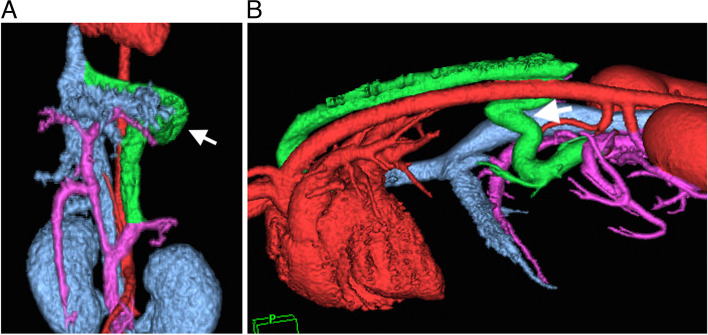
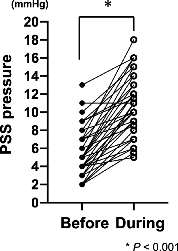
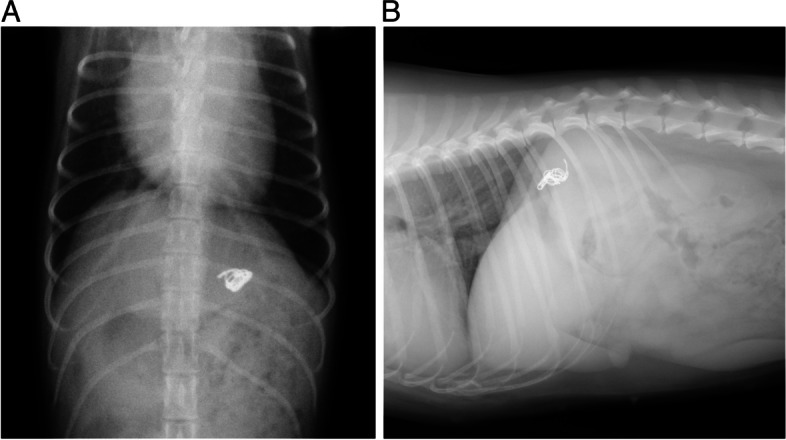
Results: In most cases, preoperative median fasting and postprandial serum total bile acid (TBA) concentrations were high (fasting, 86.5 μmol/L [ 3.7-250.0 μmol/L]; postprandial, 165.5 μmol/L [ 1.5-565.0 μmol/L]). CT revealed that 30 dogs had left gastrophrenic shunt; eight had left gastroazygos shunt; and one each had left gastrocaval, splenocaval, splenophrenic, and left colocaval shunt. TRP revealed that intrahepatic portal vascularity was clearly detectable in all dogs. The median values of pPSS before and during the balloon occlusion were 4.8 mmHg [2.0-13.0 mmHg] and 8.6 mmHg [5.0-18.0 mmHg], respectively. The median number and diameter of coils used were 2 coils [1 - 5 coils] and 8.0 mm [4.0 - 12.0 mm], respectively. The median times of irradiation and PTCE were 9 min [4-26 min] and 40 min [23-75 min], respectively. The median fasting and postprandial TBAs significantly decreased to 8.2 μmol/L [0.3-45.1 μmol/L, n = 38, p = 0.0028] and 19.8 μmol/L [0.3-106.7 μmol/L, n = 38, p = 0.0018], respectively, approximately 1 month after PTCE. The clinical success rate of PTCE without requirement for a second surgery was 95.2% (40/42 dogs). During revision surgery, one dog underwent surgical ligation and, in another dog, an ameroid constrictor was placed.
Conclusions: PTCE was clinically effective in treating single extrahepatic PSS in dogs. Preoperative CT and TRP prior to PTCE might be clinically valuable for choosing the size of embolization coils, deciding the appropriate location of coil implantation, and estimating the number of coils to be implanted. PTCE is a promising alternative to conventional surgical procedures for single extrahepatic PSS in dogs.
Keywords: Coil embolization; Dog; Extrahepatic; Portosystemic shunt.
© 2023. BioMed Central Ltd., part of Springer Nature.
Conflict of interest statement
The authors declare no competing interests.
Figures

References
-
- Lawrence D, Bellah JR, Diaz R. Results of surgical management of portosystemic shunts in dogs: 20 cases (1985–1990) J Am Vet Med Assoc. 1992;201:1750–1753. - PubMed
-
- Bostwick DR, Twedt DC. Intrahepatic and extrahepatic portal venous anomalies in dogs: 52 cases (1982–1992) J Am Vet Med Assoc. 1995;206:1181–1185. - PubMed
MeSH terms
LinkOut - more resources
Full Text Sources
