. 2023 Oct 20;42(1):272.
doi: 10.1186/s13046-023-02840-9.

A CRISPR activation screen identifies MUC-21 as critical for resistance to NK and T cell-mediated cytotoxicity

Affiliations

A CRISPR activation screen identifies MUC-21 as critical for resistance to NK and T cell-mediated cytotoxicity

Dong-Hee Lee et al. J Exp Clin Cancer Res. .

Abstract

Background: Immunotherapy has significantly advanced cancer treatments, but many patients do not respond to it, partly due to immunosuppressive mechanisms used by tumor cells. These cells employ immunosuppressive ligands to evade detection and elimination by the immune system. Therefore, the discovery and characterization of novel immunosuppressive ligands that facilitate immune evasion are crucial for developing more potent anti-cancer therapies.

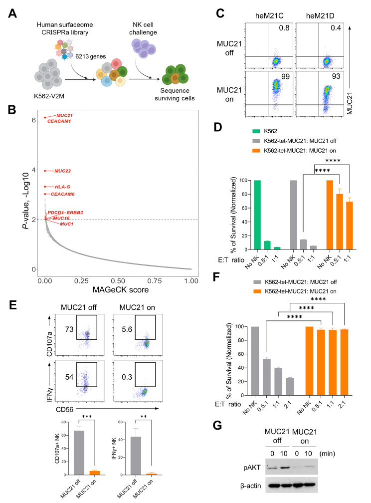
Methods: We conducted gain-of-function screens using a CRISPRa (CRISPR activation) library that covered the entire human transmembrane sub-genome to identify surface molecules capable of hindering NK-mediated cytotoxicity. The immunosuppressive role and mechanism of MUC21 were validated using NK and T cell mediated cytotoxicity assays. Bioinformatics tools were employed to assess the clinical implications of mucin-21 (MUC21) in cancer cell immunity.

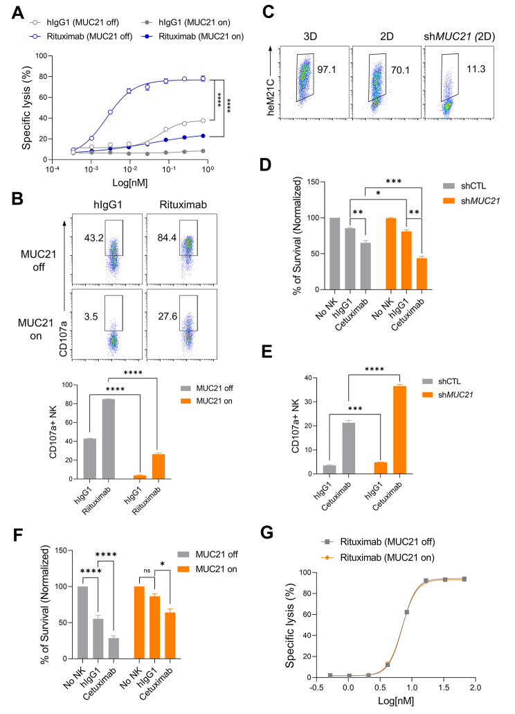
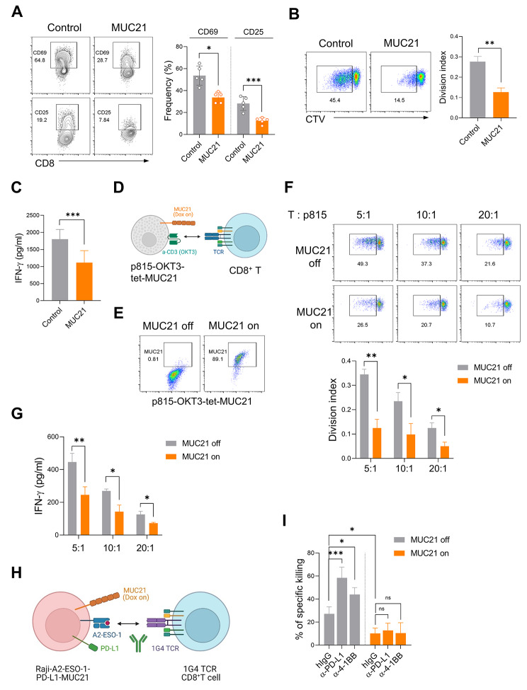
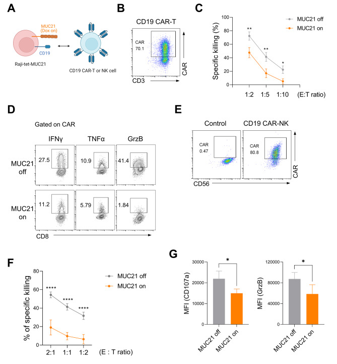
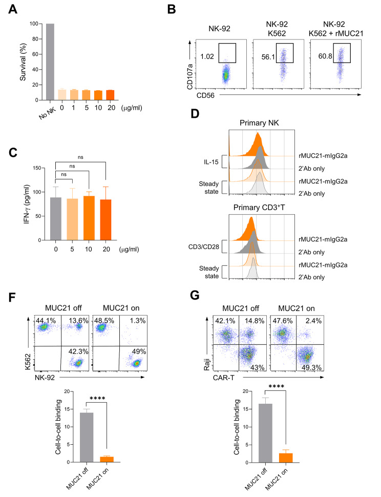
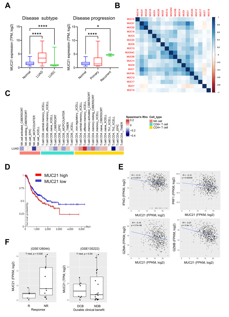
Results: Our genetic screens revealed that MUC21 expression on cancer cell surfaces inhibits both the cytotoxic activity of NK cells and antibody-dependent cellular cytotoxicity, but not affecting complement-dependent cytotoxicity. Additionally, MUC21 expression hinders T cell activation by impeding antigen recognition, thereby diminishing the effectiveness of the immune checkpoint inhibitor, anti-PD-L1. Moreover, MUC21 expression suppress the antitumor function of both CAR-T cells and CAR-NK cells. Mechanistically, MUC21 facilitates immune evasion by creating steric hindrance, preventing interactions between cancer and immune cells. Bioinformatics analysis revealed elevated MUC21 expression in lung cancer, which correlated with reduced infiltration and activation of cytotoxic immune cells. Intriguingly, MUC21 expression was higher in non-small cell lung cancer (NSCLC) tumors that were non-responsive to anti-PD-(L)1 treatment compared to responsive tumors.

Conclusions: These findings indicate that surface MUC21 serves as a potent immunosuppressive ligand, shielding cancer cells from NK and CD8+T cell attacks. This suggests that inhibiting MUC21 could be a promising strategy to improve cancer immunotherapy.

Keywords: CAR; CRISPR library screening; Cancer immunotherapy; MUC21.


Conflict of interest statement

All authors declare that they have no competing interests.

Figures

Fig. 1
Fig. 1
Identification of candidate genes conferring resistance to NK cell cytotoxicity through a surfaceome CRISPR activation screen. (A) Overview of the surfaceome-focused CRISPRa screen used in this study. A lentiviral library comprising 58,071 sgRNAs targeting the promoter regions of 6213 cell surface protein genes and 500 control sgRNAs was employed. K562-V2M cells were transduced with the sgRNA library and exposed to NK-92 cells for two days. Genomic DNA was extracted from the cells that survived, and gene abundance was determined using next-generation sequencing. (B) MAGeCK Analysis of the enrichment of sgRNA sequences in the surviving K562-V2M cells. X-Axis: MAGeCK Gene Score; Y-Axis: inverse log P value. (C) Representative FACS analysis of surface MUC21 expression in K562-tet-MUC21 cells after 24 h of culture with or without Dox (1 µg/mL). Two different clones of antibodies (clones heM21C and heM21D) targeting human MUC21 were used. (D-E) K562-tet-MUC21 cells were cultured in the presence of Dox (1 µg/mL) for 24 h, and co-incubated with NK-92 cells for 6 h. (D) NK cell-mediated killing activity was evaluated by measuring the luciferase activity of the surviving K562 cells. Parental K562 cells were used as a positive control. Ratio of effector to target cells (E:T). (E) Representative FACS analysis (above panel) of the surface CD107a and intracellular IFN-γ expression in NK-92 cells. A summary graph (below panel) showing the percentage of CD107a and IFN-γ expressing NK-92 cells in two independent experiments. (F) Dox-treated K562-tet-MUC21 cells were exposed to primary NK cells at a 1:1 E:T ratio for six hours. NK cell killing was evaluated by measuring luciferase activity in surviving K562-tet-MUC21 cells. (G) Immunoblotting for phospho-AKT expression in lysates from NK-92 cells after stimulation with paraformaldehyde-fixed K562-tet-MUC21 cells for 0 and 10 min. β-actin was used as a loading control. Statistical significance was determined by two-tailed unpaired t-tests; **P < 0.01, ***P < 0.001, ****P < 0.001
Fig. 2
Fig. 2
Surface MUC21 on cancer cells suppresses ADCC activity but has no impact on CDC. (A-B) Raji-tet-MUC21 cells were cultured with or without Dox (1 µg/mL) for 24 h. (A) Raji-tet-MUC21 cells were co-incubated with NK-92-CD16 cells at an E:T ratio of 0.5:1 for 4 h in the presence of varying concentrations of human IgG1 or rituximab. NK-92-CD16 cytotoxicity against Raji cells was measured by the luciferase activity of the surviving Raji cells. (B) Representative FACS analysis (above panel) of the surface CD107a expression of NK-92-CD16 cells cultured with Raji-tet-MUC21 cells in the presence of 10 µg/ml of hIgG1 or rituximab. A summary graph (below panel) showing the percentage of CD107a expressing NK-92 cells. (C) FACS analysis of surface MUC21 expression in wild-type NCI-H441 cultured in both 2D and 3D conditions, as well as MUC21 knockdown NCI-H441 cells (shMUC21) cultured in 2D. (D-E) The NCI-H441 cells, which were stably expressing scramble shRNA (shCTL) or shRNA targeting MUC21 (shMUC21), were cultured in 2D condition with NK-92-CD16 cells at an E:T ratio of 0.5:1 for 4 h. The co-culture was conducted in the presence of human IgG1 (0.1 µg/ml) or cetuximab (0.1 µg/ml). (D) NK-92-CD16 cytotoxicity against NCI-H441 cells was measured by luciferase activity in surviving NCI-H441 cells. (E) A summary graph of surface CD107a expression of NK-92-CD16 cells. (F) A549-tet-MUC21 cells were cultured with or without Dox (1 µg/ml) for 24 h and then co-incubated with NK-92-CD16 cells at an E:T ratio of 0.5:1 for 4 h in the presence of either human IgG1 (0.1 µg/ml) or cetuximab (0.1 µg/ml). (G) Raji-tet-MUC21 cells were cultured with or without Dox (1 µg/ml) for 24 h and incubated with indicated concentration of rituximab for 15 min followed by addition of 10% human complement. Cell viability was then measured by luciferase activity. Statistical significance was determined by a two-way ANOVA with Holm-Sidak comparisons in (A) or one-way ANOVA with Holm–Sidak multiple comparisons in (B), (D) and (E) or multiple t tests with correction for multiple comparisons using the Holm–Sidak method in (F). *P < 0.01, **P < 0.01, ***P < 0.001, ****P < 0.001; and ns, not significant
Fig. 3
Fig. 3
MUC21 attenuates T cell activation by hindering their antigen recognition. (A-C) CD8+T cells isolated from human PBMCs were co-cultured with 293FT cells that were transfected with mock (control) or MUC21 expressing plasmids for five days. The co-culture was performed in the presence of plate-coated anti-CD3 antibody (3 µg/ml). (A) Representative FACS plots (left panel) and a summary graph (right panel) showing the percentages of CD69+ or CD25+ CD8+T cells. Each dot represents an individual human sample. (B) Representative FACS plots showing proliferating CTVlow CD8+T cells (left panel) and a summary graph showing the division index of CTVlow CD8+T cells (right panel). (C) ELISA of IFN-γ secretion by CD8+T cells. (D-G) CD8+T cells were isolated from human PBMCs and co-cultured with p815 cells expressing membrane bound anti-CD3 scFv and Dox inducible MUC21 (p815-OKT3-tet-MUC21) at various E:T ratios for 3 days, in the presence or absence of Dox (1 µg/ml). (D) Schematic illustration of the p815-OKT3-tet-MUC21 artificial APC assay. (E) Representative FACS plots showing the expression of MUC21 in p815-OKT3-tet-MUC21 cells upon Dox (1 µg/ml) treatment. (F) Representative FACS plots showing proliferating CTVlow CD8+T cells (above panel) and a summary graph showing the division index of CTVlow CD8+T cells (below panel). (G) ELISA analysis of IFN-γ secretion by CD8+T cells. (H-I) 1G4 TCR-engineered CD8+T (1G4 TCR-CD8+T) cells were co-cultured with Raji cells expressing A*02:01/NY157–165 single-chain trimers, PD-L1 and Dox-inducible MUC21 (Raji-A2-ESO-1-PD-L1-MUC21) for three days, in the presence of the indicated antibodies (10 µg/ml) or Dox (1 µg/ml). (H) Schematic illustration of 1G4 TCR-engineered CD8+T cell-mediated Raji-A2-ESO-1-PD-L1-MUC21 cell killing. (I) Percentages of antigen-specific killing of Raji-A2-ESO-1-PD-L1-MUC21 cells by 1G4 TCR-CD8+T cells at an E:T ratio of 1:10 in the presence of the indicated antibodies with or without Dox. Data were compiled from three independent experiments with two replications. Statistical significance was determined by two-tailed unpaired t-test in (A), (B), (C), (F) and (G) or one-way ANOVA with Holm-Sidak multiple comparisons in (I). *P < 0.05, **P < 0.01, ***P < 0.001; and ns, not significant
Fig. 4
Fig. 4
Expression of MUC21 on the cell membrane suppresses the cytotoxic functions of anti-CD19 CAR-T and CAR-NK cells. (A-D) CD3+T cells isolated from human PBMCs were stimulated with anti-CD3/CD28 beads and transduced with anti-CD19 CAR lentivirus. CD19 CAR-T cells were co-incubated with Raji-tet-MUC21 cells at variable E:T ratios for two days in the presence or absence of Dox. (A) Schematic illustration of the killing of Raji-tet-MUC21 cells by CD19-CAR T or NK cells. (B) FACS analysis of the expression of anti-CD19 CAR on T cells using biotinylated CD19 protein. (C) The percentages of killing of Raji-tet-MUC21 cells by CD19 CAR-T cells were determined at the indicated E:T ratios. (D) Representative FACS plots showing the percentages of IFN-γ, TNF-α or Granzyme B (GrzB) expression by CD19 CAR-T cells co-incubated with Raji-tet-MUC21 cells at an E:T ratio of 1:2. (E-G) NK-92 cells were transduced with anti-CD19 CAR lentivirus. CD19 CAR-NK-92 cells were co-incubated with Raji-tet-MUC21 cells at variable E:T ratios for four hours in the presence or absence of Dox (1 µg/ml). (E) FACS analysis of the expression of anti-CD19 CAR on NK-92 cells using biotinylated CD19 protein. (F) Percentages of the specific killing of Raji-tet-MUC21 cells by CD19 CAR-NK-92 cells at the indicated E:T ratios. (G) Summary graph showing the mean fluorescence intensity (MFI) of CD107a and GrzB expression in CD19 CAR-NK-92 cells co-incubated with Raji-tet-MUC21 cells at an E:T ratio of 1:1. Data were compiled from four independent experiments with two replicates. Statistical significance was determined by a 2-way ANOVA with Holm-Sidak comparisons in (C) and (F), or two-tailed unpaired t-tests in (G). *P < 0.05, **P < 0.01, ****P < 0.0001
Fig. 5
Fig. 5
The presence of membrane-bound MUC21 on cancer cells obstructs their interaction with immune cells. (A-B) K562 cells were co-cultured with NK-92 cells at an E:T ratio of 0.5:1 for four hours in the presence of varying concentrations of rMUC21-mFc. (A) The cytotoxicity against K562 cells was measured by the luciferase activity of the surviving cells. (B) Representative flow cytometry analysis of surface CD107a expression on NK cells. (C) Human CD8+T cells were activated with anti-CD3 antibody in the presence of varying concentrations of rMUC21-mFc. ELISA of IFN-γ secretion by CD8+T cells. (D) FACS analysis showing the binding of rMUC21-mFc to resting and IL-15-stimulated primary NK cells (above), as well as resting and activated primary CD3+ T cells (below). (F) K562-tet-MUC21 cells were cultured in the presence or absence of Dox for 24 h. These cells were then co-incubated with CTV-labeled NK-92 cells for 15 min. Representative FACS plots (above) and a summary plot (below) showing the percentages of cell-to-cell conjugation. (G) Raji-tet-MUC21 cells were co-incubated with CTV-labeled CD19 CAR-T cells for 15 min. Representative FACS plots (above) and a summary plot (below) showing the percentages of the cell-to-cell binding. Data were compiled from two independent experiments. Statistical significance was determined by two-tailed unpaired t-tests. ns: not significant. ****P < 0.001
Fig. 6
Fig. 6
Negative correlation between elevated MUC21 expression and immune cytotoxicity in LUAD. (A) Expression levels of the MUC21 gene in LUAD and LUSC patients from TCGA, along with their matched normal individuals from TCGA and GTEx. This analysis considered both the disease subtype and progression. Statistical significance was assessed using one-way ANOVA and a Dunnett’s multiple comparison test. (B) Heatmap illustrating the Pearson correlation coefficients between the gene expression levels of each member of the mucin family in TCGA LUAD samples. (C) Heatmap displaying scores representing the infiltration of NK cells, CD8+T cells, and CD4+T cells based on different immune cell deconvolution methods in TCGA LUAD. (D) Progression-free survival curve of lung cancer patients (LUAD and LUSC) from TCGA plotted based on the expression of the MUC21 gene. The Kaplan-Meier curve compares the top and bottom quartiles of MUC21 expression, and significance was evaluated using log-rank test statistics. (E) Scatter plots depicting the correlations between the expression of the MUC21 gene and cytotoxicity genes (IFNG, PRF1, GZMA, and GZMB) involved in NK/T cell-mediated cytotoxic responses in TCGA LUAD. The correlation was tested using a Spearman’s rank correlation coefficient. (F) Boxplots comparing the expression of the MUC21 gene between patients who responded to anti-PD-(L)1 immune checkpoint inhibitor therapy and those who did not respond in two different lung cancer cohorts. Responders (R) or those with durable clinical benefit (DCB) achieved partial response (PR) or stable disease (SD) for more than six months. Non-responders (NR) or those with non-durable benefit (NDB) experienced progressive disease (PD) or SD for less than six months. The significance of the differences was evaluated using a Student’s t-test

References

    1. Budimir N, Thomas GD, Dolina JS, Salek-Ardakani S. Reversing T-cell exhaustion in Cancer: Lessons learned from PD-1/PD-L1 Immune Checkpoint Blockade. Cancer Immunol Res. 2022;10(2):146–53. doi: 10.1158/2326-6066.CIR-21-0515. - DOI - PubMed
    1. Leick MB, Maus MV, Frigault MJ. Clinical perspective: treatment of aggressive B cell lymphomas with FDA-Approved CAR-T cell therapies. Mol Ther. 2021;29(2):433–41. doi: 10.1016/j.ymthe.2020.10.022. - DOI - PMC - PubMed
    1. Vinay DS, Ryan EP, Pawelec G, Talib WH, Stagg J, Elkord E, Lichtor T, Decker WK, Whelan RL, Kumara H, et al. Immune evasion in cancer: mechanistic basis and therapeutic strategies. Semin Cancer Biol. 2015;35(Suppl):185–S198. doi: 10.1016/j.semcancer.2015.03.004. - DOI - PubMed
    1. Wisnovsky S, Mockl L, Malaker SA, Pedram K, Hess GT, Riley NM, Gray MA, Smith BAH, Bassik MC, Moerner WE et al. Genome-wide CRISPR screens reveal a specific ligand for the glycan-binding immune checkpoint receptor Siglec-7. Proc Natl Acad Sci U S A 2021, 118(5). - PMC - PubMed
    1. Kallingal A, Olszewski M, Maciejewska N, Brankiewicz W, Baginski M. Cancer immune escape: the role of antigen presentation machinery. J Cancer Res Clin Oncol 2023. - PMC - PubMed

MeSH terms