. 2023 Oct 20;11(1):92.
doi: 10.1186/s40364-023-00531-3.

A functional role of Ephrin type-B receptor 6 (EPHB6) in T-cell acute lymphoblastic leukemia

Affiliations

A functional role of Ephrin type-B receptor 6 (EPHB6) in T-cell acute lymphoblastic leukemia

Mattia Colucci et al. Biomark Res. .

Abstract

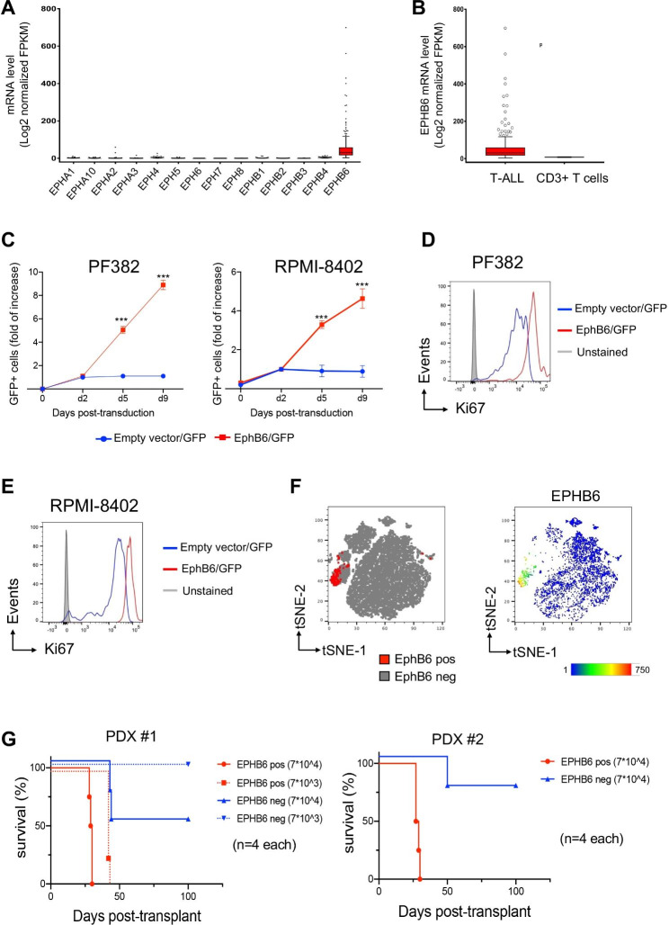
T-cell lymphoblastic acute leukemia (T-ALL) is an aggressive blood cancer, characterized by restricted cellular subsets with enriched leukemia initiating cells (LICs). Recently, Ephrin receptors (Eph) were described to be highly expressed in cancer stem cells. Here, using public RNA-Seq datasets of human T-ALL, we reported that EphB6 was the only member within the Eph family overexpressed in over 260 samples. We also found the highest level of EphB6 in a minor cell subpopulation within bulk tumors of patient-derived xenografts, obtained through the injection of primary patient biopsy material into immunocompromised NOD-Scid/IL2Rγc-/- (NSG) mice. Interestingly, this EphB6 positive (EphB6+) subset showed an enriched LIC activity after in vivo transplantation into NSG mice. Additionally, gene expression data at the single-cell level of primary patients' leukemic cells revealed that EphB6 + cells were significantly selected in minimal residual disease up to 30 days from the standard treatments and characterized by high levels of markers related to cell proliferation and poor clinical outcome, such as CCNB1 and KIF20A. Taken together, our data suggest that EphB6 supports LICs' maintenance and progression in T-ALL and, thus, targeting EphB6 + cells could be therapeutically relevant for the treatment of T-ALL patients.

Keywords: Biomarkers; EphB6; Leukemia-initiating cells (LICs); Single-cell RNA-Sequencing; T-ALL.


Conflict of interest statement

Alberto Visioli is an employee of Stemgen Spa and does not own shares in the Company. The others authors declare that they have no competing interests.

Figures

Fig. 1
Fig. 1
The constitutive expression of EPHB6 receptor in human T-ALL promotes in vitro proliferation and in vivo cell expansion. (A) Analysis of public COG TARGET dataset shows a significant enrichment of EphB6 mRNA level as compared with those of Eph receptors family in T-ALL patients (***P < 0.0001 n = 264, by one-way ANOVA) and (B) higher EphB6 expression in T-ALL cases versus CD3 + T-cells from healthy donors from the Human Protein Atlas database (***P < 0.0001 n = 20, by Mann-Whitney test). (C) Flow cytometry analysis of abundance of GFP + alive cell fraction after transduction with EPHB6 or empty (EV) lentivectors as indicated. Transduced FACS-sorted subsets of PF382 and RPMI-8402 cell lines were tracked over time at the indicated time points by flow cytometry. Alive GFP + cells were discriminated for DAPI exclusion. Means ± SD fraction of the initial transduction value are plotted for experiments performed in biological triplicate. ***, p < 0.001 (Student’s t-test).(D-E) Flow cytometry analysis of Ki67 level in PF382 (D) and RPMI-8402 (E) cell lines after transduction with EPHB6 or empty (EV) lentivectors as indicated. Transduced subsets of PF382 and RPMI-8402 cell lines were valued after three days of in vitro growth. (F) t-SNE plots based on the immunophenotyping of M71 (PDX#1) and H3255 (PDX#2), two independent clones of PDX samples by multiparametric flow cytometry. (G) Survival of recipient NSG mice after transplantation with FACS-sorted EPHB6 + cell subsets from M71 (PDX#1) and H3255 (PDX#2) PDX samples. The cell doses injected in each of 4 recipient animals are indicated in parentheses. Two separate experiments are depicted using independent PDX clones as indicated
Fig. 2
Fig. 2
EphB6 positive cells are characterized by high transcriptional levels of genes related to cell proliferation and significantly selected in minimal residual disease (MRD) of human T-ALL. (A) Hierarchical clustering of EphB6 positive and EphB6 negative subpopulations using 20 differentially expressed genes from M71 (PDX#1), H3255 (PDX#2) and F1313 (PDX#3), three independent PDX clones. A dual-color code represents markers over-(red) and under-represented (blue). (B) Flow cytometric analysis depicting the enrichment of CCNB1 and CDCA3 in EphB6-overexpressing cells (EphB6/GFP) as compared to control (Empty vector/GFP). (C) Heatmap showing the Spearman’s rank correlation between the EphB6 fractions and 15 differentially expressed genes. The higher the statistical correlation the bigger the circles. A dual-color code reports positive (blue) and negative (red) correlation and color intensity represents the increase of correlation. *P < 0.05, by Student’s t-test. (D) tSNE plots based on the single cell RNA-Sequencing (scRNA-Seq) data from 4 primary T-ALL samples, CSS12401, CSS13693, CSS15501 and CSS20705 at the diagnosis (d0) and up to 30 days after the start of therapy (MRD) without passaging into immunocompromised mice. In the map, EphB6 negative and positive leukemia cells are colored in grey and red, respectively. (E) Graphical representation of cell percentage in the EphB6 positive cell population at the diagnosis (d0) and up to 30 days after the start of therapy (MRD) according to each sample group. (F) mRNA expression level of CDCA3, CCNB1 and KIF20A genes in EphB6 positive vs. EphB6 negative cells as determined by scRNA-seq data. ***, P < 0.001 (Student’s t-test). (G) Higher KIF20A and CCNB1 genes are significantly associated with lower patients’ survival according to the COG TARGET dataset (P = 0.032 and P = 0.029 from log-rank test, n = 264). Kaplan-Meier survival plots shows the same trend for CDCA3 gene although without statistical significance (P = 0.075, n = 264)

References

    1. Pui CH, Robison LL, Look AT. Acute lymphoblastic leukaemia. Lancet. 2008;371(9617):1030–43. doi: 10.1016/S0140-6736(08)60457-2. - DOI - PubMed
    1. Guru Murthy GS, Pondaiah SK, Abedin S, Atallah E. Incidence and survival of T-cell acute lymphoblastic leukemia in the United States. Leuk Lymphoma. 2019;60(5):1171–8. doi: 10.1080/10428194.2018.1522442. - DOI - PubMed
    1. Magee JA, Piskounova E, Morrison SJ. Cancer stem cells: impact, heterogeneity, and uncertainty. Cancer Cell. 2012;21(3):283–96. doi: 10.1016/j.ccr.2012.03.003. - DOI - PMC - PubMed
    1. El Zawily A, McEwen E, Toosi B, Vizeacoumar FS, Freywald T, Vizeacoumar FJ, Freywald A. The EphB6 receptor is overexpressed in pediatric T cell acute lymphoblastic leukemia and increases its sensitivity to doxorubicin treatment. Sci Rep. 2017;7(1):14767. doi: 10.1038/s41598-017-15200-3. - DOI - PMC - PubMed
    1. Committee EN. Unified nomenclature for eph family receptors and their ligands, the ephrins. Eph Nomenclature Committee Cell. 1997;90(3):403–4. - PubMed

LinkOut - more resources