. 2023 Nov 1;4(11):1562-1579.
doi: 10.34067/KID.0000000000000276. Epub 2023 Oct 20.

Molecular Phenotyping and Mechanisms of Myocardial Fibrosis in Advanced Chronic Kidney Disease

Affiliations

Molecular Phenotyping and Mechanisms of Myocardial Fibrosis in Advanced Chronic Kidney Disease

Gayatri Narayanan et al. Kidney360. .

Abstract

Key Points:

  1. Myocardial fibrosis in hearts from patients with CKD is characterized by increased trimeric tensile collagen type I and decreased elastic collagen type III compared with hearts from hypertensive or healthy donors, suggesting a unique fibrotic phenotype.

  2. Myocardial fibrosis in CKD is driven by alterations in extracellular matrix proteostasis, including dysregulation of metalloproteinases and cross-linking enzymes.

  3. CKD-associated mineral stressors uniquely induce a fibronectin-independent mechanism of fibrillogenesis characterized by formation of trimeric collagen compared with proinflammatory/fibrotic cytokines.

Background: Myocardial fibrosis is a major life-limiting problem in CKD. Despite this, the molecular phenotype and metabolism of collagen fibrillogenesis in fibrotic hearts of patients with advanced CKD have been largely unstudied.

Methods: We analyzed explanted human left ventricular (LV) heart tissues in a three-arm cross-sectional cohort study of deceased donor patients on hemodialysis (HD, n=18), hypertension with preserved renal function (HTN, n=8), and healthy controls (CON, n=17), ex vivo. RNA-seq and protein analysis was performed on human donor hearts and cardiac fibroblasts treated with mineral stressors (high phosphate and high calcium). Further mechanistic studies were performed using primary cardiac fibroblasts, in vitro treated with mineral stressors, proinflammatory and profibrotic cytokines.

Results: Of the 43 donor participants, there was no difference in age (P > 0.2), sex (P > 0.8), or body mass index (P > 0.1) between the groups. Hearts from the HD group had extensive fibrosis (P < 0.01). All LV tissues expressed only the trimeric form of collagen type I. HD hearts expressed increased collagen type I (P < 0.03), elevated collagen type I:III ratio (P < 0.05), and decreased MMP1 (P < 0.05) and MMP2 (P < 0.05). RNA-seq revealed no significant differential gene expression of extracellular matrix proteins of interest in HD hearts, but there was significant upregulation of LH2, periostin, α-SMA, and TGF-β1 gene expression in mineral stressor–treated cardiac fibroblasts. Both mineral stressors (P < 0.009) and cytokines (P < 0.03) increased collagen type I:III ratio. Mineral stressors induced trimeric collagen type I, but cytokine treatment induced only dimeric collagen type I in cardiac fibroblasts. Mineral stressors downregulated fibronectin (P < 0.03) and MMP2 zymogen (P < 0.01) but did not significantly affect expression of periostin, MMP1, or cross-linking enzymes. TGF-β upregulated fibronectin (P < 0.01) and periostin (P < 0.02) only.

Conclusions: Myocardial fibrosis in advanced CKD hearts is characterized by increased trimeric collagen type I and dysregulated collagen metabolism, and is differentially regulated by components of uremia.


Conflict of interest statement

K. Lim reports consultancy agreements with Ambassadors Global and MBX Biosciences; ownership interest in OVIBIO Corporation, Ambassadors Global, and MBX Biosciences; advisory or leadership role in Ambassadors Global MBX Biosciences; and PI grant of Dialysis Clinics Inc (DCI) (Paul Teschan Research Fund (PTRF)), IU Health Values Fund Award. T. Lu reports ownership interest in Ovibio corporation, and CHF Foundation Funding Award. S. Moe reports consultancy agreements with Amgen, Ardelyx, Sanifit/Vifor, and Inozyme; ownership interest in Eli Lilly (stock); research funding from NIH-research grant and Keryx-research grant; honoraria from Ardelyx, Sanifit, and Inozyme; and advisory or leadership role in Editorial Board of AJ Nephrology and AJ Nutrition. G. Narayanan reports patents or royalties from Indiana University. A. Halim reports NIH NRSA Fellowship from NIH T32 AR065971, ownership interest in OVIBIO Corporation and patents or royalties from Indiana University. D. Zehnder reports research funding from Abbott Laboratories and Amgen and honoraria from Abbott Laboratories and Amgen. The remaining authors have nothing to disclose.

Figures

None
Graphical abstract
Figure 1
Figure 1
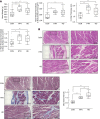
Characteristic of human whole heart and left ventricular sections. Gross characterization and histological analysis of whole and left ventricular human hearts. Representative images utilized 10× (left images) and 20× (right images) objectives. (A) HD and HTN hearts had significantly higher heart weight (HD: P = 0.0003, HTN: P < 0.0001) and heart weight normalized to BSA (HD: P < 0.0001, HTN: P < 0.0019) compared with CON hearts. HD and HTN hearts had significantly higher left ventricular (LV) wall thickness (HD: P = 0.0033, HTN: P < 0.0007) compared with CON hearts. Only HD hearts had significantly higher normalized LV wall thickness to BSA compared with CON hearts (P < 0.0123). (B) Hematoxylin and eosin staining of representative left ventricular sections of hearts from a patient on hemodialysis (HD), a patient with hypertension (HTN), and a healthy control donor (CON). HD hearts exhibited disorganized myofibers compared with CON hearts. Masson's trichrome staining of the same tissue sections from the same heart donors. Blue staining represents collagen staining. Quantification of aniline blue stain intensity from Masson's trichrome staining as measurement of fibrosis in hearts from HD (n=13), HTN (n=8), and CON (n=11) donors. P values: *<0.05, **<0.01. Statistical significance determined by one-way ANOVA and pairwise t test.
Figure 2
Figure 2
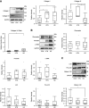
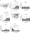
HD hearts exhibit dysregulated collagen synthesis, cross-linking, and turnover. (A) Representative immunoblots of collagen type I, collagen type III, and GAPDH of HD (n=13), HTN (n=8), and CON (n=11) hearts. Trimeric collagen type I appears at 400 kDa, whereas dimeric collagen type I at 250 kDa was not detectable in any group. HD and HTN hearts had a significant increase in collagen type I compared with CON hearts. HD exhibited a significant decrease in collagen type III compared with HTN and CON hearts. HD and HTN hearts had a significantly higher collagen type I/III ratio compared with CON hearts. (B) Representative immunoblots of fibronectin, periostin, α-smooth muscle actin (α-SMA), and GAPDH of HD (n=13), HTN (n=8), and CON (n=11) hearts. There was no significant difference in fibronectin, periostin, and α-SMA expression across all groups. (C) Representative immunoblots of lysyl hydroxylase 2 (LH2), proenzyme lysyl oxidase (pro-LOX), proteolytically processed pro-LOX (mature LOX), and GAPDH of HD (n=13), HTN (n=9), and CON (n=13) hearts. HD hearts had significantly decreased levels of mature LOX compared to CON hearts. There was no significant difference in LH2 and pro-LOX expression across all groups. (D) Representative immunoblots of metalloproteinases 1 (MMP1), 2 (MMP2), and 9 (MMP9), and GAPDH of HD (n=13), HTN (n=9), and CON (n=13) hearts. HD hearts had a significant decrease in MMP1 and MMP2 compared with CON hearts. P values: *<0.05, **<0.01. Statistical significance was determined by one-way ANOVA and pairwise t test.
Figure 2
Figure 2
HD hearts exhibit dysregulated collagen synthesis, cross-linking, and turnover. (A) Representative immunoblots of collagen type I, collagen type III, and GAPDH of HD (n=13), HTN (n=8), and CON (n=11) hearts. Trimeric collagen type I appears at 400 kDa, whereas dimeric collagen type I at 250 kDa was not detectable in any group. HD and HTN hearts had a significant increase in collagen type I compared with CON hearts. HD exhibited a significant decrease in collagen type III compared with HTN and CON hearts. HD and HTN hearts had a significantly higher collagen type I/III ratio compared with CON hearts. (B) Representative immunoblots of fibronectin, periostin, α-smooth muscle actin (α-SMA), and GAPDH of HD (n=13), HTN (n=8), and CON (n=11) hearts. There was no significant difference in fibronectin, periostin, and α-SMA expression across all groups. (C) Representative immunoblots of lysyl hydroxylase 2 (LH2), proenzyme lysyl oxidase (pro-LOX), proteolytically processed pro-LOX (mature LOX), and GAPDH of HD (n=13), HTN (n=9), and CON (n=13) hearts. HD hearts had significantly decreased levels of mature LOX compared to CON hearts. There was no significant difference in LH2 and pro-LOX expression across all groups. (D) Representative immunoblots of metalloproteinases 1 (MMP1), 2 (MMP2), and 9 (MMP9), and GAPDH of HD (n=13), HTN (n=9), and CON (n=13) hearts. HD hearts had a significant decrease in MMP1 and MMP2 compared with CON hearts. P values: *<0.05, **<0.01. Statistical significance was determined by one-way ANOVA and pairwise t test.
Figure 3
Figure 3
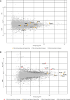
Differential gene expression analysis of human hearts and cardiac fibroblasts. (A) RNA-seq analysis of left ventricular human heart tissues from hemodialysis (HD, n=13) and healthy control (CON, n=4) hearts. Fibrosis-related genes of interest and reference genes are highlighted in red. There was no significant difference in expression of the genes of interest. Significantly differentially expressed genes (FDR<0.05) are shaped as triangular points versus circular points. (B) RNA-seq analysis human primary cardiac fibroblasts treated with 5 mM phosphate and 5 mM calcium (n=3) and 0.9 mM phosphate and 1.8 mM calcium controls (n=3). Fibrosis-related genes of interest and reference genes are highlighted in red. Significantly differentially expressed genes (FDR<0.05) are shaped as triangular points versus circular points. Periostin (POSTN), lysl hydroxylase 2 (PLOD2), TGF-β1 (TGFB1), and α-smooth muscle actin (ACTA2) were significantly upregulated in cardiac fibroblasts treated with high phosphate and high calcium. CPM, counts per million; FC, fold change.
Figure 4
Figure 4
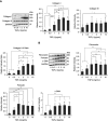
Elevated phosphate can induce trimerization of collagen type I and downregulate fibrillogenesis component, fibronectin, in cardiac fibroblasts. (A) Representative immunoblots of collagen type I, collagen type III, and GAPDH of cardiac fibroblasts treated with varying concentrations of β-glycerophosphate (n=6 each treatment). Trimeric collagen type I appears at 400 kDa and dimeric collagen type I appears at 250 kDa. 1.8 and 2.7 mM phosphate treatment exhibited significantly increased collagen type I dimer compared with 0.9 mM phosphate control levels. Trimeric collagen type I was significantly increased only at 3.8 mM phosphate compared with control. Collagen type III levels significantly decrease at 3.8 mM phosphate compared with 0.9 mM control. 2.7 and 3.8 mM phosphate treatment resulted in a higher collagen type I:III ratio compared with 0.9 mM control. (B) Representative immunoblots of fibronectin, periostin, α-SMA, and GAPDH of cardiac fibroblasts treated with varying concentrations of β-glycerophosphate (n=4–6). Fibronectin levels significantly decrease with an increase in phosphate levels compared with 0.9 mM control. There was no significant difference in periostin or α-SMA expression across all treatments. P values: * or # <0.05, ** or ## <0.01. Statistical significance was determined by one-way ANOVA and pairwise t test.
Figure 5
Figure 5
Elevated calcium can induce trimerization of collagen type I and downregulate fibrillogenesis component, fibronectin, in cardiac fibroblasts. (A) Representative immunoblots of collagen type I, collagen type III, and GAPDH of cardiac fibroblasts treated with varying concentrations of calcium chloride (n=6 each treatment). Trimeric collagen type I appears at 400 kDa and dimeric collagen type I appears at 250 kDa. 2.4, 3, and 4 mM calcium treatment exhibited significantly increased collagen type I dimer compared with 0.9 mM phosphate control levels. 5 mM calcium significantly increased collagen type I, decreased collagen type III, and overall increased collagen type I:III ratio compared with 0.9 mM control. (B) Representative immunoblots of fibronectin, periostin, α-smooth muscle actin (α-SMA), and GAPDH of cardiac fibroblasts treated with varying concentrations of calcium chloride (n=3–6). Fibronectin levels significantly decrease with elevated calcium levels at 4 and 5 mM compared with 0.9 mM control. There was no significant difference in periostin or α-SMA expression across all treatments. P values: * or # <0.05, ** or ## <0.01. Statistical significance determined by one-way ANOVA and pairwise t test.
Figure 6
Figure 6
Mineral stressors promote intramolecular collagen cross-linking and fibrotic ratio of collagen type I:III while downregulating fibronectin and collagen turnover. (A) Representative immunoblots of collagen type I, collagen type III, and GAPDH of cardiac fibroblasts treated with combined calcium chloride and β-glycerophosphate totaling 2 mM calcium and 3.8 mM phosphate (n=6). Trimeric collagen type I appears at 400 kDa and dimeric collagen type I appears at 250 kDa. Combined elevated calcium and phosphate treatment significantly increased trimeric collagen type I, decreased collagen type III, and overall higher collagen type I:III ratio compared with 1.8 mM calcium and 0.9 mM phosphate control media. (B) Representative immunoblots of fibronectin, periostin, α-smooth muscle actin (α-SMA), and GAPDH of cardiac fibroblasts treated with combined calcium chloride and β-glycerophosphate totaling 2 mM calcium and 3.8 mM phosphate (n=6). Combined elevated calcium and phosphate treatment significantly decreased fibronectin and increased α-SMA compared with control. There was no significant difference in periostin expression. (C) Representative immunoblots of lysyl hydroxylase 2 (LH2), proenzyme lysyl oxidase (pro-LOX), proteolytically processed pro-LOX (mature LOX), and GAPDH of cardiac fibroblasts treated with combined calcium chloride and β-glycerophosphate totaling 2 mM calcium and 3.8 mM phosphate (n=6). There was no difference in cross-linking enzyme expression. (D) Representative immunoblots of metalloproteinase-1 (MMP1) and proenzyme metalloproteinase-2 (Pro-MMP2), and GAPDH of cardiac fibroblasts treated with combined calcium chloride and β-glycerophosphate totaling 2 mM calcium and 3.8 mM phosphate (n=6). Combined elevated calcium and phosphate treatment significantly decreased pro-MMP2 compared with control. There was no significant difference in MMP1 expression. P values: **<0.01. Statistical significance determined by one-way ANOVA and pairwise t test.
Figure 7
Figure 7
TGF-β increases production of both collagen types I and III and upregulates collagen fibrillogenesis. (A) Representative immunoblots of collagen type I, collagen type III, and GAPDH of cardiac fibroblasts treated with varying concentrations of TGF-β (n=5–7 each treatment). Only dimeric collagen type I (250 kDa) was observed. Increase in TGF-β led to an increase in dimeric collagen type I, collagen type III, and overall higher collagen type I:III ratio compared with control. (B) Representative immunoblots of fibronectin, periostin, α-SMA, and GAPDH of cardiac fibroblasts treated with varying concentrations of TGF-β (n=5–7). Increase in TGF-β led to a significant increase in fibronectin starting at 1 ng/ml compared with control. Increase in TGF-β also led to a significant increase in periostin and α-SMA starting at 0.5 ng/ml compared with control. P values: *<0.05, **<0.01. Statistical significance determined by one-way ANOVA and pairwise t test.
Figure 8
Figure 8
TNF-α selectively increased production of collagen type I but not type III with no effect on fibrillogenesis factors. (A) Representative immunoblots of collagen type I, collagen type III, and GAPDH of cardiac fibroblasts treated with varying concentrations of TNF-α (n=5–7 each treatment). Only dimeric collagen type I (250 kDa) was observed. Increase in TNF-α led to an increase in dimeric collagen type I and a higher collagen type I:III ratio without a significant change in collagen type III compared with control. (B) Representative immunoblots of fibronectin, periostin, α-SMA, and GAPDH of cardiac fibroblasts treated with varying concentrations of TNF-α (n=5–7). Increase in TNF-α led to a significant decrease in α-SMA starting at 5 ng/ml compared with control. There was a slight significant increase in fibronectin from 1 to 30 ng/ml TNF-α. There was no significant difference in periostin expression across all treatments. P values: *<0.05, **<0.01. Statistical significance determined by one-way ANOVA and pairwise t test.

Comment in

References

    1. Romero-Gonzalez G, Gonzalez A, Lopez B, Ravassa S, Diez J. Heart failure in chronic kidney disease: the emerging role of myocardial fibrosis. Nephrol Dial Transplant. 2022;37(5):817–824. doi:10.1093/ndt/gfaa284 - DOI - PubMed
    1. Hayer MK Radhakrishnan A Price AM, et al. . Defining myocardial abnormalities across the stages of chronic kidney disease: a cardiac magnetic resonance imaging study. JACC Cardiovasc Imaging. 2020;13(11):2357–2367. doi:10.1016/j.jcmg.2020.04.021 - DOI - PMC - PubMed
    1. Panizo S Martinez-Arias L Alonso-Montes C, et al. . Fibrosis in chronic kidney disease: pathogenesis and consequences. Int J Mol Sci. 2021;22(1):408. doi:10.3390/ijms22010408 - DOI - PMC - PubMed
    1. Edwards NC, Hayer MK, Ferro CJ, Townend JN, Steeds RP. CKD associated cardiomyopathy: molecular mechanisms, imaging modalities, disease evolution and interventions. In: Rangaswami J, Lerma EV, Ronco C, eds. Cardio-Nephrology: Confluence of the Heart and Kidney in Clinical Practice. Springer International Publishing; 2017:45–58.
    1. Yamamoto K Masuyama T Sakata Y, et al. . Myocardial stiffness is determined by ventricular fibrosis, but not by compensatory or excessive hypertrophy in hypertensive heart. Cardiovasc Res. 2002;55(1):76–82. doi:10.1016/s0008-6363(02)00341-3 - DOI - PubMed

Publication types