Review
. 2024;19(7):959-980.
doi: 10.2174/011574888X263597231001164351.

A Monocytic Barrier to the Humanization of Immunodeficient Mice

Affiliations
Review

A Monocytic Barrier to the Humanization of Immunodeficient Mice

Emily J Du et al. Curr Stem Cell Res Ther. 2024.

Abstract

Mice with severe immunodeficiencies have become very important tools for studying foreign cells in an in vivo environment. Xenotransplants can be used to model cells from many species, although most often, mice are humanized through the transplantation of human cells or tissues to meet the needs of medical research. The development of immunodeficient mice is reviewed leading up to the current state-of-the-art strains, such as the NOD-scid-gamma (NSG) mouse. NSG mice are excellent hosts for human hematopoietic stem cell transplants or immune reconstitution through transfusion of human peripheral blood mononuclear cells. However, barriers to full hematopoietic engraftment still remain; notably, the survival of human cells in the circulation is brief, which limits overall hematological and immune reconstitution. Reports have indicated a critical role for monocytic cells - monocytes, macrophages, and dendritic cells - in the clearance of xenogeneic cells from circulation. Various aspects of the NOD genetic background that affect monocytic cell growth, maturation, and function that are favorable to human cell transplantation are discussed. Important receptors, such as SIRPα, that form a part of the innate immune system and enable the recognition and phagocytosis of foreign cells by monocytic cells are reviewed. The development of humanized mouse models has taken decades of work in creating more immunodeficient mice, genetic modification of these mice to express human genes, and refinement of transplant techniques to optimize engraftment. Future advances may focus on the monocytic cells of the host to find ways for further engraftment and survival of xenogeneic cells.

Keywords: Mice; SCID; blood cells; dendritic cells; humanized mice; macrophages; myeloid cells; transplantation..


Conflict of interest statement

CONFLICT OF INTEREST

Dr. Marcus Muench is on the editorial advisory board of the journal CSCRT.

Figures
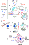
Fig. (1).
Fig. (1).
Effects of the NOD/ShiLtSz genetic background on monocytic cells. The growth, maturation, and functional defects of NOD monocytic progenitors, macrophages, and DCs are summarized. Altered cytokine responsiveness diminishes macrophage development in vitro (A). Stimulated peritoneal macrophages produce less cytokines (B), have altered responses to IFN-γ, and are more resistant to apoptosis (C). Increased GM-CSF production is observed in the bone marrow of NOD mice (D). However, DC development in response to GM-CSF and other cytokines is diminished in vitro (E). Similar to their macrophage counterparts, stimulated NOD DCs exhibit altered cytokine production and phenotype (F). Phagocytosis of apoptotic cells by NOD macrophages was also found to be reduced (G). (A higher resolution / colour version of this figure is available in the electronic copy of the article).
Fig. (2).
Fig. (2).
Human hematopoietic chimerism in three strains of immunodeficient mice after stem cell transplant. Mice were transplanted with human fetal bone marrow and analyzed 12 weeks later. Violin plots show the frequency of human β2-microglobulin+ cells among whole bone marrow, light-density splenocytes, light-density liver cells, light density blood (PBMC), and whole blood. NSG mice were compared as hosts to two mouse strains expressing human-specific cytokines. Violin plots compare engraftment among the three mouse strains for each tissue (n=5). Note % engraftment is on a log scale. These data were previously published in [71]. (A higher resolution / colour version of this figure is available in the electronic copy of the article).
Fig. (3).
Fig. (3).
Rapid clearance of human erythrocytes from the circulation of NSG mice. Mice were transfused with a mixture of human and mouse red cells. Donor mouse erythrocytes were obtained from mice that expressed enhanced green fluorescence protein so that donor cells could be distinguished from the host’s own red cells. The starting ratio of human:mouse erythrocytes transfused was >4:1, which was already greatly reduced by the first measured time point after transfusion (5 minutes). The frequencies of mouse erythrocytes remained relatively stable over the course of the experiment compared to the rapid decline in circulating human erythrocytes. Note that a natural log transformation of the measurement times is used to better visualize early measurements (5, 60, 120, and 180 minutes) while also allowing for the display of the two last time points (20 and 48 hours). These data were previously published in [12]. (A higher resolution / colour version of this figure is available in the electronic copy of the article).
Fig. (4).
Fig. (4).
Mechanisms by which mouse monocytic cells identify human cells for removal. Mouse macrophages receive inhibitory signals from CD47 expressed on most cells (A). Human CD47 does not provide as strong of a signal through SIRPα receptor on macrophages as mouse CD47, which may result in phagocytosis. Opsonization of human cells with complement 3 components (C3) can also lead to phagocytosis by macrophages (B). Mouse macrophages may also bind human cells based on carbohydrate moieties, such as sialic acid binding by CD169, which may foster phagocytosis of the human cells (C). (A higher resolution / colour version of this figure is available in the electronic copy of the article).
Fig. (5).
Fig. (5).
Human erythrocytes are larger than mouse erythrocytes. Flow cytometry was used to compare forward light scatter-area (FSC-A) characteristics of mouse and human RBCs. The overlay histogram data show the larger size of the human cells (A). The mean fluorescence intensity ± standard deviation of FSC-A intensity is shown as a bar plot for both cell types (B). Human and mouse red cells were mixed for analysis with the human cells identified using CD235a staining and mouse cells identified by enhanced green fluorescence protein expression derived from a transgenic marker expressed in all tissues. Mouse RBCs were purified from whole blood prior to mixing with human cells. Data are from [12]. (A higher resolution / colour version of this figure is available in the electronic copy of the article).

References

    1. Goyama S, Wunderlich M, Mulloy JC. Xenograft models for normal and malignant stem cells. Blood 2015; 125(17): 2630–40. 10.1182/blood-2014-11-570218 - DOI - PubMed
    1. Abarrategi A, Mian SA, Passaro D, Rouault-Pierre K, Grey W, Bonnet D. Modeling the human bone marrow niche in mice: From host bone marrow engraftment to bioengineering approaches. J Exp Med 2018; 215(3): 729–43. 10.1084/jem.20172139. - DOI - PMC - PubMed
    1. Mehler VJ, Burns C, Moore ML. Concise review: Exploring immunomodulatory features of mesenchymal stromal cells in humanized mouse models. Stem Cells 2019; 37(3): 298–305. 10.1002/stem.2948 - DOI - PMC - PubMed
    1. Alves da Costa T, Lang J, Torres RM, Pelanda R. The development of human immune system mice and their use to study tolerance and autoimmunity. J Transl Autoimmun 2019; 2: 100021. 10.1016/j.jtauto.2019.100021 - DOI - PMC - PubMed
    1. Tyagi AM, Yu M, Darby TM, et al. The microbial metabolite butyrate stimulates bone formation via t regulatory cell-mediated regulation of WNT10B expression. Immunity 2018; 49(6): 1116–1131.e7. 10.1016/j.immuni.2018.10.013 - DOI - PMC - PubMed

Substances