. 2023 Dec;50(6):214.
doi: 10.3892/or.2023.8651. Epub 2023 Oct 20.

Licochalcone A induces cell cycle arrest and apoptosis via suppressing MAPK signaling pathway and the expression of FBXO5 in lung squamous cell cancer

Affiliations

Licochalcone A induces cell cycle arrest and apoptosis via suppressing MAPK signaling pathway and the expression of FBXO5 in lung squamous cell cancer

Xiaoli Fan et al. Oncol Rep. 2023 Dec.

Abstract

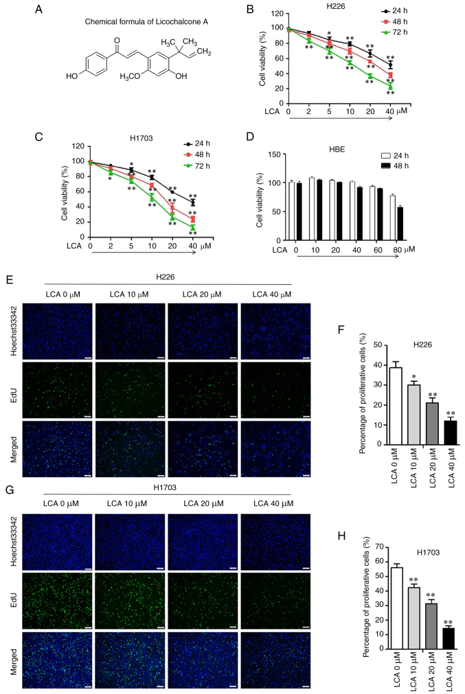
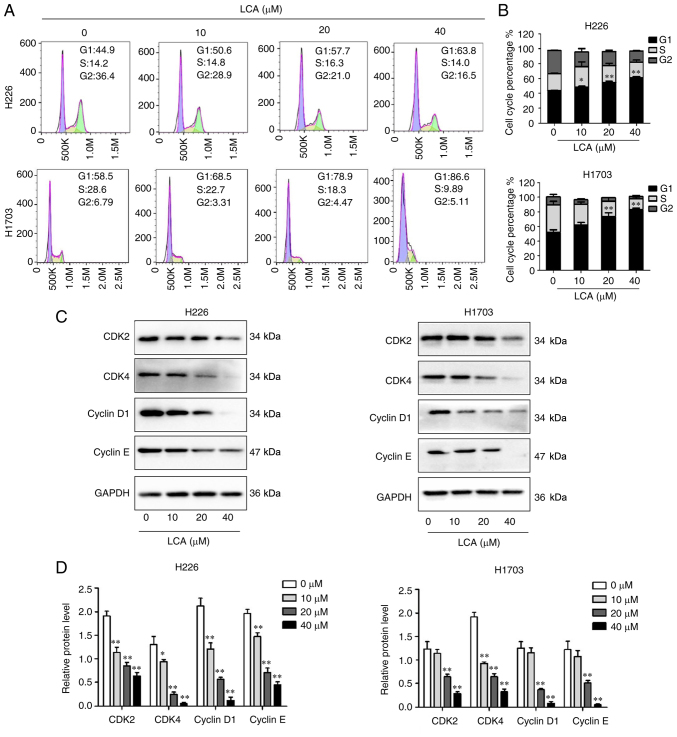
Lung squamous cell carcinoma (LSCC) is a highly heterogeneous malignancy with high mortality and few therapeutic options. Licochalcone A (LCA, PubChem ID: 5318998) is a chalcone extracted from licorice and possesses anticancer and anti‑inflammatory activities. The present study aimed to elucidate the anticancer effect of LCA on LSCC and explore the conceivable molecular mechanism. MTT assay revealed that LCA significantly inhibited the proliferation of LSCC cells with less cytotoxicity towards human bronchial epithelial cells. 5‑ethynyl‑2'‑deoxyuridine (EdU) assay demonstrated that LCA could reduce the proliferation rate of LSCC cells. The flow cytometric assays indicated that LCA increased the cell number of the G1 phase and induced the apoptosis of LSCC cells. LCA downregulated the protein expression of cyclin D1, cyclin E, CDK2 and CDK4. Meanwhile, LCA increased the expression level of Bax, cleaved poly(ADP‑ribose)polymerase‑1 (PARP1) and caspase 3, as well as downregulated the level of Bcl‑2. Proteomics assay demonstrated that LCA exerted its antitumor effects via inhibiting mitogen‑activated protein kinase (MAPK) signaling pathways and the expression of F‑box protein 5 (FBXO5). Western blot analysis showed that LCA decreased the expression of p‑ERK1/2, p‑p38MAPK and FBXO5. In the xenograft tumors of LSCC, LCA significantly inhibited the volumes and weight of tumors in nude mice with little toxicity in vital organs. Therefore, the present study demonstrated that LCA effectively inhibited cell proliferation and induced apoptosis in vitro, and suppressed xenograft tumor growth in vivo. LCA may serve as a future therapeutic candidate of LSCC.

Keywords: F‑box protein 5; apoptosis; licochalcone A; lung squamous cell carcinoma; proteomics.


Conflict of interest statement

The authors declare that they have no competing interests.

Figures

Figure 1.
Figure 1.
LCA inhibits lung squamous cell carcinoma cell viability and proliferation. (A) Chemical structure of LCA. (B and C) The viability of H226 and H1703 cells after treatment with LCA was detected by MTT. (D) The viability of HBE cells after treatment with LCA was detected by MTT. (E and F) EdU staining assay was used to detect cell proliferative ability of H226 cells (scale bar, 200 µm). (G and H) EdU staining assay was used to detect cell proliferative ability of H1703 cells (scale bar, 200 µm). The experiments were repeated at least three times (n=3). *P<0.05 and **P<0.01 vs. 0 µM. LCA, licochalcone A; EdU, 5-ethynyl-2′-deoxyuridine.
Figure 2.
Figure 2.
The effect of LCA on cell cycle in lung squamous cell carcinoma cells. (A and B) Analysis of the cell cycle proportion of H226 and H1703 cells after treated with LCA (0, 10, 20 and 40 µM) by flow cytometry. (C and D) The protein expression of CDK2, CDK4, cyclin D1 and cyclin E in H226 and H1703 cells determined by western blot analysis after treatment with LCA. The intensity of the bands was quantified by ImageJ and GAPDH was used as control. The experiments were repeated at least three times (n=3). *P<0.05 and **P<0.01 vs. 0 µM. LCA, licochalcone A.
Figure 3.
Figure 3.
The effect of LCA on cell apoptosis in lung squamous cell carcinoma cells. (A and B) The apoptotic rates of H226 and H1703 cells after treatment with LCA were detected by flow cytometry. (C and D) The level of apoptosis-related proteins of H226 and H1703 cells determined by western blot analysis. The experiments were repeated at least three times (n=3). *P<0.05 and **P<0.01 vs. 0 µM. LCA, licochalcone A; cle-, cleaved; PARP1, poly(ADP-ribose)polymerase-1.
Figure 4.
Figure 4.
Proteomic analysis of control and the LCA-treated LSCC cells. (A) Distribution of peptide length. (B) Distribution of the sample abundance. (C) Pearson's correlation analysis of the protein expression patterns. (D) Principal component analysis. (E and F) The histogram and volcano plots demonstrated the 1101 DEPs including 616 upregulated and 485 downregulated after LCA treatment in LSCC cells. (G) The heatmap revealed the different expression patterns of the differentially expressed genes. (H) Kyoto Encyclopedia of Genes and Genomes pathway analysis. LCA, licochalcone A; LSCC, lung squamous cell carcinoma; ctrl, control; DEPs, differentially expressed proteins.
Figure 5.
Figure 5.
Network pharmacology analysis of the therapeutic target of LCA against LSCC. (A) Venn diagram revealed 17 overlapping genes for LCA target genes and LSCC-related genes. (B) The bubble chart illustrated enriched Kyoto Encyclopedia of Genes and Genomes pathways related to the therapeutic target. (C) Protein-protein interaction analysis revealed interaction of the 17 overlapping genes. (D-F) Western blot analysis results revealed the protein expression of p38 MAPK, p-p38 MAPK, ERK1/2 and p-ERK1/2 after treatment with LCA (0, 10, 20 and 40 µM) in LSCC cells. The experiments were repeated at least three times (n=3). **P<0.01 vs. control. LCA, licochalcone A; LSCC, lung squamous cell carcinoma; p-, phosphorylated.
Figure 6.
Figure 6.
Bioinformatics analysis of FBXO5 based on TCGA. (A) The differential expression of FBXO5 between tumor and adjacent normal tissues across all TCGA tumors. Distributions of gene expression levels are displayed using box plots. The statistical significance computed by the Wilcoxon test is annotated by the number of stars (*P<0.05, **P<0.01 and ***P<0.001). (B) Expression of FBXO5 in LSCC based on sample types across TCGA samples (P<0.001). (C) Expression of FBXO5 in LSCC based on individual cancer stages (P<0.01). (D and E) Western blot analysis results revealed the protein expression of FBXO5 after LCA treatment in LSCC cells. (F and G) Visualization of LCA and FBXO5 molecules. F: The molecular surface form of van der Waals; G: Molecule-protein binding site. (H) Western blot analysis results revealed the protein expression of FBXO5 in control and si-FBXO5-transfected H1703 cells. (I) Western blot analysis results demonstrated the protein expression of CDK2, CDK4, cyclin D1 and cyclin E in control and si-FBXO5-transfected H1703 cells. The experiments were repeated at least three times (n=3). *P<0.05 and **P<0.01 vs. control. FBXO5, F-box protein 5; TCGA, The Cancer Genome Atlas; LSCC, lung squamous cell carcinoma; LCA, licochalcone A; siRNA, small interfering RNA; LUSC, lung squamous cell carcinoma.
Figure 7.
Figure 7.
LCA suppresses lung squamous cell carcinoma xenograft tumor growth in vivo (n=5). (A) Representative images of xenograft tumor models and tumor tissues. (B) The wet weight of the mice xenograft tumors. (C) The tumor volumes of the mice xenograft tumors. (D) The body weight of the xenograft tumor mice. (E) The organ weight of the xenograft tumor mice. (F) The hematoxylin and eosin staining representative pictures of vital organs of the xenograft tumors mice (scale bar, 200 µm). (G and H) The levels of FBXO5, p-p38 MAPK and p-ERK1/2 in tumor tissue determined by western blot analysis. The experiments were repeated at least three times (n=3). *P<0.05 and **P<0.01 vs. control. LCA, licochalcone A; FBXO5, F-box protein 5; CDDP, cisplatin; p-, phosphorylated. The body weight of the xenograft tumor mice.

Similar articles

Cited by

References

    1. Sung H, Ferlay J, Siegel RL, Laversanne M, Soerjomataram I, Jemal A, Bray F. Global cancer statistics 2020: GLOBOCAN estimates of incidence and mortality worldwide for 36 cancers in 185 countries. CA Cancer J Clin. 2021;71:209–249. doi: 10.3322/caac.21660. - DOI - PubMed
    1. Siegel RL, Miller KD, Jemal A. Cancer statistics, 2019. CA Cancer J Clin. 2019;69:7–34. doi: 10.3322/caac.21551. - DOI - PubMed
    1. Cheng TY, Cramb SM, Baade PD, Youlden DR, Nwogu C, Reid ME. The international epidemiology of lung cancer: Latest trends, disparities, and tumor characteristics. J Thorac Oncol. 2016;11:1653–1671. doi: 10.1016/j.jtho.2016.05.021. - DOI - PMC - PubMed
    1. Herbst RS, Morgensztern D, Boshoff C. The biology and management of non-small cell lung cancer. Nature. 2018;553:446–454. doi: 10.1038/nature25183. - DOI - PubMed
    1. Derman BA, Mileham KF, Bonomi PD, Batus M, Fidler MJ. Treatment of advanced squamous cell carcinoma of the lung: A review. Transl Lung Cancer Res. 2015;4:524–532. - PMC - PubMed

MeSH terms