Review
. 2023 Oct 12;2(1):e000548.
doi: 10.1136/bmjmed-2023-000548. eCollection 2023.

Polycystic ovary syndrome: pathophysiology and therapeutic opportunities

Affiliations
Review

Polycystic ovary syndrome: pathophysiology and therapeutic opportunities

Jiawen Dong et al. BMJ Med. .

Abstract

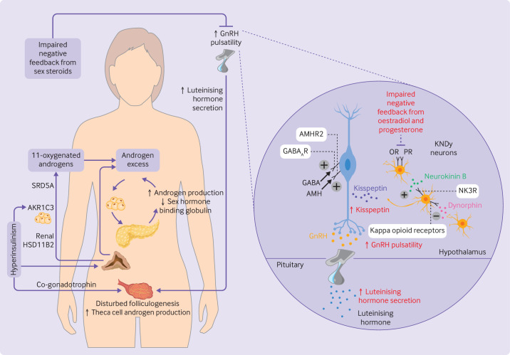
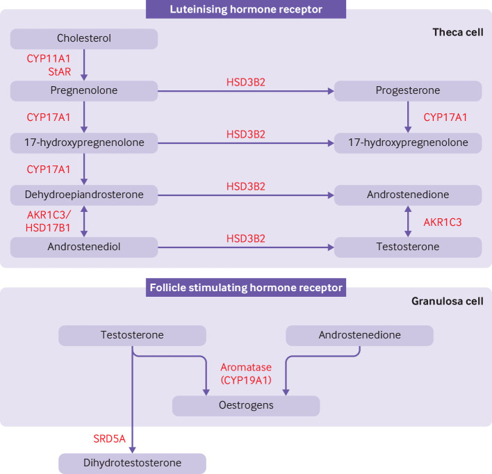
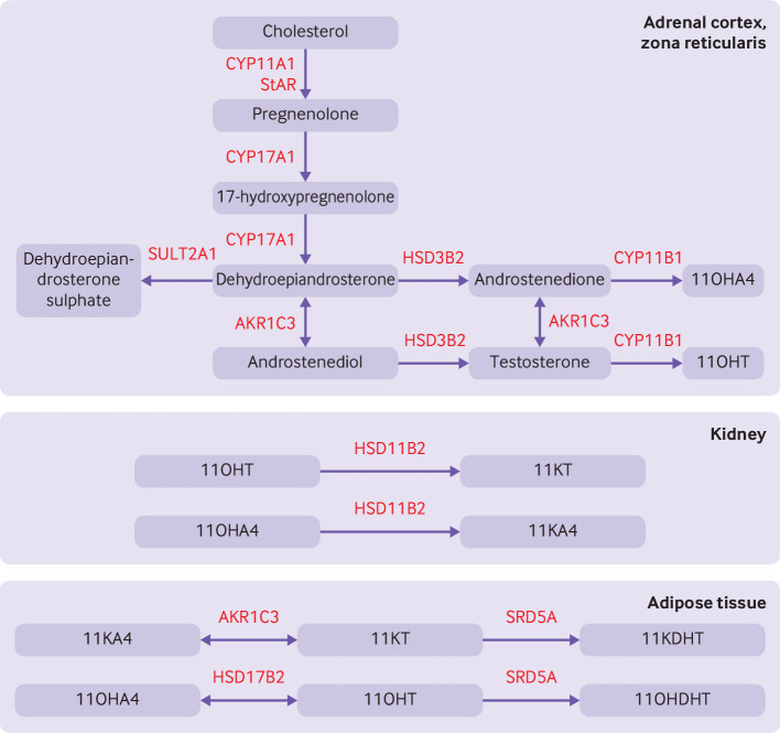
Polycystic ovary syndrome is characterised by excessive levels of androgens and ovulatory dysfunction, and is a common endocrine disorder in women of reproductive age. Polycystic ovary syndrome arises as a result of polygenic susceptibility in combination with environmental influences that might include epigenetic alterations and in utero programming. In addition to the well recognised clinical manifestations of hyperandrogenism and ovulatory dysfunction, women with polycystic ovary syndrome have an increased risk of adverse mental health outcomes, pregnancy complications, and cardiometabolic disease. Unlicensed treatments have limited efficacy, mostly because drug development has been hampered by an incomplete understanding of the underlying pathophysiological processes. Advances in genetics, metabolomics, and adipocyte biology have improved our understanding of key changes in neuroendocrine, enteroendocrine, and steroidogenic pathways, including increased gonadotrophin releasing hormone pulsatility, androgen excess, insulin resistance, and changes in the gut microbiome. Many patients with polycystic ovary syndrome have high levels of 11-oxygenated androgens, with high androgenic potency, that might mediate metabolic risk. These advances have prompted the development of new treatments, including those that target the neurokinin-kisspeptin axis upstream of gonadotrophin releasing hormone, with the potential to lessen adverse clinical sequelae and improve patient outcomes.

Keywords: Endocrinology; Medicine; Metabolic diseases; Obstetrics; Pharmacology; Reproductive medicine.


Conflict of interest statement

Competing interests: We have read and understood the BMJ Policy on declaration of interests and declare the following interests: AR has undertaken educational activities funded by Pfizer and Diurnal, and received grant funding from the Waterloo Foundation. AR has participated as principal investigator in clinical trials funded by Neurocrine Biosciences, Sparrow Pharmaceuticals, Diurnal, and Ascendis.

Figures

Figure 1
Figure 1
Pathophysiology and neuroendocrine disruption of the hypothalamo-pituitary-gonadal axis in polycystic ovary syndrome. (Left) Increased pulsatility of gonadotrophin releasing hormone (GnRH) causes increased secretion of luteinising hormone, consequent disrupted folliculogenesis, and increased production of ovarian androgens. Adrenal androgens are also increased, including 11-oxygenated androgens which are activated peripherally by renal 11β-hydroxysteroid dehydrogenase type 2 (HSD11B2) and aldo-keto reductase 1C3 (AKR1C3) in adipocytes. Steroid-5α-reductase (SRD5A) converts 11-ketotestosterone to 11-ketodihydrotestosterone. Excess levels of androgens stimulate deposition of abdominal adipose tissue which subsequently increases insulin resistance and hyperinsulinism. Hyperinsulinism stimulates AKR1C3 activity, increases androgen production from the ovaries (by its action as a co-gonadotrophin) and adrenal cortex, reduces production of hepatic sex hormone binding globulin, and inhibits progesterone mediated negative feedback onto GnRH neurons, worsening androgen excess in a vicious cycle. (Right) Kisspeptin, neurokinin B, and dynorphin A neurons (KNDy neurons) act in a paracrine and autocrine way to regulate release of kisspeptin onto GnRH neurons and consequent GnRH pulsatility. Neurokinin B binds to neurokinin 3 receptors (NK3R) to stimulate release of kisspeptin whereas dynorphin binds to kappa opioid receptors to inhibit kisspeptin release. γ-aminobutyric acid (GABA) and anti-müllerian hormone (AMH) bind to GABAA receptors (GABAAR) and AMH receptor type 2 (AMHR2), respectively, to stimulate GnRH pulsatility. Impaired negative feedback from oestradiol and progesterone is seen at the level of the hypothalamus. Neuroendocrine abnormalities in the control of these components are shown in red. OR=oestrogen receptor; PR=progesterone receptor
Figure 2
Figure 2
Classical pathway of androgen synthesis. Luteinising hormone stimulates the classical pathway of androgen synthesis in ovarian theca cells. Cholesterol is transported to the inner mitochrondrial membrane by steroidogenic acute regulatory protein (StAR). A cleavage system of the cytochrome P450 enzyme, CYP11A1, ferrodoxin, and ferrodoxin reductase converts cholesterol to pregnenolone. Expression of CYP11A1 is stimulated by activation of the luteinising hormone receptor. Pregnenolone is transported to smooth endoplasmic reticulum where it is converted to 17-hydroxypregnenolone and subsequently to dehydroepiandrosterone by the 17-hydroxylase and 17,20-lyase subunit of the CYP17A1 enzyme, respectively. Dehydroepiandrosterone is then converted to androstenedione or androstenediol and subsequently to testosterone by a combination of 3β-hydroxysteroid dehydrogenase type II (HSD3B2) and aldo-keto reductase type 1C3 (AKR1C3). 17β-hydroxysteroid dehydrogenase 1 (HSD17B1) also catalyses the conversion of dehydroepiandrosterone to androstenediol. HSD3B2 converts pregnenolone and 17-hydroxypregnenolone to progesterone and 17-hydroxyprogesterone, respectively, which are substrates for a back door alternative pathway of androgen synthesis. Androstenedione and testosterone diffuse into granulosa cells where they are converted to oestrogens by the action of aromatase (CYP19A1), under the control of follicle stimulating hormone receptor activation. Testosterone can be converted to dihydrotestosterone by steroid 5α-reductase (SRD5A) in peripheral tissues
Figure 3
Figure 3
Pathway for 11-oxygenated androgen synthesis, which begins in the adrenal cortex. Androstenedione and testosterone are produced by the classical pathway (figure 2). Dehydroepiandrosterone is diverted to downstream androgens or sulphonated to dehydroepiandrosterone sulphate by the sulphotransferase, SULT2A1. Androstenedione and testosterone are hydroxylated by 11β-hydroxylase (CYP11B) to produce abundant 11β-hydroxyandrostenedione (11OHA4) and smaller amounts of 11β-hydroxytestosterone (11OHT). Renal 11β-hydroxysteroid dehydrogenase type 2 (HSD11B2) converts 11OHT to 11-ketotestosterone (11KT) and 11OHA4 to 11-ketoandrostenedione (11KA4). In adipose tissue, 11KA4 is metabolised to 11KT and 11-ketodihydrotestosterone (11DHKT) by aldo-keto reductase type 1C3 (AKR1C3) and steroid-5α-reductase (SRD5A), respectively. 11OHA4 is metabolised to 11OHT and 11β-hydroxydihydrotestosterone (11OHDHT) by 17β-hydroxysteroid dehydrogenase 2 (HSD17B2) and SRD5A, respectively. 11KT and 11KDHT are potent agonists of the androgen receptor whereas 11OHT and 11OHDHT have milder potency. StAR=steroidogenic acute regulatory protein; HSD3B2=3β-hydroxysteroid dehydrogenase type II; CYP11A1, CYP17A1, CYP11B1=cytochrome P450 enzymes

References

    1. Allen LA, Shrikrishnapalasuriyar N, Rees DA. Long-term health outcomes in young women with polycystic ovary syndrome: A narrative review. Clin Endocrinol (Oxf) 2022;97:187–98. 10.1111/cen.14609 - DOI - PubMed
    1. Gibson-Helm M, Teede H, Dunaif A, et al. . Delayed diagnosis and a lack of information associated with dissatisfaction in women with polycystic ovary syndrome. J Clin Endocrinol Metab 2017;102:604–12. 10.1210/jc.2016-2963 - DOI - PMC - PubMed
    1. Teede HJ, Tay CT, Laven JJE, et al. . Recommendations from the 2023 international evidence-based guideline for the assessment and management of polycystic ovary syndrome. J Clin Endocrin Metab 2023;108:2447–69. 10.1210/clinem/dgad463 - DOI - PMC - PubMed
    1. Garg A, Patel B, Abbara A, et al. . Treatments targeting neuroendocrine dysfunction in polycystic ovary syndrome (PCOS). Clin Endocrinol (Oxf) 2022;97:156–64. 10.1111/cen.14704 - DOI - PubMed
    1. O’Reilly MW, Kempegowda P, Jenkinson C, et al. . 11-oxygenated C19 steroids are the predominant androgens in polycystic ovary syndrome. J Clin Endocrinol Metab 2017;102:840–8. 10.1210/jc.2016-3285 - DOI - PMC - PubMed

LinkOut - more resources