. 2023 Sep 18;14(16):3066-3077.
doi: 10.7150/jca.86611. eCollection 2023.

Mifepristone inhibits hepatoma growth by enhancing the GR-HSP60-survivin interaction to facilitate survivin degradation

Affiliations

Mifepristone inhibits hepatoma growth by enhancing the GR-HSP60-survivin interaction to facilitate survivin degradation

Ya-Hui Huang et al. J Cancer. .

Abstract

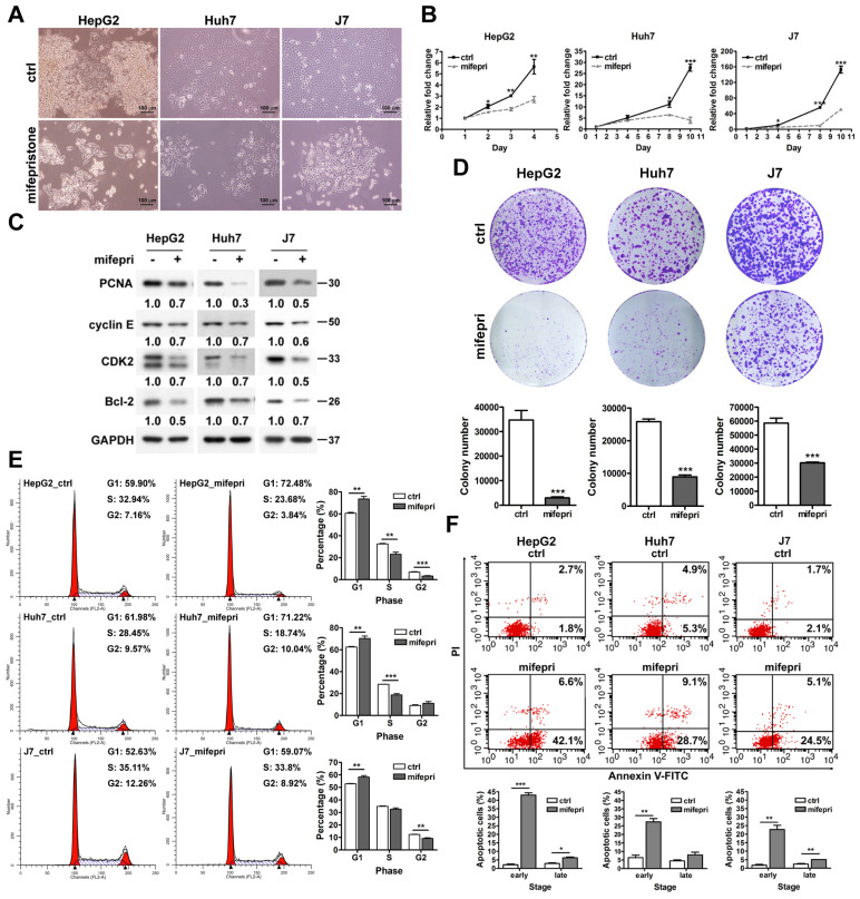
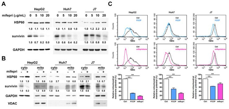
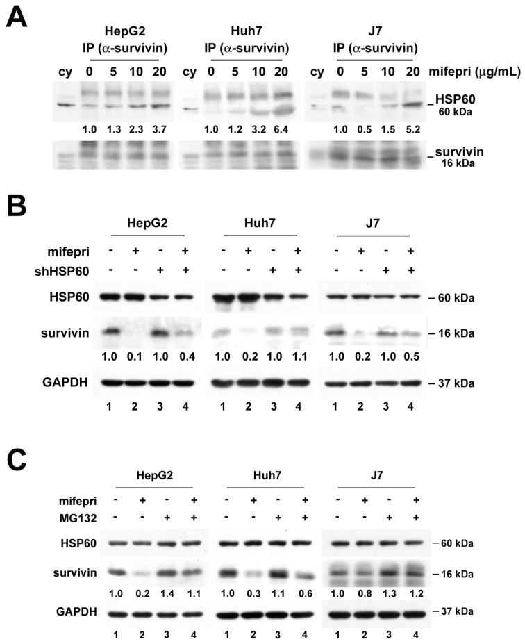
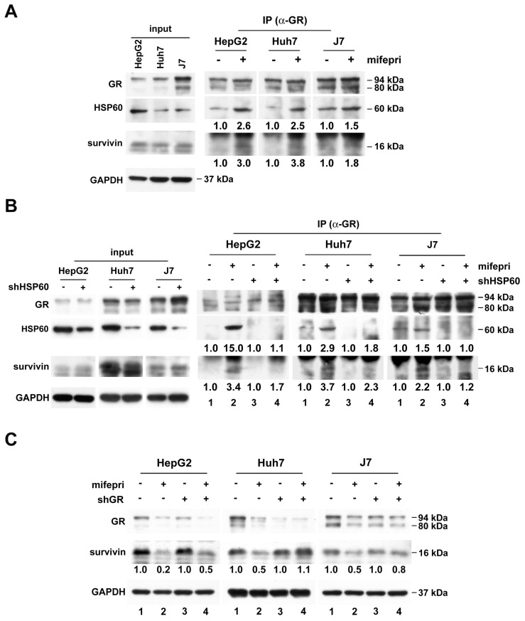
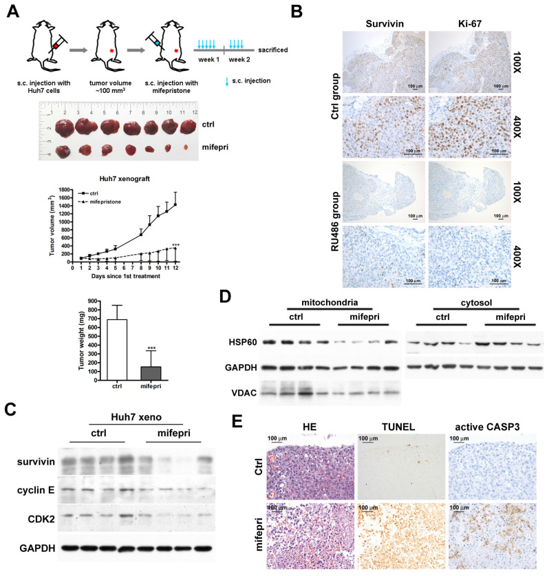
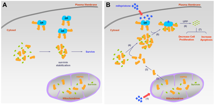
Silencing of heat shock protein 60 (HSP60) suppresses the growth of hepatocellular carcinoma (HCC). Mifepristone inhibits HSP60 mRNA expression in Chlamydophila-infected epithelial cells. The aim of this study was to determine whether mifepristone could inhibit the growth of HCC cells by affecting the functions of HSP60. The effect of mifepristone on cell viability was examined by flow cytometry and a cell proliferation assay. Protein-protein interactions were examined using the immunoprecipitation assay. The anti-tumor effect of mifepristone was evaluated using a xenograft model. Our results indicated that mifepristone induces cell cycle arrest at the G1 phase and early-stage apoptosis in HCC cells. Instead of reducing the total amount of HSP60, mifepristone induced the release of mitochondrial HSP60 into the cytosol by causing a loss of ΔΨm, thereby enhancing glucocorticoid receptor (GR)-HSP60-survivin complex formation as well as survivin degradation. Animal models have confirmed the growth inhibitory effects of mifepristone on HCC, including changes in the abundance of HSP60 in mitochondria and cytosol, decreased survivin and Ki-67-positive cells, as well as increased cell apoptosis. In conclusion, the inhibition of HCC growth by mifepristone may be achieved by altering the subcellular distribution of HSP60 to enhance the formation of cytosolic GR-HSP60-survivin complexes in the cells, leading to the degradation of survivin.

Keywords: HSP60; glucocorticoid receptor; hepatocellular carcinoma; mifepristone; survivin.


Conflict of interest statement

Competing Interests: The authors have declared that no competing interest exists.

Figures

Figure 1
Figure 1
Effect of mifepristone on the growth of HCC cells. (A) Cell morphology of HCC cell lines (HepG2, Huh7, and J7) in the absence (ctrl) or presence of mifepristone (20 μg/mL) for 48 h. (B) MTT cell proliferation assay performed with or without mifepristone (mifepri, 10 μg/mL). (C) Immunoblot assay: HCC cells were treated with or without mifepristone (mifepri, 10 μg/mL) for 24 h. (D) Colony formation assay for HCC cells after mifepristone (mifepri) treatment. Top panel: representative image; Bottom panel: quantitative assessment of the number of colonies. (E-F) Cell cycle (E) and cell apoptosis (F) were analyzed using flow cytometry. The histogram displays the percentage of cells in each cell cycle phase (E) and the percentage of apoptotic cells in the early and late stages (F). Detailed conditions and procedures for mifepristone treatment are described in the Materials and Methods section. The data represent the mean ± SD from three independent experiments and were analyzed using Student's t-test. Statistical significance is indicated as *P < 0.05; **P < 0.01; ***P < 0.001 compared to the control (ctrl).
Figure 2
Figure 2
Effect of mifepristone on HSP60 and survivin in HCC cells. (A) Immunoblot assay was performed to detect the expression of HSP60 and survivin in HepG2, Huh7, and J7 cells treated with various concentrations of mifepristone (mifepri) for 72 h. GAPDH served as a loading control. The protein expression levels of genes in the group without mifepristone are used as references for calculating the fold differences among groups with different concentrations of mifepristone. (B) Cytosolic and mitochondrial fractions were extracted from cells with (+) or without (-) mifepristone (mifepri) treatment (10 μg/mL) for 48 h (J7) or 72 h (HepG2 and Huh7). Immunoblot assays were conducted to examine the levels of HSP60 and survivin. GAPDH served as a loading control, and Voltage-dependent anion channel (VDAC) was used as a mitochondrial marker. (C) Mitochondrial membrane potential was assessed through flow cytometry. HCC cells were stained with TMRE in the presence (red) or absence (black) of mifepristone (mifepri), while FCCP (blue) was used as a positive control. The top panel shows TMRE intensity distribution, and the bottom panel presents statistical results. The data present the mean ± SD from at least three independent experiments and were analyzed using Student's t-test. Statistical significance is indicated as ***P < 0.001 compared to the control (Ctrl).
Figure 3
Figure 3
Mifepristone-induced downregulation of survivin in HCC cells requires the involvement of HSP60. (A) Cytosolic extracts from HepG2, Huh7, and J7 cells were incubated with increasing concentrations of mifepristone (mifepri; 5, 10, 20 μg/mL) for 4 h. The extracts were then subjected to immunoprecipitation using an anti-survivin antibody, followed by immunoblotting with an anti-HSP60 antibody. The protein expression levels of HSP60 in the group without mifepristone are used as references for calculating the fold differences among groups with different concentrations of mifepristone. (B) HepG2, Huh7, and J7 cells with (+) or without (-) HSP60 knockdown were treated with mifepristone (mifepri; 20 μg/mL) for 24 hours. The expression levels of HSP60, survivin, and GAPDH were detected using immunoblot assays. The survivin expression in lanes 1 and 3, without mifepristone treatment, serves as the reference for calculating the fold differences in the presence of mifepristone (lane 1 vs. lane 2 and lane 3 vs. lane 4). (C) Following treatment with (+) or without (-) mifepristone (mifepri), HepG2, Huh7, and J7 cells were treated with MG132 for 4-19 h to extract total protein. Immunoblot assays were performed to detect the expression of HSP60, survivin, and GAPDH. The survivin expression in lane 1 is used as the reference for calculating the fold differences among lanes 2, 3, and 4.
Figure 4
Figure 4
GR participates in mifepristone-trigged survivin degradation in HCC cells. (A-B) Immunoprecipitation was performed using cytosolic extracts from untreated HCC cells (A) and cells with (+) or without (-) shHSP60 expression (B). After incubation of cytosolic extracts with (+) or without (-) mifepristone (mifepri) for 4 h, they were immunoprecipitated with anti-GR antibodies to examine the amounts of GR, HSP60, and survivin by immunoblot. The left panels (A and B) are represent input controls for cytosolic extracts. (C) Total protein was extracted from GR silencing (shGR) HCC cells with or without mifepristone (mifepri) treatment for the detection of GR and survivin by immunoblot. GAPDH served as a loading control. The lane in the group without mifepristone treatment serves as the reference for calculating the fold differences in the presence of mifepristone.
Figure 5
Figure 5
Suppression of tumor growth by administration of mifepristone in a Huh7 xenograft mouse model. (A) Top: Schematic representation of the establishment of Huh7 xenograft mouse models and treatment with mifepristone. Middle: Final xenograft tumors and tumor volume for the control (ctrl) and mifepristone (mifepri) groups. Bottom: Tumor weight for the control (ctrl) and mifepristone (mifepri) groups. “***”, P < 0.001. (B) Immunohistochemical images showing survivin and Ki-67 (a marker for cell proliferation) staining at magnifications of 100× and 400×. (C) Immunoblot assay. Protein expression levels of survivin, cyclin E, and CDK2 in the control (ctrl) and mifepristone (mifepri) groups. (D) Changes in HSP60 expression in mitochondria and cytosol of xenograft tumors from the control (ctrl) and mifepristone (mifepri) groups. GAPDH is used as a loading control, and VDAC is a mitochondrial marker. (E) H&E staining (HE), TUNEL assay, and immunohistochemical staining of active caspase-3 (CASP3) for xenograft tumors.
Figure 6
Figure 6
Schematic diagram of the molecular mechanism on mifepristone in inhibiting HCC cell growth. This figure illustrates the roles of GR, HSP60, and survivin in HCC cells in the presence and absence of mifepristone, showcasing the molecular mechanism behind its inhibitory effects. (A) Under normal physiological conditions, HSP60-GR and HSP60-survivin complexes are localized in the cytosol. HSP60 binds to survivin, thereby stabilizing survivin for cell survival. (B) In the presence of mifepristone, it not only binds to GR but also reduces the activity of ΔΨm, leading to the release of HSP60 from mitochondria to the cytosol (steps 1 to 2). The accumulation of cytosolic HSP60 enhances the formation of HSP60-survivin complexes, facilitating interaction with GR (steps 3 to 5). Subsequently, survivin is degraded through the ubiquitin-proteasome pathway (UPP) (step 6). Ultimately, this process results in decreased cell proliferation and increased apoptosis (step 7).

Similar articles

Cited by

References

    1. Khalaf N, Ying J, Mittal S, Temple S, Kanwal F, Davila J. et al. Natural History of Untreated Hepatocellular Carcinoma in a US Cohort and the Role of Cancer Surveillance. Clin Gastroenterol Hepatol. 2017;15:273–81. e1. - PubMed
    1. Huang YH, Lin KH, Yu JS, Wu TJ, Lee WC, Chao CC. et al. Targeting HSP60 by subcutaneous injections of jetPEI/HSP60-shRNA destabilizes cytoplasmic survivin and inhibits hepatocellular carcinoma growth. Mol Carcinog. 2018;57:1087–101. - PubMed
    1. Ito T, Shiraki K, Sugimoto K, Yamanaka T, Fujikawa K, Ito M. et al. Survivin promotes cell proliferation in human hepatocellular carcinoma. Hepatology. 2000;31:1080–5. - PubMed
    1. Ishida K, Yamazaki T, Motohashi K, Kobayashi M, Matsuo J, Osaki T. et al. Effect of the steroid receptor antagonist RU486 (mifepristone) on an IFNgamma-induced persistent Chlamydophila pneumoniae infection model in epithelial HEp-2 cells. J Infect Chemother. 2012;18:22–9. - PubMed
    1. Baulieu EE. RU 486 (mifepristone). A short overview of its mechanisms of action and clinical uses at the end of 1996. Ann N Y Acad Sci. 1997;828:47–58. - PubMed