

Elucidating the role played by bone marrow in visceral leishmaniasis
- PMID: 37860064
- PMCID: PMC10582953
- DOI: 10.3389/fcimb.2023.1261074
Elucidating the role played by bone marrow in visceral leishmaniasis
Abstract
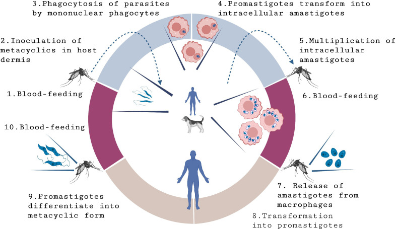
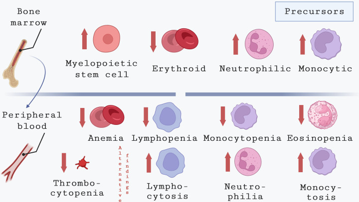
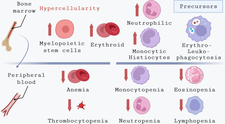
Leishmaniasis is a widespread group of infectious diseases that significantly impact global health. Despite high prevalence, leishmaniasis often receives inadequate attention in the prioritization of measures targeting tropical diseases. The causative agents of leishmaniasis are protozoan parasites of the Leishmania genus, which give rise to a diverse range of clinical manifestations, including cutaneous and visceral forms. Visceral leishmaniasis (VL), the most severe form, can be life-threatening if left untreated. Parasites can spread systemically within the body, infecting a range of organs, such as the liver, spleen, bone marrow and lymph nodes. Natural reservoirs for these protozoa include rodents, dogs, foxes, jackals, and wolves, with dogs serving as the primary urban reservoir for Leishmania infantum. Dogs exhibit clinical and pathological similarities to human VL and are valuable models for studying disease progression. Both human and canine VL provoke clinical symptoms, such as organ enlargement, fever, weight loss and abnormal gamma globulin levels. Hematologic abnormalities have also been observed, including anemia, leukopenia with lymphocytosis, neutropenia, and thrombocytopenia. Studies in dogs have linked these hematologic changes in peripheral blood to alterations in the bone marrow. Mouse models of VL have also contributed significantly to our understanding of the mechanisms underlying these hematologic and bone marrow abnormalities. This review consolidates information on hematological and immunological changes in the bone marrow of humans, dogs, and mice infected with Leishmania species causing VL. It includes findings on the role of bone marrow as a source of parasite persistence in internal organs and VL development. Highlighting gaps in current knowledge, the review emphasizes the need for future research to enhance our understanding of VL and identify potential targets for novel diagnostic and therapeutic approaches.
Keywords: Leishmania; bone marrow; canine visceral leishmaniasis; human visceral leishmaniasis; visceral leishmaniasis.
Copyright © 2023 Veras, de Santana, Brodskyn, Fraga, Solcà, De Menezes, Leite and Teixeira.
Conflict of interest statement
The authors declare that the research was conducted in the absence of any commercial or financial relationships that could be construed as a potential conflict of interest. The author(s) declared that they were an editorial board member of Frontiers, at the time of submission. This had no impact on the peer review process and the final decision.
Figures
References
-
- Abreu R., Carvalho M das G., Carneiro C. M., Giunchetti R. C., Teixeira-Carvalho A., Martins-Filho O. A., et al. (2011). Influence of clinical status and parasite load on erythropoiesis and leucopoiesis in dogs naturally infected with Leishmania (Leishmania) chagasi. PloS One 6 (5), 1–9. doi: 10.1371/journal.pone.0018873 - DOI - PMC - PubMed
-
- Agrawal Y., Sinha A. K., Upadhyaya P., Kafle S. U., Rijal S., Khanal B. (2013). Hematological profile in visceral leishmaniasis. Int. J. Infect. Microbiol. 2 (2), 39–44. doi: 10.3126/ijim.v2i2.8320 - DOI
Publication types
MeSH terms
LinkOut - more resources
Full Text Sources
Medical