. 2023 Oct 4:13:1134063.
doi: 10.3389/fonc.2023.1134063. eCollection 2023.

Identification and validation of PCSK9 as a prognostic and immune-related influencing factor in tumorigenesis: a pan-cancer analysis

Affiliations

Identification and validation of PCSK9 as a prognostic and immune-related influencing factor in tumorigenesis: a pan-cancer analysis

Chao Sun et al. Front Oncol. .

Abstract

Introduction: Proprotein convertase subtilisin/kexin-9 (PCSK9) has been primarily studied in the cardiovascular field however, its role in cancer pathophysiology remains incompletely defined. Recently, a pivotal role for PCSK9 in cancer immunotherapy was proposed based on the finding that PCSK9 inhibition was associated with enhancing the antigen presentation efficacy of target programmed cell death-1 (PD-1). Herein, we provide results of a comprehensive pan-cancer analysis of PCSK9 that assessed its prognostic and immunological functions in cancer.

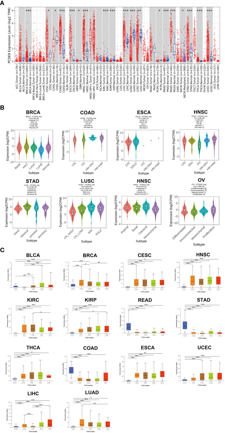
Methods: Using a variety of available online cancer-related databases including TIMER, cBioPortal, and GEPIA, we identified the abnormal expression of PCSK9 and its potential clinical associations in diverse cancer types including liver, brain and lung. We also validated its role in progression-free survival (PFS) and immune infiltration in neuroblastoma.

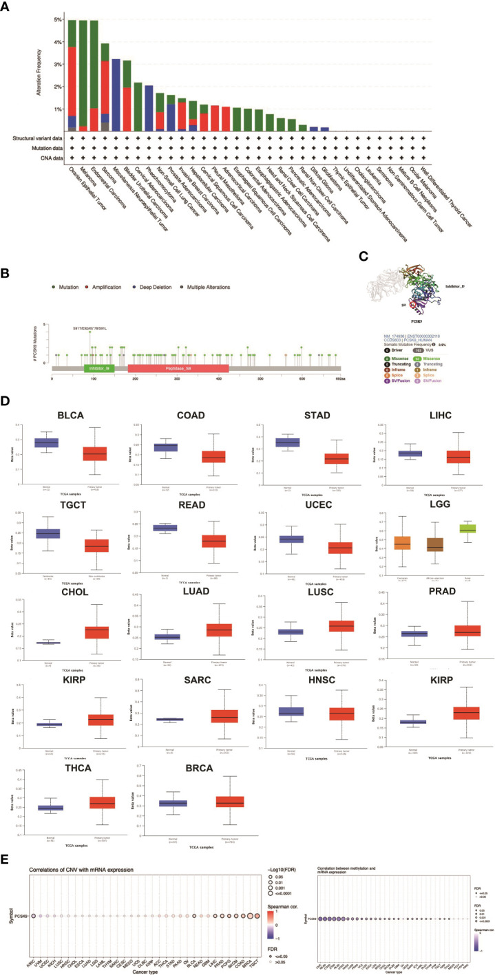
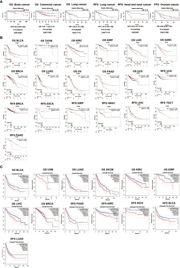
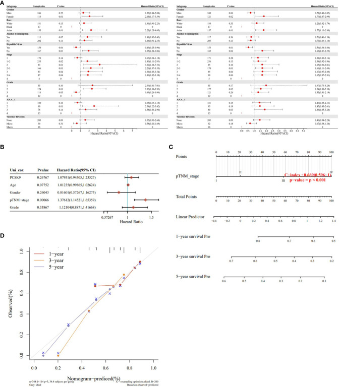
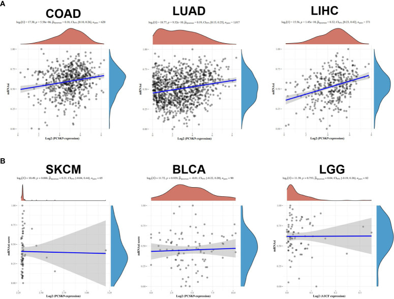
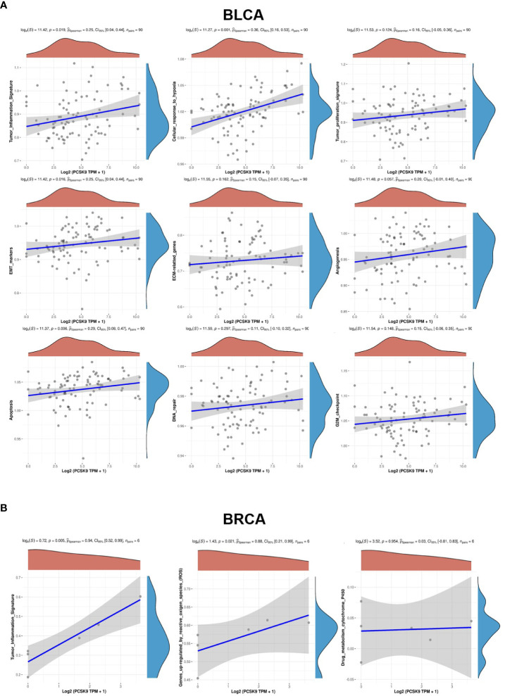
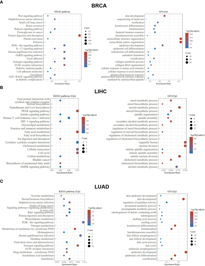
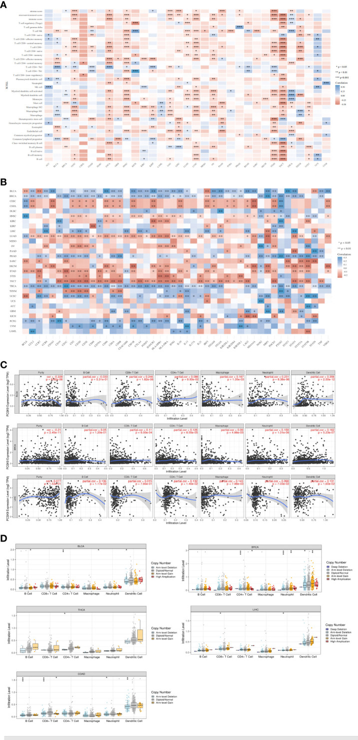
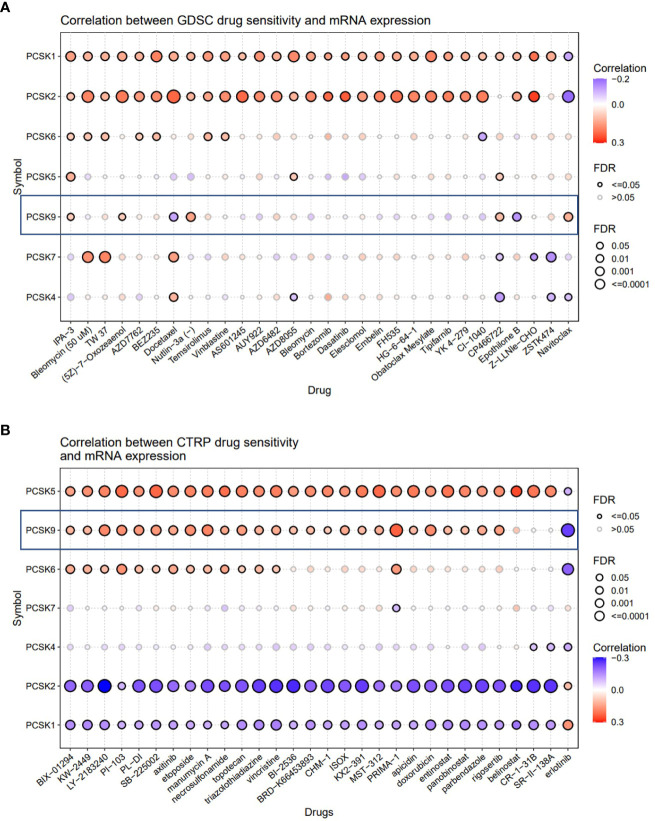
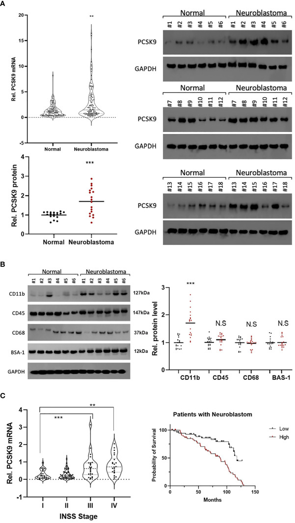
Results: Overall, the pan-cancer survival analysis revealed an association between dysregulated PCSK9 and poor clinical outcomes in various cancer types. Specifically, PCSK9 was extensively genetically altered across most cancer types and was consistently found in different tumor types and substages when compared with adjacent normal tissues. Thus, aberrant DNA methylation may be responsible for PCSK9 expression in many cancer types. Focusing on liver hepatocellular carcinoma (LIHC), we found that PCSK9 expression correlated with clinicopathological characteristics following stratified prognostic analyses. PCSK9 expression was significantly associated with immune infiltrate since specific markers of CD8+ T cells, macrophage polarization, and exhausted T cells exhibited different PCSK9-related immune infiltration patterns in LIHC and lung squamous cell carcinoma. In addition, PCSK9 was connected with resistance of drugs such as erlotinib and docetaxel. Finally, we validated PCSK9 expression in clinical neuroblastoma samples and concluded that PCSK9 appeared to correlate with a poor PFS and natural killer cell infiltration in neuroblastoma patients.

Conclusion: PCSK9 could serve as a robust prognostic pan-cancer biomarker given its correlation with immune infiltrates in different cancer types, thus potentially highlighting a new direction for targeted clinical therapy of cancers.

Keywords: immune infiltrate; pan-cancer; pcsk9; prognosis; tumorigenesis.


Conflict of interest statement

The authors declare that the research was conducted in the absence of any commercial or financial relationships that could be construed as a potential conflict of interest

Figures

Figure 1
Figure 1
(A) PCSK9 expression levels in different cancer types. Increased or decreased expression of PCSK9 compared with normal tissues across different cancer types from the TCGA database in TIMER (*p < 0.05, **p < 0.01, ***p < 0.001). (B) Pan-cancer PCSK9 expression in different subtypes. (A–H), Pan-cancer differential expression of PCSK9 in different cancer subtypes in the indicated tumor types from GEPIA. (C) Correlation between PCSK9 expression and the main pathological WHO stages for BLCA, BRCA, CESC, HNSC, KIRC, KIRP, READ, STAD, THCA, COAD, ESCA, UCEC, LIHC, and LIHC (A–N) from the UALCAN database (*p < 0.05, **p < 0.01, ***p < 0.001).
Figure 2
Figure 2
DNA methylation and mutation features of PCSK9 across cancer types. (A) The alteration frequency with different types of mutations was examined using the cBioPortal database. (B, C) The mutation site with the highest alteration frequency (E92Afs*78) in the 3D structure of PCSK9. (D) Promoter methylation level of PCSK9 across cancers. The results were obtained from the UALCAN database and GSCA database. (E) The correlation between PCSK9 expression and copy number variations (CNV) are shown from the GSCA database.
Figure 3
Figure 3
(A) Survival analyses of PCSK9 expression across cancers (based on PrognoScan). OS (n = 67) in brain cancer cohort GSE16581. OS (n = 177) in colorectal cancer cohort GSE17536. OS (n = 56) in lung cancer cohort GSE17710. RFS (n = 56) in lung cancer cohort GSE17710. RFS (n = 28) in head and neck cancer cohort GSE2837. PFS (n = 110) in ovarian cancer cohort GSE17260. OS, overall survival; RFS, relapse free survival; PFS, progression free survival. (B) Kaplan-Meier survival curves of survival comparing high and low expression of PCSK9 in the Kaplan-Meier Plotter database. Overall survival differences between groups in BLCA, THYM, KIRC, KIRP, LIHC, SARC, BRCA, LUAD, OV, PAAD, PCPG, and UCS. (M–T) Relapse-free interval difference between groups in UCS, BRCA, ESCA, KIRP, HNSC, LIHC, TGCT, and PAAD. (C) Kaplan-Meier survival curves of survival comparing high and low expression of PCSK9 in the GEPIA database. (A–H) Overall survival differences between groups in BLCA, UVM, LUAD, SKCM, KIRC, KIRP, LIHC, and BRCA. (I–M) Disease-free interval difference between groups in PAAD, KIRC, KICH, BLCA, and LUAD.
Figure 4
Figure 4
Survival analysis of PCSK9 expression in different clinicopathologic features in hepatocellular carcinoma. (A) Correlation of PCSK9 mRNA expression with OS in LIHC. (B) Correlation of PCSK9 mRNA expression with PFS in LIHC. OS, overall survival; PFS, progression free survival. (C, D) Development of a nomogram prediction model using PCSK9 expression levels and clinicopathological parameters for survival prediction.
Figure 5
Figure 5
Associations of stemness indices with the PCSK9 expression in various cancers. (A) Positive correlations between mRNAsi and PCSK9 in COAD, LUAD and LIHC (p < 0.05). (B) No correlation between mRNAsi and PCSK9 expression in SKCM, BLCA and LGG (p > 0.05).
Figure 6
Figure 6
GVSA analysis of PCSK9 in various tumors. (A) BLCA, (B) BRCA.
Figure 7
Figure 7
PCSK9-related KEGG pathway analysis and GO enrichment analysis. (A–C) KEGG pathway analysis and GO enrichment analysis of biological processes based on the PCSK9-interacted and PCSK9-correlated genes in BRCA, LIHC and LUAD.
Figure 8
Figure 8
Correlation of PCSK9 expression with immune infiltration and various subsets of immune cells. (A) Correlation of PCSK9 expression with the levels of infiltrating immune cells based on xCell (*p < 0.05, **p < 0.01). (B) Correlation between PCSK9 expression and immunosuppressive factors or immune stimulatory factors (*p < 0.05, **p < 0.01). (C) Correlation between PCSK9 expression and infiltration scores of six immune infiltrates, including B cells, CD4+ T cells, CD8+ T cells, dendritic cells, macrophages, and neutrophils, in BRCA, BLCA, and LIHC. (D) The association between PCSK9 copy number variations and immune infiltrates in LIHC, BLCA, BRCA, COAD, and THCA (i–v, *p < 0.05, **p < 0.01, ***p < 0.001).
Figure 9
Figure 9
Correlation analysis between PCSK9 expression and drug sensitivity. (A) Correlation between PCSK9 and sensitivity of the top 10 anticancer drugs in GDSC database. (B) Difference of drug sensitivity between PCSK9-related expression groups in CTRP database.
Figure 10
Figure 10
(A) Preliminary experimental verification of PCSK9 expression in neuroblastoma (NB). The mRNA and protein expression levels of PCSK9 in paired NB and adjacent non-tumor tissues by qRT-PCR (n=18; Figure 1A ) and western blotting (n=18; Figures 1B, C ) **p < 0.01, ***p < 0.001; (B) The association between NK cell-relevant immune checkpoints (CD11b, CD45, CD68) and PCSK9 expression in NB was detected by western blot in 18 NB tissue samples (***p < 0.001). (C) The association between PCSK9 expression in different clinical stages and survival analysis of patients. WHO stage III and IV showed highly expressed PCSK9 that correlated with poor progression-free survival (PFS, HR=1.51, 95% CI 1.25–1.71, p < 0.05).

Similar articles

Cited by

References

    1. Han Y, Liu D, Li L. PD-1/PD-L1 pathway: current researches in cancer. Am J Cancer Res (2020) 10(3):727–42. - PMC - PubMed
    1. Bellmunt J, de Wit R, Vaughn DJ, Fradet Y, Lee JL, Fong L, et al. . Pembrolizumab as second-line therapy for advanced Urothelial carcinoma. N Engl J Med (2017) 376(11):1015–26. doi: 10.1056/NEJMoa1613683 - DOI - PMC - PubMed
    1. Tang T, Huang X, Zhang G, Hong Z, Bai X, Liang T. Advantages of targeting the tumor immune microenvironment over blocking immune checkpoint in cancer immunotherapy. Signal Transduct Target Ther (2021) 6(1):72. doi: 10.1038/s41392-020-00449-4 - DOI - PMC - PubMed
    1. Wu T, Dai Y. Tumor microenvironment and therapeutic response. Cancer Lett (2017) 387:61–8. doi: 10.1016/j.canlet.2016.01.043 - DOI - PubMed
    1. Darvin P, Toor SM, Sasidharan Nair V, Elkord E. Immune checkpoint inhibitors: recent progress and potential biomarkers. Exp Mol Med (2018) 50(12):1–11. doi: 10.1038/s12276-018-0191-1 - DOI - PMC - PubMed