

Measurement of maximal muscle contraction force induced by high-frequency magnetic stimulation: a preliminary study on the identification of the optimal stimulation site
- PMID: 37860217
- PMCID: PMC10545052
- DOI: 10.11336/jjcrs.12.27
Measurement of maximal muscle contraction force induced by high-frequency magnetic stimulation: a preliminary study on the identification of the optimal stimulation site
Abstract
Tsubahara A, Kamiue M, Ito T, Kishimoto T, Kurozumi C. Measurement of maximal muscle contraction force induced by high-frequency magnetic stimulation: a preliminary study on the identification of the optimal stimulation site. Jpn J Compr Rehabil Sci 2021; 12: 27-31.
Purpose: To identify the optimal stimulation site and technique for inducing strong muscle contraction using a high-frequency magnetic stimulator.
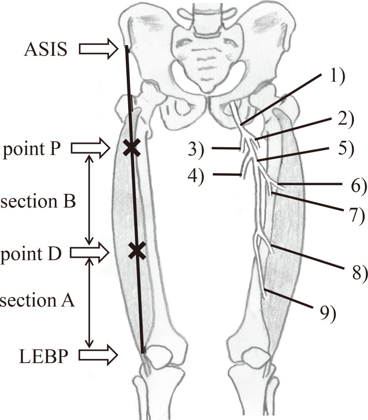
Methods: High-frequency magnetic stimulation was administered to the right vastus lateralis (VL) of eight healthy adults at maximal intensity within the range of tolerable pain. The stimulation sites were as follows: section A, the area between the lateral edge of the base of the patella (LEBP) and the distal one-third of the thigh (point D); section B, the area between point D and the proximal one-third of the thigh (point P). Isometric maximal muscle contraction forces induced by magnetic stimulation (Stim-MCF) were compared between the two sections.
Results: The Stim-MCF was significantly higher in section B than in section A. Additionally, the sites susceptible to stimulation were confined to a narrow area near point D in section A and the central part between points D and P in section B. The degree of pain was very low in both sections.
Conclusion: The optimal site for magnetic stimulation of the VL was limited to the central part of the thigh. In addition to the superficial proximal sub-branch, the deep proximal sub-branch and/or deeply clustered motor nerve endings may have been stimulated. Our results suggested that moving the probe was a useful way to identify the site that elicited the strongest muscle contraction force.
Keywords: high-frequency magnetic stimulation; motor points; muscle contraction force; quadriceps femoris; strengthening.
©Kaifukuki Rehabilitation Ward Association 2021.
Conflict of interest statement
COI: We disclose that a joint research contact has been concluded between the Kawasaki University of Medical Welfare and the OG Wellness Technologies Co., Ltd. We asked the company to manufacture an equipment and received a total of ¥ 2,398,000 as a research fund over three years.
Figures


References
-
- Aranceta J, Pérez-Rodrigo C, Gondra J, Orduna J.. Community-based programme to promote physical activity among elderly people: the GeroBilbo study. J Nutr Health Aging 2001; 5: 238-42. - PubMed
-
- Lee H, Kim I-G, Sung C, Jeon T-B, Cho K, Ha Y-C, et al.. Exercise training increases skeletal muscle strength independent of hypertrophy in older adults aged 75 years and older. Geriatr Gerontol Int 2019; 19: 265-70. - PubMed
LinkOut - more resources
Full Text Sources