. 2023 Nov 1;79(Pt 11):992-1009.
doi: 10.1107/S2059798323007672. Epub 2023 Oct 20.

The impact of molecular variants, crystallization conditions and the space group on ligand-protein complexes: a case study on bacterial phosphotriesterase

Affiliations

The impact of molecular variants, crystallization conditions and the space group on ligand-protein complexes: a case study on bacterial phosphotriesterase

Orly Dym et al. Acta Crystallogr D Struct Biol. .

Abstract

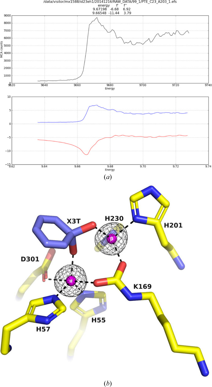
A bacterial phosphotriesterase was employed as an experimental paradigm to examine the effects of multiple factors, such as the molecular constructs, the ligands used during protein expression and purification, the crystallization conditions and the space group, on the visualization of molecular complexes of ligands with a target enzyme. In this case, the ligands used were organophosphates that are fragments of the nerve agents and insecticides on which the enzyme acts as a bioscavenger. 12 crystal structures of various phosphotriesterase constructs obtained by directed evolution were analyzed, with resolutions of up to 1.38 Å. Both apo forms and holo forms, complexed with the organophosphate ligands, were studied. Crystals obtained from three different crystallization conditions, crystallized in four space groups, with and without N-terminal tags, were utilized to investigate the impact of these factors on visualizing the organophosphate complexes of the enzyme. The study revealed that the tags used for protein expression can lodge in the active site and hinder ligand binding. Furthermore, the space group in which the protein crystallizes can significantly impact the visualization of bound ligands. It was also observed that the crystallization precipitants can compete with, and even preclude, ligand binding, leading to false positives or to the incorrect identification of lead drug candidates. One of the co-crystallization conditions enabled the definition of the spaces that accommodate the substituents attached to the P atom of several products of organophosphate substrates after detachment of the leaving group. The crystal structures of the complexes of phosphotriesterase with the organophosphate products reveal similar short interaction distances of the two partially charged O atoms of the P-O bonds with the exposed β-Zn2+ ion and the buried α-Zn2+ ion. This suggests that both Zn2+ ions have a role in stabilizing the transition state for substrate hydrolysis. Overall, this study provides valuable insights into the challenges and considerations involved in studying the crystal structures of ligand-protein complexes, highlighting the importance of careful experimental design and rigorous data analysis in ensuring the accuracy and reliability of the resulting phosphotriesterase-organophosphate structures.

Keywords: bioscavengers; crystallization conditions; expression systems; organophosphates; phosphotriesterases; space groups; zinc ions.


Figures

Figure 1
Figure 1
View of a typical PTE structure (PDB entry 1hzy; Benning et al., 2001 ▸). (a) The (β/α)8 TIM-barrel fold is shown as a cartoon, with helices in red, sheets in yellow, coils in green, the α-Zn2+ (buried) and β-Zn2+ (exposed) ions as magenta spheres and a single bridging water shown as a cyan sphere. The six residues that bind to the two Zn2+ ions are shown as stick representations, with C atoms colored yellow, N atoms blue and O atoms red. The N- and C-terminal residues are labeled N and C, respectively. (b) Close-up view of the active site of the apo PTE structure. The buried α-Zn2+ ion is directly bound to His55, His57 and Asp301, while the exposed β-Zn2+ ion is bound to His201 and His230. The carbamate functional group bound to Lys169 interacts with both Zn2+ ions. Coloring is as in (a).
Figure 2
Figure 2
PTE variants. (a) Schematic presentation of the maltose-binding protein (MBP) fused before the factor Xa cleavage motif (IEGR) and the octapeptide spacer sequence, 26ISEFITNS33, followed by the mature PTE protein sequence starting with Gly34. Factor Xa cleaves after the arginine residue of the cleavage motif, leaving the 26ISEFITNS33 linker attached to the PTE, Gly34–Ser365. (b) Sequence alignment of wt PTE, PDB entry 1hzy, A53, C23, C23M, A53_T, C23_T and C23M_T. The last three bear the octapeptide tag. Secondary-structure elements of PDB entry 1hzy are labeled above the alignments: α-helices and 310-helices (shown with the symbol η) are indicated by coils and β-strands by arrows. Residues conserved in all variants are in red. Multiple sequence alignment was performed using MultAlin (Corpet, 1988 ▸) and the figure was created using ESPript (Robert & Gouet, 2014 ▸).
Figure 3
Figure 3
Methylphosphonates that were crystallized with the PTE variants. (a) Methylphosphonic acid, (b) O-ethyl methylphosphonic acid, (c) O-ethyl-O-(N,N-diisopropylaminoethyl) methylphosphonate (the oxo analogue of VX), (d) O-isopropyl methylphosphonic acid. In all four structures the CH3 moiety is pointing away from the viewing plane and thus is not seen. The OPs are shown as stick figures with C atoms colored yellow, N atoms blue, O atoms red and P atoms orange.
Figure 4
Figure 4
Comparison of the active-site regions of PTEs containing different numbers of Zn2+ ions. (a) PDB entry 1pta, which is devoid of Zn2+ ions. (b) A53_1, containing one Zn2+ ion. (c) PDB entry 1hzy, containing two Zn2+ ions. Zn2+ ions are shown in magenta and the bridging water is in cyan.
Figure 5
Figure 5
Cartoon tube diagrams of the backbones of the monomer structures of apo A53_2 (beige) and apo A53_1 (green). A region displaying sizeable conformational differences (residues 60–79) is circled by a black dashed line. This region participates in dimer formation in the A53_2, A53_3 and A53_4 structures, which are very similar and differ significantly from that of A53_1. The missing regions in A53_1, i.e. residues 203–210, 254–274 and 314–320, are colored red and circled by red dashed lines. The two Zn2+ ions in A53_2 are shown as magenta spheres and the residues which bind them are displayed as sticks.
Figure 6
Figure 6
Ribbon representation of the A53_3 variant. It shows the octapeptide tag on subunit B penetrating the active-site region of the symmetry-related subunit A. The tag is shown in magenta. The active-site residues of the symmetrically related chain A are shown in yellow, with those residues within 5 Å of the tag shown in cyan. The green electron density corresponds to an omit map with the octapeptide omitted (contoured at 3σ). The cyclic compound X3E, which was presumably carried over from the protein expression and purification process, is seen in the active site, and the black electron density corresponds to a 2F oF c map (contoured at 1σ). The two Zn2+ ions are shown as magenta spheres.
Figure 7
Figure 7
Active-site region of A53_4. Acrylic acid (AA; blue sticks) is clearly seen bound at the active site. The green electron density corresponds to an omit map with AA and the two Zn2+ ions omitted (contoured at 3σ). The black electron density corresponds to a 2F oF c map (contoured at 1σ). The two Zn2+ ions are shown as magenta spheres.
Figure 8
Figure 8
(a) Scan of the C23M_1 crystal at a range of energies showing a peak corresponding to the zinc absorption edge (top). Scattering factors (f′ and f′′) are plotted as a function of energy (bottom). (b) An anomalous omit electron-density map of the active-site region of C23M_1, contoured at 6σ, is shown in black. The two Zn2+ ions are shown as magenta spheres and were omitted in calculating the electron density.
Figure 9
Figure 9
Electron-density omit map of the active-site region of C23M_1. The two Zn2+ ions and the electron density of an unidentified six-membered ring ligand, labeled X3T, were omitted in calculating the electron densities. The 2F oF c omit map, contoured at 1σ, is shown in black. The F oF c omit map, contoured at 3σ, is shown in green. The two Zn2+ ions are shown as magenta spheres.
Figure 10
Figure 10
Electron-density omit map of the active-site region of A53_2. The two Zn2+ ions and the electron density of an unidentified six-membered ring, labeled X3B, were omitted in calculating the electron densities. The 2F o − F c omit map, contoured at 1σ, is shown in black. The F oF c omit map, contoured at 3σ, is shown in green. The two Zn2+ ions are shown as magenta spheres.
Figure 11
Figure 11
Electron-density omit maps (F oF c) of the active-site regions of PTEs. The two Zn2+ ions and the OPs were omitted from the calculations. (a) Methylphosphonate was observed in the C23M_2 structure. (b) O-Ethyl methylphosphonic acid was observed in C23_5. However, the electron density could only account for the first C atom of the OCH2CH3 substituent. (c) O-Ethyl methylphosphonic acid, the hydrolysis product of O-ethyl-O-(N,N-diisopropylaminoethyl) methylphosphonate, was observed in the C23_4 structure. (d) O-Isopropyl methylphosphonic acid was observed in C23_2. The interatomic distances observed between the O atoms of the P—O bond in all OP acid products are 1.9–2.0 Å. The F oF c omit map, contoured at 3σ, is shown in green. The six residues that bind to the two Zn2+ ions are shown as stick representations, with C atoms colored yellow, N atoms blue, O atoms red and P atoms orange.
Figure 12
Figure 12
Schematic depiction of the putative TS for the hydrolysis of an O-alkyl methylphosphonate. The nucleophile is a hydroxide ion that originates from a water molecule that bridges the two Zn2+ ions. LG represents the leaving group. The geometries displayed are for the purposes of illustration and discussion.

References

    1. Agirre, J., Atanasova, M., Bagdonas, H., Ballard, C. B., Baslé, A., Beilsten-Edmands, J., Borges, R. J., Brown, D. G., Burgos-Mármol, J. J., Berrisford, J. M., Bond, P. S., Caballero, I., Catapano, L., Chojnowski, G., Cook, A. G., Cowtan, K. D., Croll, T. I., Debreczeni, J. É., Devenish, N. E., Dodson, E. J., Drevon, T. R., Emsley, P., Evans, G., Evans, P. R., Fando, M., Foadi, J., Fuentes-Montero, L., Garman, E. F., Gerstel, M., Gildea, R. J., Hatti, K., Hekkelman, M. L., Heuser, P., Hoh, S. W., Hough, M. A., Jenkins, H. T., Jiménez, E., Joosten, R. P., Keegan, R. M., Keep, N., Krissinel, E. B., Kolenko, P., Kovalevskiy, O., Lamzin, V. S., Lawson, D. M., Lebedev, A. A., Leslie, A. G. W., Lohkamp, B., Long, F., Malý, M., McCoy, A. J., McNicholas, S. J., Medina, A., Millán, C., Murray, J. W., Murshudov, G. N., Nicholls, R. A., Noble, M. E. M., Oeffner, R., Pannu, N. S., Parkhurst, J. M., Pearce, N., Pereira, J., Perrakis, A., Powell, H. R., Read, R. J., Rigden, D. J., Rochira, W., Sammito, M., Sánchez Rodríguez, F., Sheldrick, G. M., Shelley, K. L., Simkovic, F., Simpkin, A. J., Skubak, P., Sobolev, E., Steiner, R. A., Stevenson, K., Tews, I., Thomas, J. M. H., Thorn, A., Valls, J. T., Uski, V., Usón, I., Vagin, A., Velankar, S., Vollmar, M., Walden, H., Waterman, D., Wilson, K. S., Winn, M. D., Winter, G., Wojdyr, M. & Yamashita, K. (2023). Acta Cryst. D79, 449–461.
    1. Aharoni, A., Amitai, G., Bernath, K., Magdassi, S. & Tawfik, D. S. (2005). Chem. Biol. 12, 1281–1289. - PubMed
    1. Arkaban, H., Barani, M., Akbarizadeh, M. R., Pal Singh Chauhan, N., Jadoun, S., Dehghani Soltani, M. & Zarrintaj, P. (2022). Polymers, 14, 1259. - PMC - PubMed
    1. Benning, M. M., Hong, S. B., Raushel, F. M. & Holden, H. M. (2000). J. Biol. Chem. 275, 30556–30560. - PubMed
    1. Benning, M. M., Kuo, J. M., Raushel, F. M. & Holden, H. M. (1994). Biochemistry, 33, 15001–15007. - PubMed