. 2023 Nov 6;20(11):5901-5909.
doi: 10.1021/acs.molpharmaceut.3c00736. Epub 2023 Oct 20.

Transferrin Receptor-Targeted Iduronate-2-sulfatase Penetrates the Blood-Retinal Barrier and Improves Retinopathy in Mucopolysaccharidosis II Mice

Affiliations

Transferrin Receptor-Targeted Iduronate-2-sulfatase Penetrates the Blood-Retinal Barrier and Improves Retinopathy in Mucopolysaccharidosis II Mice

Atsushi Imakiire et al. Mol Pharm. .

Abstract

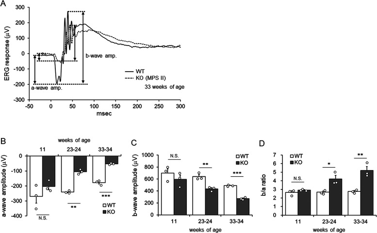
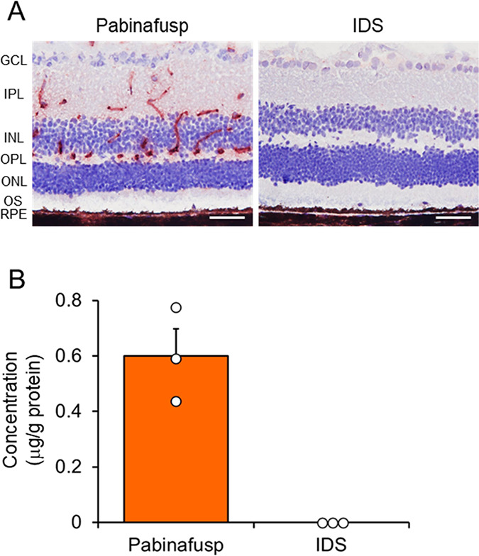
Mucopolysaccharidoses (MPSs) make up a group of lysosomal storage diseases characterized by the aberrant accumulation of glycosaminoglycans throughout the body. Patients with MPSs display various signs and symptoms, such as retinopathy, which is also observed in patients with MPS II. Unfortunately, retinal disorders in MPS II are resistant to conventional intravenous enzyme-replacement therapy because the blood-retinal barrier (BRB) impedes drug penetration. In this study, we show that a fusion protein, designated pabinafusp alfa, consisting of an antihuman transferrin receptor antibody and iduronate-2-sulfatase (IDS), crosses the BRB and reaches the retina in a murine model of MPS II. We found that retinal function, as assessed by electroretinography (ERG) in MPS II mice, deteriorated with age. Early intervention with repeated intravenous treatment of pabinafusp alfa decreased heparan sulfate deposition in the retina, optic nerve, and visual cortex, thus preserving or even improving the ERG response in MPS II mice. Histological analysis further revealed that pabinafusp alfa mitigated the loss of the photoreceptor layer observed in diseased mice. In contrast, recombinant nonfused IDS failed to reach the retina and hardly affected the retinal disease. These results support the hypothesis that transferrin receptor-targeted IDS can penetrate the BRB, thereby ameliorating retinal dysfunction in MPS II.

Keywords: blood-retinal barrier; electroretinography; mucopolysaccharidosis II; pabinafusp alfa; retinopathy; transferrin receptor.


Conflict of interest statement

The authors declare the following competing financial interest(s): A. Imakiire, H. Morimoto, H.S., T.M., E.Y., A. Inoue, H. Morioka, T.K., A.M., R.S. R.K., T.H., H.S., and K.M. are employees and/or stockholders of JCR Pharmaceuticals Co., Ltd.

Figures

Figure 1
Figure 1
ERG responses of WT and MPS II mice at different ages. (A) Raw ERG traces from representative WT and Ids-KO (MPS II) mice at 33 weeks of age. Scotopic ERG responses were recorded at 10 cd s/cm2. Amplitudes of a-wave and b-wave were indicated by double arrows. (B–D) Amplitudes of photoreceptor-generated a-wave (B), amacrine/bipolar cell-generated b-wave (C), and b/a ratio (D). Bars indicate the mean with SEM (n = 3). Open circles indicate values for individual mice. *P < 0.05, **P < 0.01, and ***P < 0.001 (WT vs KO, unpaired t test). N.S., not significant; WT, wild-type mice; and KO, Ids-KO (MPS II) mice.
Figure 2
Figure 2
Retinal delivery of pabinafusp alfa. (A) Immunostaining of the human IDS enzyme in the retina after intravenous administration of 2 mg/kg of pabinafusp alfa or nonfused IDS to MPS II mice. Red-brown signals indicate positive staining for IDS. Counterstaining was performed by hematoxylin. Scale bars: 50 μm. (B) Quantification of IDS enzyme concentration by electrochemiluminescence. Bars indicate mean with SEM (n = 3). Open circles indicate values for individual mice. Retinas were isolated 17 h after administration. Pabinafusp, pabinafusp alfa; IDS, nonfused recombinant human IDS; GCL, ganglion cell layer; IPL, inner plexiform layer; INL, inner nuclear layer; OPL, outer plexiform layer; ONL, outer nuclear layer; OS, outer segment; and RPE, retinal pigment epithelium.
Figure 3
Figure 3
Effect of pabinafusp alfa on the HS concentration in tissues involved in visual function. HS concentration in the retina (A), RPE/choroid/sclera complex (B), optic nerve (C), and visual cortex of the brain (D). HS concentration was measured 1 week after the final (40th) dose administration. Bars indicate the mean with SEM (n = 5–7). Open circles indicate values for individual mice. The numbers on the X-axis indicate the dose in mg/kg. ###P < 0.001 (WT vs KO, unpaired t test), *P < 0.05, **P < 0.01, and ***P < 0.001 (between treatment groups, Tukey-Kramer test). N.S., not significant; WT, wild-type mice; KO, vehicle-treated hTfR-KI/Ids-KO (MPS II) mice; Pabinafusp, pabinafusp alfa; and IDS, nonfused recombinant human IDS.
Figure 4
Figure 4
Baseline measurement of ERG before the initiation of treatment. (A,B) ERG responses from representative WT (A) and KO (MPS II, B) mice at 10 cd s/cm2. Raw ERG traces are shown. Double arrows indicate a-wave and b-wave amplitudes. (C–E) Amplitude of a-wave (C), b-wave (D), and b/a ratio (E) of the ERG. Data are from mice before initiation of the treatment (baseline). Bars indicate the mean with SEM (n = 14 for WT and 12 for KO). Open circles indicate values for individual mice. ###P < 0.001 (WT vs KO, unpaired t test), N.S., not significant; WT, wild-type mice; KO, vehicle-treated hTfR-KI/Ids-KO (MPS II) mice; Pabinafusp, pabinafusp alfa; and IDS, nonfused recombinant human IDS.
Figure 5
Figure 5
Effect of a chronic treatment with pabinafusp alfa on retinal function. (A) ERG responses of representative mice resulting from different intensity stimuli after a 38-week treatment. (B–D) Amplitude of a-wave (B), b-wave (C), and b/a ratio (D) of ERG at 10 cd s/cm2 after treatment. Bars indicate the mean with SEM (n = 12–14). Open circles indicate values for individual mice. The numbers on the X-axis indicate the dose in mg/kg. ###P < 0.001 (WT vs KO, unpaired t test), *P < 0.05, **P < 0.01, and ***P < 0.001 (between treatment groups, Tukey-Kramer test). N.S., not significant; WT, wild-type mice; KO, vehicle-treated hTfR-KI/Ids-KO (MPS II) mice; Pabinafusp, pabinafusp alfa; and IDS, nonfused recombinant human IDS.
Figure 6
Figure 6
Histological analyses of the retina after the treatment. (A) H&E-staining of the retina. White double arrows indicate the thickness of the outer nuclear layer (ONL). Scale bars, 50 μm. (B) Thickness of the ONL at 18 positions on both sides of the retina. Values are the means ± SEM (n = 6–7). (C) Mean ONL thickness. Bars indicate the mean with SEM (n = 6–7). (D) Total cell number in the retina. Bars indicate the mean with SEM (n = 5). Open circles indicate values for individual mice (C and D). ###P < 0.001 (WT vs KO, unpaired t test), *P < 0.05, **P < 0.01, and ***P < 0.001 (between treatment groups, Tukey-Kramer test). N.S., not significant; WT, wild-type mice; KO, vehicle-treated hTfR-KI/Ids-KO (MPS II) mice; Pabinafusp, pabinafusp alfa; and IDS, nonfused recombinant human IDS.

References

    1. Neufeld E. F.; Muenzer J.. The mucopolysaccharidoses. In The Metabolic & Molecular Bases of Inherited Disease; Scriver C. R., Beaudet A. L., Sly W. S., Valle D., Eds.; McGraw Hill: New York, USA, 2001; pp 3421–3452.
    1. Verma S.; Pantoom S.; Petters J.; Pandey A. K.; Hermann A.; Lukas J. A molecular genetics view on Mucopolysaccharidosis Type II. Mutat. Res. Rev. Mutat. Res. 2021, 788, 10839210.1016/j.mrrev.2021.108392. - DOI - PubMed
    1. Tylki-Szymańska A. Mucopolysaccharidosis type II, Hunter’s syndrome. Pediatr. Endocrinol. Rev. 2014, 12 (Suppl 1), 107–113. - PubMed
    1. Hopwood J. J.; Bunge S.; Morris C. P.; Wilson P. J.; Steglich C.; Beck M.; Schwinger E.; Gal A. Molecular basis of mucopolysaccharidosis type II: mutations in the iduronate-2-sulphatase gene. Hum. Mutat. 1993, 2, 435–442. 10.1002/humu.1380020603. - DOI - PubMed
    1. Parini R.; Deodato F. Intravenous Enzyme Replacement Therapy in Mucopolysaccharidoses: Clinical Effectiveness and Limitations. Int. J. Mol. Sci. 2020, 21, 2975.10.3390/ijms21082975. - DOI - PMC - PubMed

Publication types