. 2023 Oct;3(10):e924.
doi: 10.1002/cpz1.924.

Colorimetric Synchronization of Drosophila Larvae

Affiliations

Colorimetric Synchronization of Drosophila Larvae

Taylor Hailstock et al. Curr Protoc. 2023 Oct.

Abstract

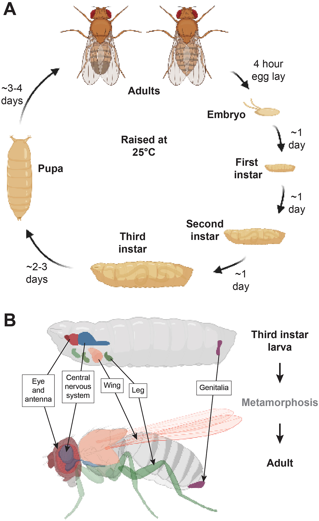
The rapid succession of events during development poses an inherent challenge to achieve precise synchronization required for rigorous, quantitative phenotypic and genotypic analyses in multicellular model organisms. Drosophila melanogaster is an indispensable model for studying the development and function of higher order organisms due to extensive genome homology, tractability, and its relatively short lifespan. Presently, nine Nobel prizes serve as a testament to the utility of this elegant model system. Ongoing advancements in genetic and molecular tools allow for the underlying mechanisms of human disease to be investigated in Drosophila. However, the absence of a method to precisely age-match tissues during larval development prevents further capitalization of this powerful model organism. Drosophila spends nearly half of its life cycle progressing through three morphologically distinct larval instar stages, during which the imaginal discs, precursors of mature adult external structures (e.g., eyes, legs, wings), grow and develop distinct cell fates. Other tissues, such as the central nervous system, undergo massive morphological changes during larval development. While these three larval stages and subsequent pupal stages have historically been identified based on the number of hours post egg-laying under standard laboratory conditions, a reproducible, efficient, and inexpensive method is required to accurately age-match larvae within the third instar. The third instar stage is of particular interest, as this developmental stage spans a 48-hr window during which larval tissues switch from proliferative to differentiation programs. Moreover, some genetic manipulations can lead to developmental delays, further compounding the need for precise age-matching between control and experimental samples. This article provides a protocol optimized for synchronous staging of Drosophila third instar larvae by colorimetric characterization and is useful for age-matching a variety of tissues for numerous downstream applications. We also provide a brief discussion of the technical challenges associated with successful application of this protocol. © 2023 Wiley Periodicals LLC. Basic Protocol: Synchronization of third instar Drosophila larvae.

Keywords: Drosophila; age-match; development; larval; staging.


Conflict of interest statement

CONFLICT OF INTEREST STATEMENT:

The authors declare no conflicts of interest.

Figures

Figure 1.
Figure 1.. Lifespan of Drosophila and development of adult structures from larval imaginal discs.
(A) Diagram showing the progression of the Drosophila life cycle and typical developmental timing (25 °C). (B) Cartoon shows several color-coded primordial structures within a third instar larva and corresponding structures within an adult. Images were generated using BioRender.com.
Figure 2.
Figure 2.. The three stages of a third instar larva.
Cartoons show the passage of blue colored food through the larval intestinal tract. Beneath each schematic is a representative image of the corresponding stage from samples processed using this protocol. Bars: 1mm. Cartoons were created using BioRender.com.
Figure 3.
Figure 3.. orb2-dependent microcephaly in larval brains.
(A-B) Images show control and orb2 null third instar larval brains stained with DAPI. The optic lobe is outlined (dashed line). Bars: 40 μm (C) Graph shows quantification of brain volumes from age-matched (partial gut clearance) samples, where each dot represents a measurement of a single optic lobe from N=30 brains per genotype normalized to the wild-type (WT) control. Loss of orb2 results in significantly smaller brain volumes by t-test analysis. Data shown are adapted from (Robinson et al., 2022).
Figure 4.
Figure 4.. EcRLBD reveals an EcR repression-to-activation switch in third instar larval wing discs.
Optical (z-stack) projections of third instar larval wing discs immunostained with anti-β-galactosidase (LacZ; green) to detect EcRE-lacZ expression in control (en>RFP; A-C) or en>EcRLBD (D-F) discs. RFP reporters are displayed in red and mark the posterior compartment. Images are representative from N=16 control dark, 19 partial, and 7 clear versus N=6 EcRLBD dark, 15 partial, and 7 clear third instar larvae. Note how in the control samples, LacZ expression increases over time. In contrast, expression of EcRLBD results in posterior enrichment of LacZ in early (dark) larvae, while anterior enrichment is observed during middle and late (partial and clear, respectively) third larval instar development. Data shown are adapted from (Wardwell-Ozgo et al., 2022). [*Copyeditor: Please double check with authors regarding the permission to use the adapted figures 3 and 4. I asked them and here is their response: “These figures contain data uploaded to Biorxiv by authors of this protocol, accessible under a CC-BYNC 4.0 license.” If this is the case, it should be mentioned in the Acknowledgements section and/or in the legends]

References

    1. Andres AJ, & Thummel CS (1994). Methods for quantitative analysis of transcription in larvae and prepupae. Methods Cell Biol, 44, 565–573. doi:10.1016/s0091-679x(08)60932-2 - DOI - PubMed
    1. Ashburner M (1967). Patterns of puffing activity in the salivary gland chromosomes of Drosophila. I. Autosomal puffing patterns in a laboratory stock of Drosophila melanogaster. Chromosoma, 21(4), 398–428. doi:10.1007/bf00336950 - DOI - PubMed
    1. Beermann W, & Clever U (1964). CHROMOSOME PUFFS. Sci Am, 210, 50–58. doi:10.1038/scientificamerican0464-50 - DOI - PubMed
    1. Cabernard C, & Doe CQ (2009). Apical/basal spindle orientation is required for neuroblast homeostasis and neuronal differentiation in Drosophila. Dev Cell, 17(1), 134–141. doi:10.1016/j.devcel.2009.06.009 - DOI - PubMed
    1. Coronado-Zamora M, Salvador-Martínez I, Castellano D, Barbadilla A, & Salazar-Ciudad I (2019). Adaptation and Conservation throughout the Drosophila melanogaster Life-Cycle. Genome Biol Evol, 11(5), 1463–1482. doi:10.1093/gbe/evz086 - DOI - PMC - PubMed

LinkOut - more resources