. 2024 Jan 12;14(1):76-89.
doi: 10.1158/2159-8290.CD-23-0964.

Xaluritamig, a STEAP1 × CD3 XmAb 2+1 Immune Therapy for Metastatic Castration-Resistant Prostate Cancer: Results from Dose Exploration in a First-in-Human Study

Affiliations

Xaluritamig, a STEAP1 × CD3 XmAb 2+1 Immune Therapy for Metastatic Castration-Resistant Prostate Cancer: Results from Dose Exploration in a First-in-Human Study

William K Kelly et al. Cancer Discov. .

Abstract

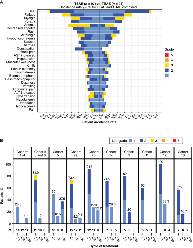
Xaluritamig (AMG 509) is a six-transmembrane epithelial antigen of the prostate 1 (STEAP1)-targeted T-cell engager designed to facilitate lysis of STEAP1-expressing cancer cells, such as those in advanced prostate cancer. This first-in-human study reports monotherapy dose exploration for patients with metastatic castration-resistant prostate cancer (mCRPC), primarily taxane pretreated. Ninety-seven patients received ≥1 intravenous dose ranging from 0.001 to 2.0 mg weekly or every 2 weeks. MTD was identified as 1.5 mg i.v. weekly via a 3-step dose. The most common treatment-related adverse events were cytokine release syndrome (CRS; 72%), fatigue (45%), and myalgia (34%). CRS occurred primarily during cycle 1 and improved with premedication and step dosing. Prostate-specific antigen (PSA) and RECIST responses across cohorts were encouraging [49% PSA50; 24% objective response rate (ORR)], with greater frequency at target doses ≥0.75 mg (59% PSA50; 41% ORR). Xaluritamig is a novel immunotherapy for prostate cancer that has shown encouraging results supporting further development.

Significance: Xaluritamig demonstrated encouraging responses (PSA and RECIST) compared with historical established treatments for patients with late-line mCRPC. This study provides proof of concept for T-cell engagers as a potential treatment for prostate cancer, validates STEAP1 as a target, and supports further clinical investigation of xaluritamig in prostate cancer. See related commentary by Hage Chehade et al., p. 20. See related article by Nolan-Stevaux et al., p. 90. This article is featured in Selected Articles from This Issue, p. 5.


Figures

Figure 1. A, Frequency and highest-grade AEs occurring in ≥20% of patients treated with xaluritamig across all cohorts, TEAE vs. TRAEs (defined by the investigator as having reasonable possibility of being caused by xaluritamig). B, Incidence and grade of CRS (32) by cycle and dose schedule. Cohort 10 was excluded (0.1–1.0 mg), as dosing schedule was adjusted for the remaining patients after initial patients with a 10-fold dose increase in 1 step experienced DLTs. ALT, alanine aminotransferase; AST, aspartate aminotransferase.
Figure 1.
A, Frequency and highest-grade AEs occurring in ≥20% of patients treated with xaluritamig across all cohorts, TEAE vs. TRAEs (defined by the investigator as having reasonable possibility of being caused by xaluritamig). B, Incidence and grade of CRS (32) by cycle and dose schedule. Cohort 10 was excluded (0.1–1.0 mg), as dosing schedule was adjusted for the remaining patients after initial patients with a 10-fold dose increase in 1 step experienced DLTs. ALT, alanine aminotransferase; AST, aspartate aminotransferase.
Figure 2. A, Mean (SD) xaluritamig serum concentration vs. time profile by individual cohorts for C1. Black dotted lines represent the lower end of the minimum predicted efficacious exposure based on EC90 for xaluritamig-mediated cell killing in vitro (74 ng/mL) and the upper end of the minimum predicted efficacious exposure based on IC50 for xaluritamig-mediated mouse tumor growth inhibition in vivo (259 ng/mL). Cohort 7c was dosed weekly for C1 and then switched to every 2 weeks starting C2 and beyond, which is not captured in this analysis. At the time of the data cut, there were 94 patients with at least one postbaseline xaluritamig concentration. T-cell pharmacodynamic biomarker response through the first infusion period is dose dependent. Peripheral lymphocyte margination (B), CD8+CD69+ activated T cells as a percentage of total CD8+ T cells (C), and induction of secreted cytokines at the indicated time points after infusion (D). Lines represent median fold change or difference of population percentage from C1D1 predose values ± median average deviation. Transparent points depict individual observations. Sample sizes within each priming dose group at each time point are shown as a strip annotation across the top of each figure. EC, effective concentration; IC, inhibitory concentration.
Figure 2.
A, Mean (SD) xaluritamig serum concentration vs. time profile by individual cohorts for C1. Black dotted lines represent the lower end of the minimum predicted efficacious exposure based on EC90 for xaluritamig-mediated cell killing in vitro (74 ng/mL) and the upper end of the minimum predicted efficacious exposure based on IC50 for xaluritamig-mediated mouse tumor growth inhibition in vivo (259 ng/mL). Cohort 7c was dosed weekly for C1 and then switched to every 2 weeks starting C2 and beyond, which is not captured in this analysis. At the time of the data cut, there were 94 patients with at least one postbaseline xaluritamig concentration. T-cell pharmacodynamic biomarker response through the first infusion period is dose dependent. Peripheral lymphocyte margination (B), CD8+CD69+ activated T cells as a percentage of total CD8+ T cells (C), and induction of secreted cytokines at the indicated time points after infusion (D). Lines represent median fold change or difference of population percentage from C1D1 predose values ± median average deviation. Transparent points depict individual observations. Sample sizes within each priming dose group at each time point are shown as a strip annotation across the top of each figure. EC, effective concentration; IC, inhibitory concentration.
Figure 3. Clinical activity of xaluritamig in evaluable patients. A, Best PSA percentage change from baseline. Asterisk indicates confirmed PSA responders, and dashed lines indicate PSA50 and PSA90 declines. B, Best percentage change in size of tumor target lesions. Dashed line indicates 30% reduction in tumor SLD from baseline. C, Example of patient showing response by PSA and radiographic assessments: CT scan and PSA curve over time of a heavily pretreated 65-year-old patient with stage IV prostate adenocarcinoma. Patient was enrolled into Cohort 11 (3-step 1.5 mg target dose of xaluritamig). CT scans showed three target lesions (two liver, one lymph node) and multiple nontarget lesions in the liver as well as two lymph nodes during screening. Patient achieved 99% PSA decline from baseline on C7D1 and PR (37.3% reduction of target lesions) after 2 cycles, which was confirmed at 16 weeks and maintained after 24 weeks. AEs occurred during C1 of treatment and included recurrent CRS, tinea faciei (both grade 1), rash, and worsening of back pain (both grade 2). During further treatment cycles, rash (grade 1), myalgia, and hyperkalemia (both grade 2) were reported. Patient remains on treatment at the time of publication. Red arrows indicate sites of tumor. D, Time on treatment for patients in high-dose cohorts. PSA and RECIST responses [RECIST evaluable (gray bars) and non–RECIST evaluable (white bars)] are presented for patients in high-dose cohorts. Patients whose treatment was ongoing are noted by an arrowhead. Double parallel lines (//) represent patients who have extended beyond 48 weeks: one patient is ongoing treatment at 90 weeks, one is ongoing treatment at 84 weeks, and one ended treatment at 58 weeks. NE, not evaluable; PD, progressive disease; SD, stable disease; SLD, sum of longest diameters.
Figure 3.
Clinical activity of xaluritamig in evaluable patients. A, Best PSA percentage change from baseline. Asterisk indicates confirmed PSA responders, and dashed lines indicate PSA50 and PSA90 declines. B, Best percentage change in size of tumor target lesions. Dashed line indicates 30% reduction in tumor SLD from baseline. C, Example of patient showing response by PSA and radiographic assessments: CT scan and PSA curve over time of a heavily pretreated 65-year-old patient with stage IV prostate adenocarcinoma. Patient was enrolled into Cohort 11 (3-step 1.5 mg target dose of xaluritamig). CT scans showed three target lesions (two liver, one lymph node) and multiple nontarget lesions in the liver as well as two lymph nodes during screening. Patient achieved 99% PSA decline from baseline on C7D1 and PR (37.3% reduction of target lesions) after 2 cycles, which was confirmed at 16 weeks and maintained after 24 weeks. AEs occurred during C1 of treatment and included recurrent CRS, tinea faciei (both grade 1), rash, and worsening of back pain (both grade 2). During further treatment cycles, rash (grade 1), myalgia, and hyperkalemia (both grade 2) were reported. Patient remains on treatment at the time of publication. Red arrows indicate sites of tumor. D, Time on treatment for patients in high-dose cohorts. PSA and RECIST responses [RECIST evaluable (gray bars) and non–RECIST evaluable (white bars)] are presented for patients in high-dose cohorts. Patients whose treatment was ongoing are noted by an arrowhead. Double parallel lines (//) represent patients who have extended beyond 48 weeks: one patient is ongoing treatment at 90 weeks, one is ongoing treatment at 84 weeks, and one ended treatment at 58 weeks. NE, not evaluable; PD, progressive disease; SD, stable disease; SLD, sum of longest diameters.

Comment in

References

    1. Siegel RL, Miller KD, Wagle NS, Jemal A. Cancer statistics, 2023. CA Cancer J Clin 2023;73:17–48. - PubMed
    1. Cancer Fact Sheets, Prostate . Lyon (France), Geneva (Switzerland): International Agency for Research on Cancer, World Health Organization; 2020 [cited 2023 Jul 3]. Available from: https://gco.iarc.fr/today/data/factsheets/cancers/27-Prostate-fact-sheet....
    1. Wadosky KM, Koochekpour S. Molecular mechanisms underlying resistance to androgen deprivation therapy in prostate cancer. Oncotarget 2016;7:64447–70. - PMC - PubMed
    1. Cancer Stat Facts: Prostate Cancer; [about 4 screens]. [cited 2023 Jul 3]. Available from: https://seer.cancer.gov/statfacts/html/prost.html.
    1. Turco F, Gillessen S, Cathomas R, Buttigliero C, Vogl UM. Treatment landscape for patients with castration-resistant prostate cancer: patient selection and unmet clinical needs. Res Rep Urol 2022;14:339–50. - PMC - PubMed

Publication types

MeSH terms