. 2023 Oct 24;120(43):e2308600120.
doi: 10.1073/pnas.2308600120. Epub 2023 Oct 20.

Identification of a carbonic anhydrase-Rubisco complex within the alpha-carboxysome

Affiliations

Identification of a carbonic anhydrase-Rubisco complex within the alpha-carboxysome

Cecilia Blikstad et al. Proc Natl Acad Sci U S A. .

Abstract

Carboxysomes are proteinaceous organelles that encapsulate key enzymes of CO2 fixation-Rubisco and carbonic anhydrase-and are the centerpiece of the bacterial CO2 concentrating mechanism (CCM). In the CCM, actively accumulated cytosolic bicarbonate diffuses into the carboxysome and is converted to CO2 by carbonic anhydrase, producing a high CO2 concentration near Rubisco and ensuring efficient carboxylation. Self-assembly of the α-carboxysome is orchestrated by the intrinsically disordered scaffolding protein, CsoS2, which interacts with both Rubisco and carboxysomal shell proteins, but it is unknown how the carbonic anhydrase, CsoSCA, is incorporated into the α-carboxysome. Here, we present the structural basis of carbonic anhydrase encapsulation into α-carboxysomes from Halothiobacillus neapolitanus. We find that CsoSCA interacts directly with Rubisco via an intrinsically disordered N-terminal domain. A 1.98 Å single-particle cryoelectron microscopy structure of Rubisco in complex with this peptide reveals that CsoSCA binding is predominantly mediated by a network of hydrogen bonds. CsoSCA's binding site overlaps with that of CsoS2, but the two proteins utilize substantially different motifs and modes of binding, revealing a plasticity of the Rubisco binding site. Our results advance the understanding of carboxysome biogenesis and highlight the importance of Rubisco, not only as an enzyme but also as a central hub for mediating assembly through protein interactions.

Keywords: CO2 fixation; carbonic anhydrase; carboxysome; cryoelectron microscopy; protein–protein interactions.


Conflict of interest statement

The authors declare no competing interest.

Figures
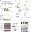
Fig. 1.
Fig. 1.
An intrinsically disordered, poorly conserved N-terminal peptide is essential and sufficient for CsoSCA encapsulation. (A) Schematic of the cso operon (carboxysome operon) in H. neapolitanus. The 10-gene set consists of Rubisco large and small subunits, the scaffolding protein CsoS2, the carbonic anhydrase CsoSCA, and six shell proteins (CsoS4A/B, CsoS1A/B/C, and CsoSD1). In the native organism, CsoS1D is transcribed from an adjacent locus, while in the synthetic pHnCB10 plasmid, all genes are in a single operon. (B) Surface representation structure of the CsoSCA dimer from H. neapolitanus (pdb: 2FGY). The N-terminal domain (dark green) consists of a ~50-aa-long unstructured peptide followed by a folded α-helical domain with unknown function. The middle domain (light green) contains the active site. The C-terminal domain (white) appears to be a gene duplication of the catalytic domain but lacks essential active site residues. (C) Maximum-likelihood phylogenetic tree of CsoSCA. Cyanobacterial homologs are colored in green and proteobacteria homologous in an orange/brown gradient. Scale bar, 0.1 substitutions per site. (D) Disorder score of four representative CsoSCA homologous calculated using DISOPRED3 and conservation calculated from multiple sequence alignment. (E) Complementation of full-length csoSCA rescues growth of a csoSCA knock-out in H. neapolitanus, while complementation with an NTD-truncated variant, ΔNTD1-49CsoSCA, fails to rescue growth. (F) Western blot analysis detecting C-terminally flag-tagged CsoSCA in lysate (L) and enriched carboxysomes (CB) fractions of carboxysomes produced heterologously in Escherichia coli. Synthetic carboxysomes consist of the full cso operon (Fig. 1A), with either wild-type CsoSCA or an N-terminal truncated variant (ΔNTD1-37CsoSCA or ΔNTD1-49CsoSCA). CsoSCA is not detected in carboxysomes with N-terminal truncated CsoSCA variants. (G) Fusing the unstructured NTD of CsoSCA (37 or 53 residues) to sfGFP targets the fusion protein to synthetic carboxysomes produced heterologously in E. coli, while untagged sfGFP does not target to carboxysomes. The control with full-length CsoSCA-sfGFP also produces fluorescent carboxysomes. The panel shows fluorescence of purified carboxysomes, western blot analysis against flag-tagged sfGFP and SDS-PAGE of lysate (L) and purified carboxysomes (CB). (F and G) The L sample contains detergent for lysing the cells (B-PER II) resulting in a small band shift on the SDS-PAGE, explaining the slightly lower CsoSCA and NTD1-37/NTD1-53 band in L compared to CB.
Fig. 2.
Fig. 2.
CsoSCA interacts with Rubisco via its N-terminal peptide. (A) Biolayer interferometry binding screen with CsoSCA against carboxysome proteins. Binding of CsoSCA (green) was assayed against Rubisco (yellow), the shell proteins; CsoS1A (purple), CsoS1B (light purple), CsoS1D (blue), and CsoS2B (light blue) and against the scaffolding protein CsoS2B (magenta). BLI responses showed binding to Rubisco, while none of the other carboxysome proteins showed detectable binding. (B) BLI response from binding affinity measurement of CsoSCA-MBP against immobilized Rubisco. CsoSCA concentration ranged from 62.5 to 2.0 nM in a 1:2 dilution series. (C) BLI response from binding affinity measurements of Rubisco against immobilized NTD1-53-sfGFP (light green). Rubisco concentration ranged from 250 to 3.9 nM in a 1:2 dilution series.
Fig. 3.
Fig. 3.
Structure of Rubisco with bound NTD1-50 CsoSCA peptide. (A) Cryo-EM map of Rubisco bound to a peptide corresponding to the first 50 residues of CsoSCA (NTD1-50). The Rubisco–NTD1-50 cocomplex is colored by subunit with color key inset. (B) Close-up of the region boxed in A of the NTD1-50 peptide shown as sticks and transparent surface and Rubisco subunits shown as opaque surfaces. (C) Same view as in B with NTD1-50 peptide and interacting Rubisco residues shown as sticks. NTD1-50 peptide density is shown as a gray mesh contoured to 2σ. (DF) Detailed polar interactions between residues of NTD1-50 peptide and Rubisco are shown as sticks with interaction depicted as dashed lines with distances in Ångströms.
Fig. 4.
Fig. 4.
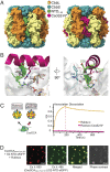
CsoSCA and CsoS2 bind at the same site on Rubisco. (A) Surface representation of Rubisco with CsoSCA’s NTD1-50 peptide bound and Rubisco with CsoS2 peptide bound (pdb: 6uew). (B) Zoomed view of the binding site showing the different conformations of the CsoSCA and CsoS2 peptides. Peptides are shown as cartoons and detailed residues as sticks. Rubisco bound with the NTD1-50 peptide is colored according to color key Inset in A and Rubisco (both subunits) bound with the CsoS2-N* peptide (pdb: 6uew) is colored white. The white transparent surface represents the Rubisco structure which binds CsoSCA. Polar interactions are depicted as dashed lines and cation–pi stacking as dashed triangles with distances in Ångströms. (C) The BLI response shows that the CsoS2 peptide fused to Rubisco (Rubisco–CsoS2-N*) passivates binding of CsoSCA to Rubisco. (D) Alexa Fluor 647, sfGFP, and merged fluorescence as well as phase contrast images of protein condensates formed from a solution of Rubisco, CsoS2-NTD-sfGFP, and Alexa Fluor 647 labeled CsoSCA-MBP showing that CsoSCA recruits into Rubisco–CsoS2 protein condensates.
Fig. 5.
Fig. 5.
Updated model for carboxysome assembly. (A) Schematic model of α-carboxysome assembly in which CsoSCA is recruited to the carboxysome via interactions with Rubisco. The model involves 1) initial molecular associations (specific order not known), 2) Cargo nucleation, 3) cargo growth with local phase separation, and finally 4) shell closure forming fully assembled carboxysomes. Current knowledge does not allow us to distinguish between whether CsoSCA associates with Rubisco during the initial association, step 1, or whether association occurs in the phase-separated condensate, step 3 (or both). CsoSCA is depicted as a dimer; however, present data cannot conclude whether CsoSCA is dimeric or hexameric. The fully assembled carboxysome in step 4 shows a stoichiometrically accurate—with respect to cargo proteins—version of the α-carboxysome. (B) Average number of cargo proteins present in an α-carboxysome (36), number of binding sites per oligomeric form of cargo protein, and total number of binding sites per carboxysome.

Comment in

Similar articles

Cited by

References

    1. Supuran C. T., Carbonic anhydrases: Novel therapeutic applications for inhibitors and activators. Nat. Rev. Drug Discov. 7, 168–181 (2008). - PubMed
    1. Merlin C., Masters M., McAteer S., Coulson A., Why is carbonic anhydrase essential to Escherichia coli? J. Bacteriol. 185, 6415–6424 (2003). - PMC - PubMed
    1. Badger M. R., Price G. D., The role of carbonic anhydrase in photosynthesis. Annu. Rev. Plant Physiol. Plant Mol. Biol. 45, 369–392 (1994).
    1. Andersson I., Catalysis and regulation in Rubisco. J. Exp. Bot. 59, 1555–1568 (2008). - PubMed
    1. Bathellier C., et al. , Ribulose 1,5-bisphosphate carboxylase/oxygenase activates O2 by electron transfer. Proc. Natl. Acad. Sci. U.S.A. 117, 24234–24242 (2020). - PMC - PubMed

Publication types

MeSH terms