Review
. 2024 Feb 12:86:71-97.
doi: 10.1146/annurev-physiol-042022-030946. Epub 2023 Oct 20.

Mechanosensing by Vascular Endothelium

Affiliations
Review

Mechanosensing by Vascular Endothelium

Xin Rui Lim et al. Annu Rev Physiol. .

Abstract

Mechanical forces influence different cell types in our bodies. Among the earliest forces experienced in mammals is blood movement in the vascular system. Blood flow starts at the embryonic stage and ceases when the heart stops. Blood flow exposes endothelial cells (ECs) that line all blood vessels to hemodynamic forces. ECs detect these mechanical forces (mechanosensing) through mechanosensors, thus triggering physiological responses such as changes in vascular diameter. In this review, we focus on endothelial mechanosensing and on how different ion channels, receptors, and membrane structures detect forces and mediate intricate mechanotransduction responses. We further highlight that these responses often reflect collaborative efforts involving several mechanosensors and mechanotransducers. We close with a consideration of current knowledge regarding the dysregulation of endothelial mechanosensing during disease. Because hemodynamic disruptions are hallmarks of cardiovascular disease, studying endothelial mechanosensing holds great promise for advancing our understanding of vascular physiology and pathophysiology.

Keywords: G protein–coupled receptors; Piezo1; endothelial cells; ion channels; mechanosensing; mechanosensors; mechanotransduction; shear stress.


Figures

Figure 1:
Figure 1:
Hemodynamic forces and endothelial mechanosensing. (a) Blood movement in the vasculature generates hemodynamic forces. Shear stress is triggered by blood flow and represents the frictional force parallel to the vascular wall. Hydrostatic pressure is the perpendicular force exerted on the vascular wall, and circumferential stretch reflects the vessel wall stretching in the circumferential direction. (b) Blood flow patterns can be either laminar or disturbed, and these patterns are linked to atheroprotection or atherogenesis, respectively. (c) As blood flows from larger arteries to smaller arterioles and then to capillaries, the vascular lumen size decreases and the shear stress onto endothelial cells (ECs) increases. (d) Key physiological responses to endothelial mechanosensing. Figure adapted from images created with BioRender.com.
Figure 2:
Figure 2:
Mechanosensors in endothelial cells. Endothelial cells lining the inside of blood vessels are constantly exposed to vascular mechanical forces. Mechanosensing these forces is feasible through various mechanosensors. Examples of these mechanosensors include ion channels (Piezo1, TRPV4, Kir2.1, ENaC, TREK-1), receptors (GPCR, receptor tyrosine kinase), and membrane structures (caveolae, PECAM-1, PlexinD1, glycocalyx, cadherin, integrin, cilia). Abbreviations: ENaC, epithelial sodium channel; GPCR, G protein–coupled receptor; Kir2.1, inwardly rectifying K+ channel 2.1; PECAM-1, platelet endothelial cell adhesion molecule-1; TRPV4, transient receptor potential vanilloid 4 channel. Figure adapted from images created with BioRender.com.
Figure 3:
Figure 3:
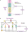
Flexibility, diversity, and cross talk between endothelial mechanosensors and mechanotransducers. (a) Example flow chart showing how different mechanosensors ultimately trigger vasodilation. Mechanotransduction occurs via one or both major pathways: NO production and release and/or EC Vm hyperpolarization. (b) Exemplary cross talk between EC mechanosensors downstream of mechano-GqPCR activation. Mechanical activation of GqPCR evokes vasodilation via NO release or hyperpolarization. GqPCR activation can directly activate PI3K/Akt, leading to eNOS activation and NO synthesis. On the other hand, GqPCR canonically facilitates PLC activity and the hydrolysis of PIP2 into DAG and IP3. The three metabolites (PIP2, DAG, and IP3) modulate the activity of mechanosensitive ion channels or their downstream effectors. PIP2 depletion inhibits Kir2.1 and TREK-1 channels but disinhibits TRPV4 activity. DAG/PKC signaling facilitates TRPV4 activity evoking Ca2+ transients that facilitate eNOS activity and/or IK/SK Ca2+-activated K+ channels; both pathways ultimately lead to vasodilation. IP3/IP3R signaling facilitates Ca2+ release from the endoplasmic reticulum, and these Ca2+ transients enhance eNOS and IK/SK activities. Abbreviations: Akt, protein kinase B; B2R, bradykinin 2 receptor; Ca2+, calcium ion; DAG, diacylglycerol; EC, endothelial cell; ENaC, epithelial sodium channel; eNOS, endothelial NO synthase; GPR68, G protein-coupled receptor 68; GqPCR, Gq protein–coupled receptor; H1R, histamine H1 receptor; IK/SK, intermediate-conductance/small-conductance; IP3, inositol 1,4,5-trisphosphate; IP3R, IP3 receptor; Kir2.1, inwardly rectifying K+ channel 2.1; NO, nitric oxide; PECAM-1, platelet endothelial cell adhesion molecule-1; PI3K, phosphatidylinositol 3-kinase; PIP2, phosphatidylinositol 4,5-bisphosphate; PKC, protein kinase C; PLC, phospholipase C; S1PR1, sphingosine-1-phosphate receptor 1; TRPV4, transient receptor potential vanilloid 4 channel; VE-cadherin, vascular endothelial-cadherin; VEGFR2, vascular endothelial growth factor receptor 2; Vm, membrane potential. Figure adapted from images created with BioRender.com.
Figure 4:
Figure 4:
Less-understood examples where endothelial mechanosensing is likely engaged. The question mark symbolizes untapped areas where endothelial mechanosensing is understudied and therefore less clear. Figure adapted from images created with BioRender.com.

Similar articles

Cited by

References

    1. Abraira VE, Ginty DD. 2013. The sensory neurons of touch. Neuron 79(4):618–39 - PMC - PubMed
    1. Hahn C, Schwartz MA. 2009. Mechanotransduction in vascular physiology and atherogenesis. Nat. Rev. 10(1):53–62 - PMC - PubMed
    1. Cecchi E, Giglioli C, Valente S, Lazzeri C, Gensini GF, et al. 2011. Role of hemodynamic shear stress in cardiovascular disease. Atherosclerosis 214(2):249–56 - PubMed
    1. Kubota Y, Kleinman HK, Martin GR, Lawley TJ. 1988. Role of laminin and basement membrane in the morphological differentiation of human endothelial cells into capillary-like structures. J. Cell Biol. 107(4):1589–98 - PMC - PubMed
    1. Przysinda A, Feng W, Li G. 2020. Diversity of organism-wide and organ-specific endothelial cells. Curr. Cardiol. Rep. 22:19. - PMC - PubMed

LinkOut - more resources