. 2023 Dec;299(12):105367.
doi: 10.1016/j.jbc.2023.105367. Epub 2023 Oct 19.

Cyclase-associated protein interacts with actin filament barbed ends to promote depolymerization and formin displacement

Affiliations

Cyclase-associated protein interacts with actin filament barbed ends to promote depolymerization and formin displacement

Nikita Alimov et al. J Biol Chem. 2023 Dec.

Abstract

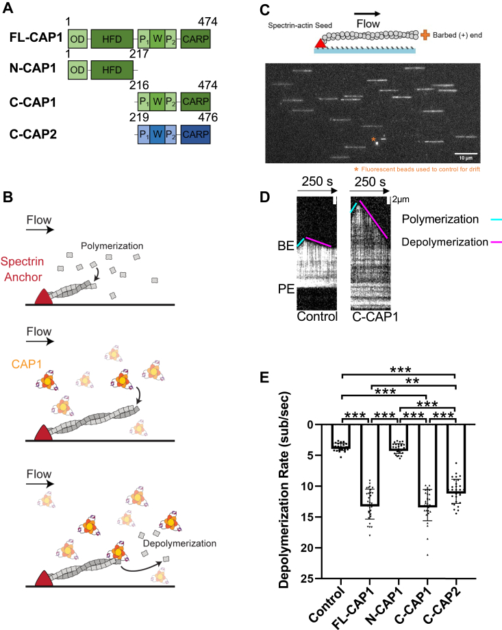
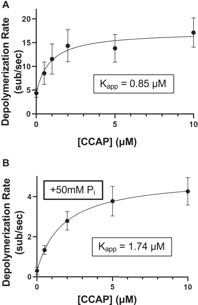
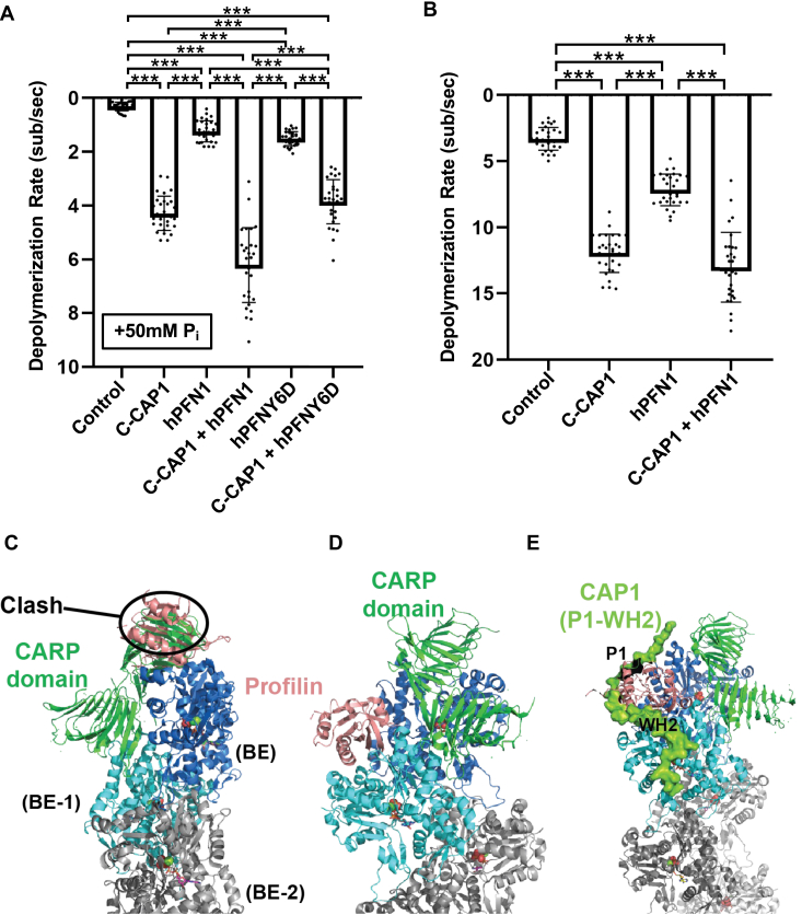
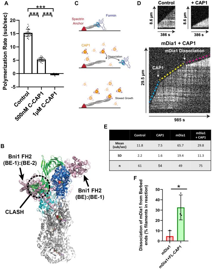
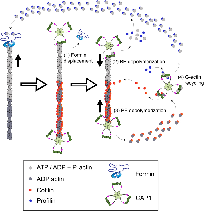
Cyclase-associated protein (CAP) has emerged as a central player in cellular actin turnover, but its molecular mechanisms of action are not yet fully understood. Recent studies revealed that the N terminus of CAP interacts with the pointed ends of actin filaments to accelerate depolymerization in conjunction with cofilin. Here, we use in vitro microfluidics-assisted TIRF microscopy to show that the C terminus of CAP promotes depolymerization at the opposite (barbed) ends of actin filaments. In the absence of actin monomers, full-length mouse CAP1 and C-terminal halves of CAP1 (C-CAP1) and CAP2 (C-CAP2) accelerate barbed end depolymerization. Using mutagenesis and structural modeling, we show that these activities are mediated by the WH2 and CARP domains of CAP. In addition, we observe that CAP collaborates with profilin to accelerate barbed end depolymerization and that these effects depend on their direct interaction, providing the first known example of CAP-profilin collaborative effects in regulating actin. In the presence of actin monomers, CAP1 attenuates barbed end growth and promotes formin dissociation. Overall, these findings demonstrate that CAP uses distinct domains and mechanisms to interact with opposite ends of actin filaments and drive turnover. Further, they contribute to the emerging view of actin barbed ends as sites of dynamic molecular regulation, where numerous proteins compete and cooperate with each other to tune polymer dynamics, similar to the rich complexity seen at microtubule ends.

Keywords: ADF; TIRF microscopy; actin; cofilin; cyclase-associated protein; turnover.


Conflict of interest statement

Conflict of interest The authors declare that they have no conflicts of interest with the contents of this article.

Figures

Figure 1
Figure 1
CAP1 and CAP2 accelerate depolymerization at the barbed ends of actin filaments.A, domain layouts for mouse CAP1 and CAP2 polypeptides used in this study. B, experimental set up of microfluidics-assisted TIRF (mf-TIRF) assays. Alexa-488–labeled actin filaments were assembled from passively absorbed spectrin-actin seeds, resulting in filaments with pointed ends anchored on the left and free barbed ends growing on the right. After filaments were polymerized to 5 to 15 μm in length, then free actin monomers were washed out, and 1 μM CAP1 polypeptides (or control buffer) were flowed into the chamber and depolymerization was immediately monitored. C, representative field of view from mf-TIRF assay showing filaments with their pointed ends anchored to spectrin-actin (left) and their growing barbed ends (right). Fluorescent TransFluoSpheres included in all reactions for drift correction. D, representative kymographs of actin filament barbed ends depolymerizing in the presence of 1 μM C-CAP1 or control buffer. Cyan line, slope of polymerization during initial filament assembly. Magenta line, slope of depolymerization at the barbed end after washing out free actin monomers and introducing C-CAP1 or control buffer. E, quantification of barbed end depolymerization rates in the presence and absence of 1 μM CAP1 or C-CAP2. The data (mean ± SD) are from three independent replicates (n = 30 filaments total per condition). One-way ANOVA followed by Tukey’s multiple comparisons test was used to determine significance between indicated conditions (∗∗p < 0.001; ∗∗∗p < 0.0001). Domains: OD, oligomerization domain; HFD, helical-folded domain; P1, polyproline 1; W, Wiskott Aldrich syndrome 2 (WH2) domain; P2, polyproline 2; CARP, CAP and RP2 (CARP) domain. BE, barbed end; C, carboxyl terminus; CAP, cyclase-associated protein; C-CAP, C-terminal half of CAP; FL, full-length; mf-TIRF, microfluidics-assisted total internal reflection fluorescence; N, amino terminus; PE pointed end.
Figure 2
Figure 2
C-CAP1 promotes depolymerization of ADP-Piactin filaments.A, rates of barbed end depolymerization at different concentrations of C-CAP1 in mf-TIRF assays. The data (mean ± SD) are from three independent replicates (n = 30 filaments total per condition). The line is a fit to a hyperbolic binding curve and was used to derive the Kapp (0.85 μM). B, rates of barbed end depolymerization at different concentrations of C-CAP1 in mf-TIRF assays in the presence of 50 mM inorganic phosphate (Pi). Data (mean ± SD) are from two independent replicates (n = 20 filaments total per condition). The line is a fit to a hyperbolic binding curve and was used to derive the Kapp (1.74 μM). CAP, cyclase-associated protein; C-CAP, C-terminal half of CAP; mf-TIRF, microfluidics-assisted total internal reflection fluorescence.
Figure 3
Figure 3
The barbed end depolymerization activity of CAP requires both its WH2 and CARP domains.A, schematics of mouse C-CAP1, C-CAP-98 (270LKHV273 > AAAA) which disrupts actin binding by the WH2 domain and P1WP2-GST which is the polyproline motif 1-Wiskott Aldrich syndrome homology domain 2 domain-polyproline motif 2 fragment dimerized by GST. Mutations marked by lightning bolts. B, rates of barbed end depolymerization for each construct (1 μM). The data (mean ± SD) are from three independent replicates (n = 30 filaments total per condition). ∗∗∗, one-way ANOVA followed by Tukey’s multiple comparisons test to determine significance between indicated conditions (p < 0.0001). C, a structural model for the interactions of C-CAP1 with the barbed end (BE) of the actin filament assembled, assembled from a cryo-EM structure of F-actin (PDB #6FHL), and the cocrystal structure of the CAP1 CARP dimer bound to two actin monomers (PDB #6FM2). One actin from the CARP domain-actin cocrystal structure was used to orient the CARP domain on the terminal barbed end actin protomer (BE, in dark blue surface representation). The CARP domain is able to fit with only a minor clash against the penultimate actin protomer (BE-1, in cyan cartoon representation). D, working model for how C-CAP accelerates depolymerization at the BE. Binding of C-CAP to the BE of the filament (state A) involves the two WH2 domains binding to the ultimate and penultimate actin subunits (dark blue and light blue) and one half of the CARP dimer binding to the ultimate subunit (dark blue). Next, interactions of the CARP and/or WH2 domains promote a conformational change in the ultimate actin subunit (orange, state B), increasing the rate of dissociation of the ultimate actin subunit from the BE, while the C-CAP dimer remains attached to the BE via its WH2 domain (state C). Finally, the original BE actin subunit (orange) is released from one half of the CARP dimer. As indicated, if the mechanism is nonprocessive, C-CAP1 then dissociates from the BE and becomes available for a new round of depolymerization. However, if the mechanism is processive, C-CAP1 remains bound to the BE by its WH2 domain after the actin subunit is released, and then the other face of the CARP dimer binds to the new ultimate actin subunit (light blue, state D). In the processive mechanism, this cycle would repeat itself until C-CAP1 dissociates from the barbed end. Domains: P1, polyproline 1; W, Wiskott Aldrich syndrome homology domain 2; P2, polyproline 2; CARP, CAP and RP2 (CARP) domain; GST, Glutathione-S-transferase. CAP, cyclase-associated protein; C-CAP, C-terminal half of CAP; WH2, Wiskott Aldrich syndrome 2.
Figure 4
Figure 4
CAP and profilin directly collaborate in promoting barbed end depolymerization.A, rates of barbed end depolymerization in the presence of 10 μM C-CAP1 and 10 μM WT PFN1 or mutant PFNY6D in mf-TIRF assays; all reactions include 50 mM inorganic phosphate (Pi). The data (mean ± SD) are from three independent replicates (n = 30 filaments total per condition) ∗∗∗, one-way ANOVA followed by Tukey’s multiple comparisons test to determine significance between indicated conditions (p < 0.0001). B, rates of barbed end depolymerization in the presence of 1 μM C-CAP1 and 10 μM human profilin (PFN1). The data (mean ± SD) are from three independent replicates (n = 30 filaments total per condition). ∗∗∗, one-way ANOVA followed by Tukey’s multiple comparisons test to determine significance between indicated conditions (p < 0.0001). C, to produce the working models shown in panels CE, we docked the CARP domain as in the figure and used a profilin-actin cocrystal structure (PDB ID#: 2BTF) to position profilin on the actin filament model, aligning the actin from the cocrystal structure to the terminal actin (BE). The first model (panel C) shows that profilin and the CARP domain cannot both bind to the ultimate actin subunit (BE), as they have a steric overlap that includes most of the volume of profilin. D, in this model, profilin binds to the penultimate actin protomer (BE-1) without overlapping with the CARP domain bound to the ultimate subunit (BE). There is a minor overlap of profilin with the ‘hydrophobic plug’ of the terminal actin protomer, but this part of actin should be dynamic when not buried in the filament and could relax out of the minor apparent steric clash. E, using AlphaFold2, we generated a working model for how the CAP1 P1-WH2 region may interact with the barbed end, with the P1 motif bound to profilin and the WH2 domain bound to actin. When aligned to the penultimate actin subunit (BE-1), the P1-WH2 region has no steric clashes with profilin, actin, or the CARP domain. CAP, cyclase-associated protein; CARP, CAP and RP2 domain; C-CAP, C-terminal half of CAP; mf-TIRF, microfluidics-assisted total internal reflection fluorescence; WH2, Wiskott Aldrich syndrome 2.
Figure 5
Figure 5
C-CAP attenuates filament growth and promotes formin dissociation from barbed ends.A, barbed end elongation rates in the presence of 2 μM G-actin with or without C-CAP1 (0.5 or 1 μM) in mf-TIRF assays. The data (mean ± SD) are from three independent replicates (n = 30 filaments total per condition). ∗∗∗, one-way ANOVA followed by Tukey’s multiple comparisons test to determine significance between indicated conditions (p < 0.0001). B, the formin FH2 domain was modeled onto the barbed end using the Bni1-FH2 domain-actin cocrystal structure (PDB ID 1Y64). The actin subunit in the cocrystal was used to orient the FH2 ‘bridge’ domain onto the terminal actin dimer, BE:BE-1, as well as the penultimate actin dimer, BE-1:BE-2 (FH2 bridge units are in pink ‘cartoon’ representation). The CARP domain of CAP1 was modeled onto the barbed end as in Figure 4 (same color scheme). In this model, the FH2 bridge at the terminal actin dimer accommodates the CARP domain while the FH2 bridge at the penultimate actin dimer has a substantial clash (indicated). C, experimental set up for testing competition between CAP and formin mDia1 at barbed ends. Filaments with free barbed ends were polymerized by exposing coverslip-anchored spectrin-actin seeds briefly to a flow containing 0.5 μM G-actin (10% Alexa-488 labeled) and 2 μM profilin. Twenty millimolar mDia1 was then introduced for 10 s to cap ∼15% of the barbed ends with mDia1 (Top), identified by their faster growth rates. After another 180 s of growth, filaments were exposed to 0.5 μM G-actin (10% Alexa-488 labeled), 2 μM profilin, and 0 or 1 μM full-length (FL) CAP1, and barbed end growth was monitored (middle and bottom). D, representative kymographs of barbed end growth. Top left, control reaction containing 0.5 μM G-actin + 2 μM profilin. Top right: filament growth in the presence of 1 μM FL-CAP1 (no mDia1). Bottom, filament that undergoes three distinct phases of growth, starting with fast growth of mDia1-capped barbed end (blue dashed line), followed by slower growth upon addition of 1 μM FL-CAP1 (yellow dashed line), and finally slower growth after mDia1 dissociates from the barbed end (magenta dashed line). E, table showing elongation rates (mean ± SD) of free barbed ends and mDia1-capped ends in the presence and absence of 1 μM FL-CAP1. The data (mean ± SD) are from three independent replicates. Total number of filaments analyzed: n = 61 (control), 54 (+CAP1), 49 (mDia1), 75 (mDia1+FL-CAP1). F, quantification of the percent (mean ± SD) of mDia1 dissociation events from barbed ends in the presence and absence of 1 μM FL-CAP1. The data (mean ± SD) are from three independent replicates. Total number of filaments analyzed: n = 111 (mDia1), 108 (+CAP1). ∗Represents statistical significance, p < 0.05, between the two groups compared using a Welch’s t test. CAP, cyclase-associated protein; C-CAP, C-terminal half of CAP; CARP, CAP and RP2 domain; mf-TIRF, microfluidics-assisted total internal reflection fluorescence.
Figure 6
Figure 6
Model summarizing new and established roles of CAP in promoting actin turnover. In this study, we have established that (1) CAP (green) promotes the displacement of formins (blue) from growing barbed ends and (2) that C-terminal domains of CAP (WH2 and CARP) interact with the barbed end (BE) to attenuate growth and promote depolymerization. Further, we have shown that CAP directly collaborates with profilin in promoting BE depolymerization through profilin interactions with the proline-rich P1 region of CAP. Previously, we and others established that (3) the N-terminal HFD domains of CAP interact with the pointed ends (PEs) of actin filaments where they synergize with cofilin (red circles) to promote PE depolymerization (54, 55), and (4) C-terminal WH2 and CARP domains of CAP recycle actin monomers, by binding to ADP-G-actin with high affinity, displacing cofilin, catalyzing nucleotide exchange (ATP for ADP) on G-actin, and handing off ATP-G-actin to profilin for new rounds of assembly (18, 49, 53, 57, 59, 60, 71). For simplicity, the ability of N-CAP to bind the sides of actin filaments (using its HFD domains) and enhance cofilin-dependent severing (18, 50, 79, 106) is not depicted in this cartoon but is likely to also contribute to F-actin disassembly. Because cellular concentrations of CAP are relatively high (6–7 μM; (43, 62)), we expect that there are sufficient CAP levels for most of these functions to be occurring simultaneously in cells. CAP, cyclase-associated protein; CARP, CAP and RP2 domain; HFD, helical folded domain; N-CAP, N-terminal half of CAP; WH2, Wiskott Aldrich syndrome 2.

References

    1. Pollard T.D., Borisy G.G. Cellular motility driven by assembly and disassembly of actin filaments. Cell. 2003;112:453–465. - PubMed
    1. Blanchoin L., Boujemaa-Paterski R., Sykes C., Plastino J. Actin dynamics, architecture, and mechanics in cell motility. Physiol. Rev. 2014;94:235–263. - PubMed
    1. Carlier M.-F., Shekhar S. Global treadmilling coordinates actin turnover and controls the size of actin networks. Nat. Rev. Mol. Cell Biol. 2017;18:389–401. - PubMed
    1. Chhabra E.S., Higgs H.N. The many faces of actin: matching assembly factors with cellular structures. Nat. Cell Biol. 2007;9:1110–1121. - PubMed
    1. Brieher W. Mechanisms of actin disassembly. Mol. Biol. Cell. 2013;24:2299–2302. - PMC - PubMed

Publication types

MeSH terms