. 2024 Aug 2;23(8):2762-2779.
doi: 10.1021/acs.jproteome.3c00244. Epub 2023 Oct 20.

Proteomic Serum Profiling of Holstein Friesian Cows with Different Pathological Forms of Bovine Paratuberculosis Reveals Changes in the Acute-Phase Response and Lipid Metabolism

Affiliations

Proteomic Serum Profiling of Holstein Friesian Cows with Different Pathological Forms of Bovine Paratuberculosis Reveals Changes in the Acute-Phase Response and Lipid Metabolism

Alejandra Isabel Navarro León et al. J Proteome Res. .

Abstract

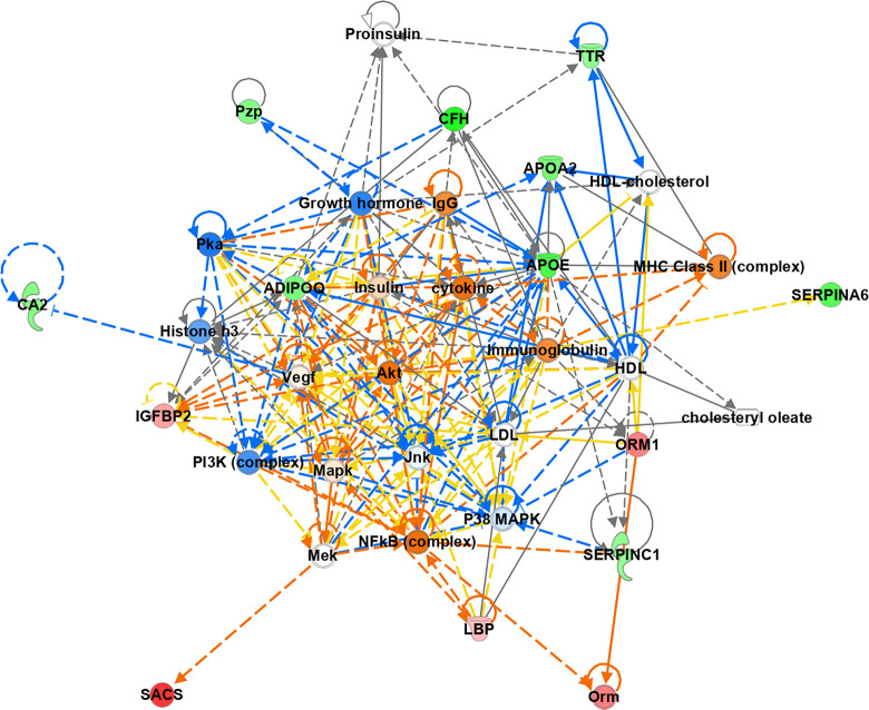
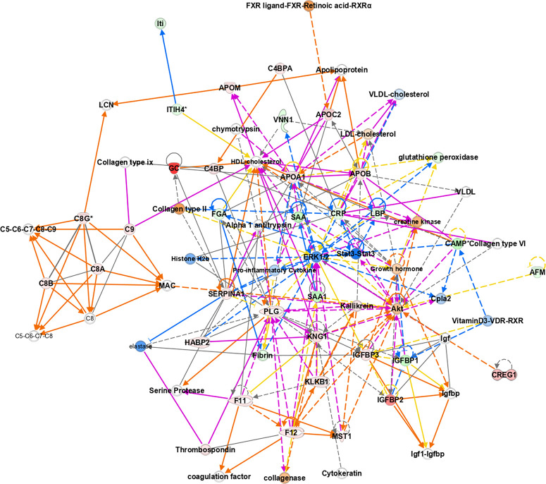
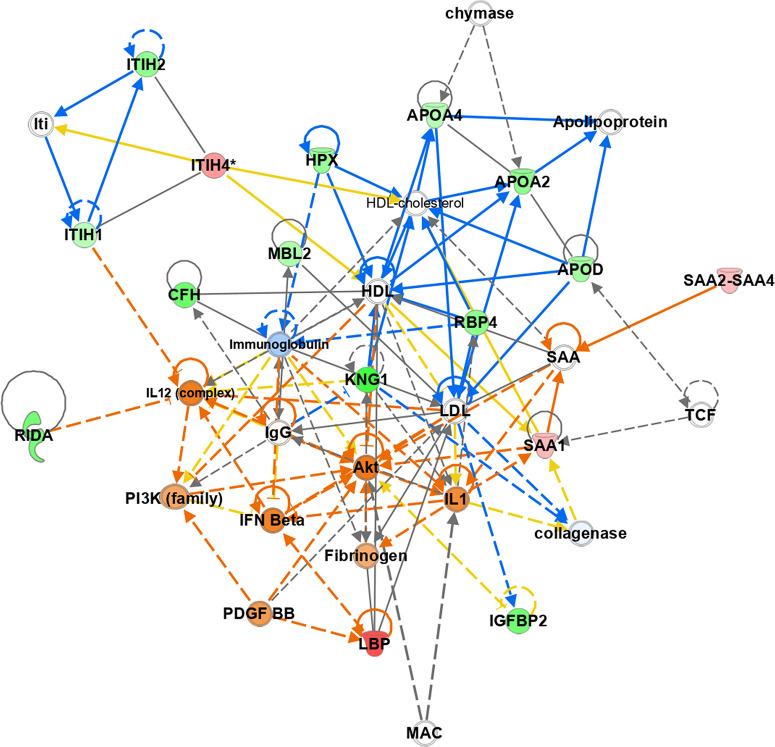
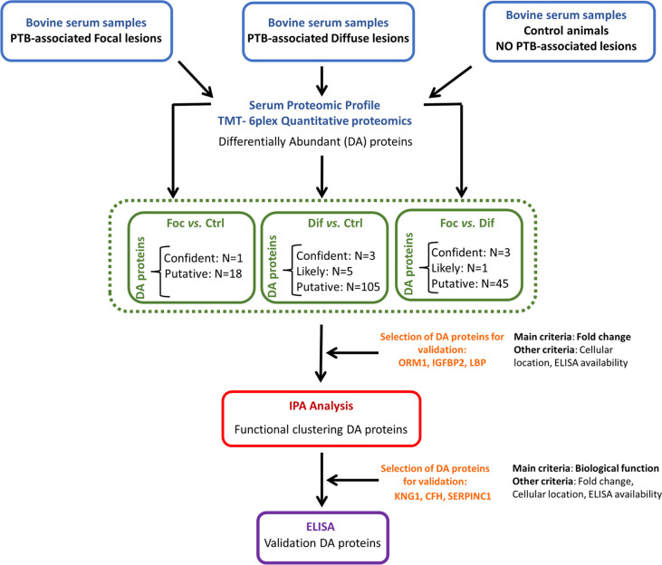
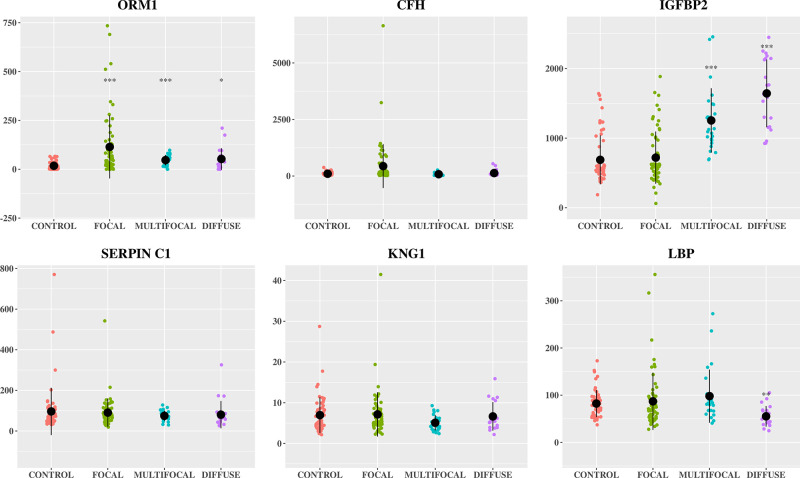
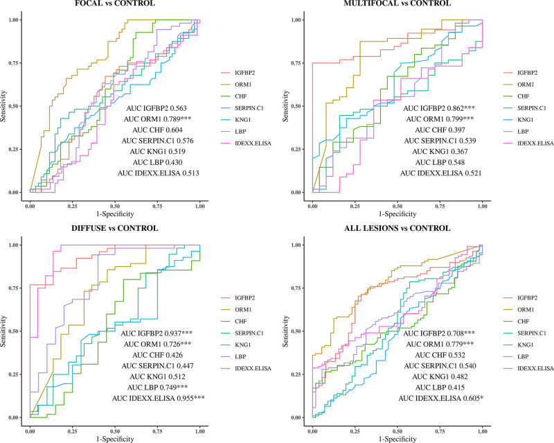
The lack of sensitive diagnostic methods to detect Mycobacterium avium subsp. paratuberculosis (Map) subclinical infections has hindered the control of paratuberculosis (PTB). The serum proteomic profiles of naturally infected cows presenting focal and diffuse pathological forms of PTB and negative controls (n = 4 per group) were analyzed using TMT-6plex quantitative proteomics. Focal and diffuse are the most frequent pathological forms in subclinical and clinical stages of PTB, respectively. One (focal versus (vs.) control), eight (diffuse vs. control), and four (focal vs. diffuse) differentially abundant (DA) proteins (q-value < 0.05) were identified. Ingenuity pathway analysis of the DA proteins revealed changes in the acute-phase response and lipid metabolism. Six candidate biomarkers were selected for further validation by specific ELISA using serum from animals with focal, multifocal, and diffuse PTB-associated lesions (n = 108) and controls (n = 56). Overall, the trends of the serum expression levels of the selected proteins were consistent with the proteomic results. Alpha-1-acid glycoprotein (ORM1)-based ELISA, insulin-like growth factor-binding protein 2 (IGFBP2)-based ELISA, and the anti-Map ELISA had the best diagnostic performance for detection of animals with focal, multifocal, and diffuse lesions, respectively. Our findings identify potential biomarkers that improve diagnostic sensitivity of PTB and help to elucidate the mechanisms involved in PTB pathogenesis.

Keywords: Mycobacterium avium subsp. paratuberculosis; acute-phase proteins; biomarkers; cattle; diagnosis; disease progression; paratuberculosis; pathogenesis; serum proteome.


Conflict of interest statement

The authors declare no competing financial interest.

Figures

Figure 1
Figure 1
Functional clustering of differentially abundant proteins in serum of animals with focal lesions versus control animals using Ingenuity Pathway Analysis (IPA). The top network “Lipid Metabolism, Molecular Transport and Small Molecule Biochemistry” (score 37) with the main protein-to-protein interactions is represented. Activation of cytokines, IgGs, immunoglobulins, protein kinase B (Akt), and the nuclear factor kappa β (NFΚB) complex are predicted. Upregulated proteins are shown in red, and downregulated proteins in green (light red and green indicate less extreme increased or decreased measurement in the data set). Orange indicates the prediction of activation of a pathway and blue prediction of inhibition (lighter orange and blue indicate that the prediction has been done with less confidence). Solid lines indicate direct connections, while dotted lines indicate indirect connections (circular arrows mean influence itself). The pointed and blunt arrowheads represent activating and inhibitory relationships, respectively. Orange lines indicate activation, blue inhibition, yellow indicates that findings are inconsistent with the state of downstream molecule, and gray indicates no predicted effect.
Figure 2
Figure 2
Functional clustering of differentially abundant proteins in serum of animals with diffuse lesions versus control animals using Ingenuity Pathway Analysis (IPA). Merged networks 1 and 2 (network 1. Humoral immune response, Inflammatory response, Hematological system development and function (score 31); and network 2. Inflammatory response, Organismal injury and abnormalities, Cell death and survival (score 31)) with four main nodes (complement system, the blood coagulation/anticoagulation cascade, metabolism of lipids and carbohydrates) are represented. Upregulated proteins are shown in red, and downregulated proteins are in green (light red and green indicate less extreme increased or decreased measurement in the data set). Orange indicates the prediction of activation of a pathway and blue prediction of inhibition (lighter orange and blue indicate that the prediction has been done with less confidence). Solid lines indicate direct connections, while dotted lines indicate indirect connections (circular arrows mean influence itself). The pointed and blunt arrowheads represent activating and inhibitory relationships, respectively. Orange lines indicate activation, blue inhibition, yellow indicate that findings are inconsistent with the state of downstream molecule, and gray indicates no predicted effect.
Figure 3
Figure 3
Functional clustering of differentially abundant proteins in serum of animals with focal lesions versus diffuse animals using Ingenuity Pathway Analysis (IPA). The top network “Carbohydrate metabolism, Lipid metabolism, Molecular transport” (score 41) with the main protein-to-protein interactions is represented. Alterations in the immune response and lipid metabolism related to the disease stage are observed. Upregulated proteins are shown in red, and downregulated proteins are in green (light red and green indicate less extreme increased or decreased measurement in the data set). Orange indicates the prediction of activation of a pathway and blue prediction of inhibition (lighter orange and blue indicate that the prediction has been done with less confidence). Solid lines indicate direct connections, while dotted lines indicate indirect connections (circular arrows mean influence itself). The pointed and blunt arrowheads represent activating and inhibitory relationships, respectively. Orange lines indicate activation, blue inhibition, yellow indicates that findings are inconsistent with the state of downstream molecule, and gray indicates no predicted effect.
Figure 4
Figure 4
Flowchart showing the analysis of bovine plasma by TMT-6plex quantitative proteomics and selection of differentially abundant proteins for validation. IPA, Ingenuity Pathway Analysis; IGFBP2, bovine insulin-like growth factor binding protein 2; ORM1, alpha-1-acid gycoprotein1; CHF, complement factor H; SERPINC1, bovine anti-thrombin III; KNG1, kininogen 1; and LBP, lipopolysaccharide binding protein.
Figure 5
Figure 5
Serum levels of ORM1, CFH, IGFBP2, SERPINC1, KNG1, and LBP in Holstein Friesian cows with focal (n = 61), multifocal (n = 25) , and diffuse (n = 22) histopathological forms of bovine paratuberculosis (PTB) and negative control animals from PTB-free farms (n = 56). The protein concentrations were quantified by specific ELISAs supplied by MyBioSource, San Diego, CA, USA. Data are represented in scatter plots with each dot representing a single animal. The mean value of each histopathological group is represented by a gross black point and the standard deviation by a vertical line. The concentrations are expressed in μg/mL for SERPINC1 and ng/mL for IGFBP2, ORM1, CFH, KNG1, and LBP (Y-axis). IGFBP2, bovine insulin-like growth factor binding protein 2; ORM1, alpha-1-acid gycoprotein1; CFH, complement Factor H; SERPINC1, bovine anti-thrombin III; KNG1, kininogen 1; and LBP, lipopolysaccharide binding protein. The asterisks indicate that differences between each histopathological group and the control are significant (* p < 0.05, ** p < 0.01, *** p < 0.001).
Figure 6
Figure 6
Receiver operator characteristic curves (ROC curves) analysis of the IGFBP2, ORM1, CFH, SERPINC1, KNG1, and LBP bovine proteins-based ELISA' results of serum samples from Holstein Friesian cows with focal (n = 61), multifocal (n = 25), and diffuse (n = 22) pathological forms of bovine paratuberculosis (PTB) and negative control animals from PTB-free farms (n = 56). All, includes animals with focal, multifocal, and diffuse lesions; vs, versus; IGFBP2, bovine insulin-like growth factor binding protein 2; ORM1, alpha-1-acid gycoprotein1; CFH, complement Factor H; SERPINC1, bovine anti-thrombin III; KNG1, kininogen 1; and LBP, lipopolysaccharide binding protein. The asterisks indicate that a specific ELISA detects significant differences (* p < 0.05, ** p < 0.01, *** p < 0.001) between the compared histopathological groups.

Similar articles

Cited by

References

    1. Benedictus G.; Dijkhuizen A. A.; Stelwagen J. Economic losses due to paratuberculosis in dairy cattle. Vet Rec 1987, 121 (7), 142–146. 10.1136/vr.121.7.142. - DOI - PubMed
    1. Kudahl A. B.; Nielsen S. S.; Sørensen J. T. Relationship between antibodies against Mycobacterium avium subsp. paratuberculosis in milk and shape of lactation curves. Pre. Vet. Med. 2004, 62 (2), 119–134. 10.1016/j.prevetmed.2003.11.008. - DOI - PubMed
    1. Ozsvari L.; Harnos A.; Lang Z.; Monostori A.; Strain S.; Fodor I. The Impact of Paratuberculosis on Milk Production, Fertility, and Culling in Large Commercial Hungarian Dairy Herds. Front. Vet. Sci. 2020, 7, 565324.10.3389/fvets.2020.565324. - DOI - PMC - PubMed
    1. Smith R. L.; Strawderman R. L.; Schukken Y. H.; Wells S. J.; Pradhan A. K.; Espejo L. A.; et al. Effect of Johne’s disease status on reproduction and culling in dairy cattle. J. Dairy Sci. 2010, 93, 3513–24. 10.3168/jds.2009-2742. - DOI - PubMed
    1. Tiwari A.; VanLeeuwen J. A.; Dohoo I. R.; Stryhn H.; Keefe G. P.; Haddad J. P. Effects of seropositivity for bovine leukemia virus, bovine viral diarrhoea virus, Mycobacterium avium subspecies paratuberculosis, and Neospora caninum on culling in dairy cattle in four Canadian provinces. Vet. Microbiol. 2005, 109 (3–4), 147–58. 10.1016/j.vetmic.2005.05.011. - DOI - PubMed

Publication types

LinkOut - more resources