. 2023 Oct 20;14(1):6653.
doi: 10.1038/s41467-023-42484-z.

Discrete hippocampal projections are differentially regulated by parvalbumin and somatostatin interneurons

Affiliations

Discrete hippocampal projections are differentially regulated by parvalbumin and somatostatin interneurons

Daniel J Lodge et al. Nat Commun. .

Abstract

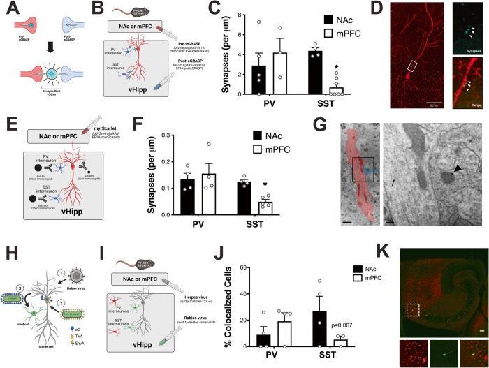
People with schizophrenia show hyperactivity in the ventral hippocampus (vHipp) and we have previously demonstrated distinct behavioral roles for vHipp cell populations. Here, we test the hypothesis that parvalbumin (PV) and somatostatin (SST) interneurons differentially innervate and regulate hippocampal pyramidal neurons based on their projection target. First, we use eGRASP to show that PV-positive interneurons form a similar number of synaptic connections with pyramidal cells regardless of their projection target while SST-positive interneurons preferentially target nucleus accumbens (NAc) projections. To determine if these anatomical differences result in functional changes, we used in vivo opto-electrophysiology to show that SST cells also preferentially regulate the activity of NAc-projecting cells. These results suggest vHipp interneurons differentially regulate that vHipp neurons that project to the medial prefrontal cortex (mPFC) and NAc. Characterization of these cell populations may provide potential molecular targets for the treatment schizophrenia and other psychiatric disorders associated with vHipp dysfunction.


Conflict of interest statement

The authors declare no competing interests.

Figures

Fig. 1
Fig. 1. Ventral hippocampal (vHipp) projections to the medial prefrontal cortex (mPFC) and nucleus accumbens (NAc) are anatomically segregated.
A To identify vHipp projection neurons, retrograde viruses expressing either myristoylated Scarlet (myrScarlet) or myristoylated Green Fluorescent Protein (myrGFP) were injected into the NAc or mPFC. B The total number of fluorescently labeled cells in the vHipp were counted and the percentage projecting to each region was determined. C The anatomical location of each cell was mapped onto a cartoon of the vHipp. A representative image of the ventral hippocampus is depicted in (D). mPFC-projecting cells are depicted in green, NAc-projecting cells are shown in red. Scale bar is 100 microns. n = 2–4 sections each from 4 mice per group. Source data are provided as a Source Data file.
Fig. 2
Fig. 2. Ventral hippocampal (vHipp) projections to the medial prefrontal cortex (mPFC) and nucleus accumbens (NAc) have unique transcriptional signatures.
A To determine gene expression in vHipp projection neurons, a retrograde virus expressing myrScarlet was injected into the mPFC or NAc. Scarlet-positive cells in the vHipp were isolated by flow cytometry and RNASequencing (RNASeq) was used to measure gene expression. B RNASeq identified 92 genes that were differentially expressed between mPFC- and NAc-projecting cells. GRN and NEDD9 are outlined in black. C Gene ontology was used to determine enriched pathways. n = 5 (mPFC-projecting cells) or 6 (NAc-projecting cells) per group. Scale bar is 50 µm. D To confirm RNASeq analysis, fluorogold was injected into the mPFC or NAc and RNAScope was used to measure expression of GRN and NEDD9 using RNAScope. E In line with the RNASeq results, GRN and NEDD9 were increased in mPFC-projecting cells compared to those that project to the NAc. Data were analyzed by Two-way ANOVA, p = 0.0369. F When total number of puncta per cell were analyzed, GRN and NEDD9 were increased in mPFC-projecting cells compared to those that project to the NAc. Data were analyzed by Two-way ANOVA, p = 0.0319. Representative images are shown in (G). * signifies a main effect of projection target. Scale bar is 10 microns. n = 3 cells each from 4 (mPFC-projecting cells, NEDD9), 5 (mPFC-projecting cells, GRN), or 6 mice (NAc-projecting cells, GRN or NEDD9) per group. All data are presented as mean values +/- SEM. Source data are provided as a Source Data file.
Fig. 3
Fig. 3. Ventral Hippocampal (vHipp) projections to the medial prefrontal cortex (mPFC) and nucleus accumbens (NAc) are differentially innervated by local interneurons.
A Schematic demonstrating enhanced GFP Recombination Across Synaptic Partners (eGRASP) technology. B The pre-eGRASP component was expressed in mPFC- or NAc-projecting pyramidal cells and post-eGRASP component was expressed in vHipp parvalbumin (PV) or somatostatin (SST) cells. C PV- and SST-positive interneurons differentially connect to vHipp pyramidal cells depending on their projection target. Data analyzed by Two-way ANOVA, p = 0.0126. * denotes significant difference from NAc-projecting pyramidal cells: SST interneurons. n = 3 cells each from 3 (mPFC-projecting pyramidal cells: PV interneurons), 4 (NAc-projecting pyramidal cells: SST interneurons), 5 (NAc-projecting pyramidal cells: PV interneurons) or 7 (mPFC-projecting pyramidal cells: SST interneurons) mice per group. Representative image showing NAc-projecting pyramidal cell: PV interneuron synapses is shown in (D). Scale bar is 50 microns. E Immunogold labeling of pyramidal cell projections and specific interneuron subtypes. F PV- and SST-positive interneurons differentially connect to vHipp pyramidal cells depending on their projection target. Data analyzed by Two-way ANOVA, p = 0.0429. * denotes significant difference from NAc-projecting pyramidal cells: SST interneurons. n = 4 dendrites each from 4 (NAc-projecting pyramidal cells: PV or SST interneurons; mPFC-projecting pyramidal cells: PV interneurons) or 5 (mPFC-projecting pyramidal cells: SST interneurons) mice per group. A representative image of mPFC-projecting pyramidal cell (red, arrows denote immunogold labeling of RFP): PV interneuron (blue, arrows denote immunogold labeling of PV) synapse is shown in (G). Scale bars are 1 µm and 200 nm. H Schematic of modified rabies virus. I The TVA receptor and optimized G protein was expressed in NAc- or mPFC-projecting pyramidal cells and the modified rabies virus was injected into the vHipp. J SST-positive interneurons show a trend toward less connectivity with vHipp pyramidal cells that project to the mPFC. n = 2-23 slices each from 3 (mPFC-projecting pyramidal cells: SST interneurons) or 4 (NAc-projecting pyramidal cells: PV or SST interneurons; mPFC-projecting pyramidal cells; PV interneurons) mice per group. Data analyzed by two-way ANOVA, p = 0.0668. A representative image of vHipp cells monosynaptically connected to NAc-projecting pyramidal cells (green) is shown in (K). SST interneurons shown in red. Scale bar is 100 microns. All data are presented as mean values +/- SEM. Source data are provided as a Source Data file.
Fig. 4
Fig. 4. Ventral Hippocampal (vHipp) projections to the medial prefrontal cortex (mPFC) and nucleus accumbens (NAc) are differentially regulated by local interneurons.
A The red-shifted ChannelRhodopsin, C1V1, and yellow fluorescent protein (YFP) were expressed in vHipp pyramidal cells and an opto-electric probe was used to record the electrical activity of fluorescently-labeled pyramidal cells before and during yellow laser activation. Emitted fluorescence and electrical activity were simultaneously recorded by the opto-electric probe as the micropositioner was used to move through the vHipp as shown in (B). C An increase in fluorescence was observed in YFP-positive cells. n = 3 (YFP + ) or 5 (YFP-) cells per group. Data analyzed by unpaired t-Test, p = 0.0009. * denotes significant difference from YFP-negative cells. D A significant increase in firing rate was observed in YFP-positive cells. n = 3 (YFP + ) or 5 (YFP-) cells per group. Data analyzed by unpaired t-test, p = 0.0093. * denotes significant difference from YFP-negative cells. Representative traces are shown in (E). Representative image of YFP-positive cells are shown in (F). Scale bar is 100 microns. G myristoylated (myrGFP) was expressed in NAc- or mPFC-projecting pyramidal cells and halorhodopsin was expressed in vHipp parvalbumin (PV) or somatostatin (SST) interneurons. An opto-electric probe shining a blue laser was used to identify GFP-positive pyramidal cells, then a yellow laser was used to activate halorhodopsin in interneuron subtypes. H Differential baseline firing rates were observed in vHipp pyramidal cells that project to the NAc and mPFC. Data analyzed by unpaired t-test, p = 0.0418. * denotes significant difference from NAc-projecting cells. n = 26 (mPFC-projecting) or 29 (NAc-projecting) cells per group. I PV- and SST-positive interneurons differentially regulate the activity of vHipp pyramidal cells depending on their projection target. Data analyzed by Kruskal–Wallis test, p = 0.0053. * denotes significant difference from control cells. Representative electrophysiological traces before and during PV interneuron inhibition are depicted in (J). Representative image of mCherry-positive PV interneurons in the hippocampus is shown in (K). Scale bar is 100 microns. n = 10 (mPFC projecting pyramidal cell: SST interneuron), 12 (NAc-projecting pyramidal cell: PV or SST interneuron, Control) or 15 (mPFC-projecting pyramidal cell: PV interneuron) cells from 5–6 mice per group. All data are presented as mean values +/- SEM. Source data are provided as a Source Data file.

References

    1. Fanselow MS, Dong HW. Are the dorsal and ventral hippocampus functionally distinct structures? Neuron. 2010;65:7–19. - PMC - PubMed
    1. Moser MB, Moser EI, Forrest E, Andersen P, Morris RG. Spatial learning with a minislab in the dorsal hippocampus. Proc. Natl Acad. Sci. USA. 1995;92:9697–9701. - PMC - PubMed
    1. Jung MW, Wiener SI, McNaughton BL. Comparison of spatial firing characteristics of units in dorsal and ventral hippocampus of the rat. J. Neurosci. 1994;14:7347–7356. - PMC - PubMed
    1. Pothuizen HH, Zhang WN, Jongen-Rêlo AL, Feldon J, Yee BK. Dissociation of function between the dorsal and the ventral hippocampus in spatial learning abilities of the rat: a within-subject, within-task comparison of reference and working spatial memory. Eur. J. Neurosci. 2004;19:705–712. - PubMed
    1. Sun W, Rebec GV. Lidocaine inactivation of ventral subiculum attenuates cocaine-seeking behavior in rats. J. Neurosci. 2003;23:10258–10264. - PMC - PubMed

Publication types

LinkOut - more resources