. 2023 Oct 20;14(1):6627.
doi: 10.1038/s41467-023-42210-9.

Structural basis for recruitment of TASL by SLC15A4 in human endolysosomal TLR signaling

Affiliations

Structural basis for recruitment of TASL by SLC15A4 in human endolysosomal TLR signaling

Xudong Chen et al. Nat Commun. .

Abstract

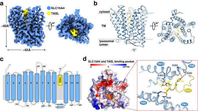
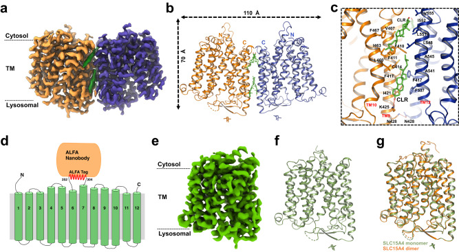
Toll-like receptors (TLRs) are a class of proteins that play critical roles in recognizing pathogens and initiating innate immune responses. TASL, a recently identified innate immune adaptor protein for endolysosomal TLR7/8/9 signaling, is recruited by the lysosomal proton-coupled amino-acid transporter SLC15A4, and then activates IRF5, which in turn triggers the transcription of type I interferons and cytokines. Here, we report three cryo-electron microscopy (cryo-EM) structures of human SLC15A4 in the apo monomeric and dimeric state and as a TASL-bound complex. The apo forms are in an outward-facing conformation, with the dimeric form showing an extensive interface involving four cholesterol molecules. The structure of the TASL-bound complex reveals an unprecedented interaction mode with solute carriers. During the recruitment of TASL, SLC15A4 undergoes a conformational change from an outward-facing, lysosomal lumen-exposed state to an inward-facing state to form a binding pocket, allowing the N-terminal helix of TASL to be inserted into. Our findings provide insights into the molecular basis of regulatory switch involving a human solute carrier and offers an important framework for structure-guided drug discovery targeting SLC15A4-TASL-related human autoimmune diseases.


Conflict of interest statement

CeMM and the Medical University of Vienna are the applicants of European priority patent applications (EP 22 203 423.3, EP 22 203 422.5, EP 22 203 421.7, status: filed) in which M.R. and G.S.-F are listed as inventors, covering small-molecule modulators of TASL and their medical use. G.S.-F. is founder and shareholder of Solgate, a company focused on SLC inhibitors. The other authors declare no competing interests.

Figures

Fig. 1
Fig. 1. Overall structure of human SLC15A4/TASL complex.
a, b 3.0-Å-resolution cryo-EM map (a) and ribbon model (b) of human SLC15A4-TASL complex. SLC15A4 and TASL are colored blue and yellow, respectively. The N-436 glycosylation is shown as sticks. c Topology diagram of human SLC15A4/TASL(1–20)-EGFP complex. TM helices and beta strands of SLC15A4 and the N-terminal sequence of TASL are schematically illustrated. The SLC15A4 and TASL are colored blue and yellow, respectively. d Left: Heatmap of the surface electrostatics of human SLC15A4 shows a TASL-binding pocket. TASL is shown as a yellow cartoon. Right: Close-up view of the binding interface between SLC15A4 and TASL. Potential hydrogen bonds are indicated with red dotted lines.
Fig. 2
Fig. 2. TASL-SLC15A4 interaction is necessary for endolysosomal TLR signaling.
a, b Confocal microscopy images of HEK293T cells co-transfected with SLC15A4-mCherry and TASL(1–20)-EGFP (WT or mutations) plasmids (a) or TASL(1–20)-EGFP and SLC15A4-mCherry (WT or mutations) plasmids (b). Localization of SLC15A4 and TASL are shown. Data are representative of three biological independent experiments. S154-mCherry, SLC15A4-mCherry. Scale bars, 5 μm. c, d Mutations of interface residues in either TASL (c) or SLC15A4 (d) abolished or diminished the TASL-SLC15A4 interaction. Data are representative of two biological independent experiments. S154, SLC15A4. WCE, whole-cell extract. e, f Mutations of interface residues in both TASL (e) and SLC15A4 (f) abolished or diminished the R848-stimulated IL-8 expression in THP1 cells. THP1 TASL-null cells were reconstituted with TASL wild type or mutants (e), whereas THP1 SLC15A4-null cells were reconstituted with SLC15A4 wild type or mutants (f). Graphs show mean ± s.d. of stimulation replicates (n = 3 biological replicates) from one experiment representative of two independent experiments. S154, SLC15A4. Source data for relevant information are provided as a Source Data file.
Fig. 3
Fig. 3. Overall structure of human SLC15A4 in apo state.
a, b 2.9-Å-resolution cryo-EM map (a) and ribbon model (b) of human SLC15A4 dimer in the apo state. The two protomers of SLC15A4 are colored orange and blue, respectively. The bound cholesterol molecules are colored green. c Close-up view of the dimer interface of SLC15A4. The side chains of the indicated interface residues are shown. d Topology diagram of the designed SLC15A4-ALFA-Nanobody construct for resolving the SLC15A4 monomer structure. The loop (253–303) between TM6 and TM7 of SLC15A4 was replaced by an ALFA tag. The TMs, the ALFA tag, and the ALFA nanobody are colored green, red, and orange, respectively. e, f 3.0-Å-resolution cryo-EM map (e) and ribbon model (f) of human SLC15A4 monomer in the apo state. SLC15A4 is colored green and the N-436 glycosylation is shown as sticks. g Structural comparison between the monomer structure (green) and the dimer structure (orange) of human SLC15A4 in the apo state.
Fig. 4
Fig. 4. Conformational changes of SLC15A4 during TASL binding.
a Heatmap of the surface electrostatics of SLC15A4 in the outward-facing apo state and the TASL-bound state. The TASL is shown as a cartoon model. b, c Close-up views of the movement of indicated TM helices and residues during the conformational change from an outward-facing state to a TASL-bound state in cytosolic view (b) and lysosomal view (c). The red arrows indicate the movement of TM helices and residues. d Structural comparison of human SLC15A4 in its outward-facing state (orange) and three SLC15A1 models [PDB accession codes: 7PMW (green), 7PMX (wheat), and 7PN1 (gray)]. e Structural comparison of human SLC15A4 in the outward-facing state (orange) and SLC15A2 model [PDB accession codes: 7NQK (teal blue)]. f Structural comparison of human SLC15A4 in the TASL-bound state (blue) and SLC15A2 in the substrate-bound state model [PDB accession codes:: 7PMY (purple)].
Fig. 5
Fig. 5. Proposed mechanism of SLC15A4−TASL signaling.
Schematic model of the TASL- and SLC15A4-dependent IRF5 activation in endolysosomal TLR signaling. Apo SLC15A4 is located on the lysosomal lumen and adopts an outward-facing conformation. During the recruitment of TASL, SLC15A4 undergoes an outward-facing to an inward-facing conformational change, leading to the opening of the TASL-binding pocket and the subsequent binding to TASL from the cytosolic side. Activation of TLR7–9 triggers the phosphorylation of the SLC15A4-TASL module, resulting in the following recruitment and activation of transcription factor IRF5. The phosphorylated IRF5 dimer then enters the nucleus to regulate the production of inflammatory cytokines. SLC15A4 can be locked in the outward-facing conformation by a selective inhibitor, which blocks the SLC15A4-TASL interaction, thus interrupting the endolysosomal TLR signaling pathway. The red and blue dashed boxes indicate the activation and inhibition process of SLC15A4, respectively.

References

    1. Akira S, Uematsu S, Takeuchi O. Pathogen recognition and innate immunity. Cell. 2006;124:783–801. doi: 10.1016/j.cell.2006.02.015. - DOI - PubMed
    1. Takeda K, Kaisho T, Akira S. Toll-like receptors. Annu. Rev. Immunol. 2003;21:335–376. doi: 10.1146/annurev.immunol.21.120601.141126. - DOI - PubMed
    1. Akira S, Takeda K. Toll-like receptor signalling. Nat. Rev. Immunol. 2004;4:499–511. doi: 10.1038/nri1391. - DOI - PubMed
    1. de Marcken M, Dhaliwal K, Danielsen AC, Gautron AS, Dominguez-Villar M. TLR7 and TLR8 activate distinct pathways in monocytes during RNA virus infection. Sci. Signal. 2019;12:eaaw1347. doi: 10.1126/scisignal.aaw1347. - DOI - PubMed
    1. Asano T, et al. X-linked recessive TLR7 deficiency in ~1% of men under 60 years old with life-threatening COVID-19. Sci. Immunol. 2021;6:eabl4348. doi: 10.1126/sciimmunol.abl4348. - DOI - PMC - PubMed

Publication types

LinkOut - more resources