. 2023 Oct 20;14(10):692.
doi: 10.1038/s41419-023-06215-y.

Control of TGFβ signalling by ubiquitination independent function of E3 ubiquitin ligase TRIP12

Affiliations

Control of TGFβ signalling by ubiquitination independent function of E3 ubiquitin ligase TRIP12

Kripa S Keyan et al. Cell Death Dis. .

Abstract

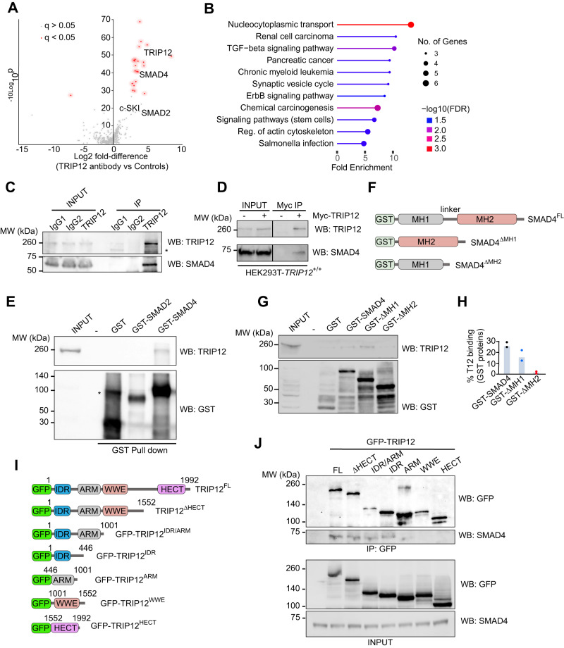
Transforming growth factor β (TGFβ) pathway is a master regulator of cell proliferation, differentiation, and death. Deregulation of TGFβ signalling is well established in several human diseases including autoimmune disorders and cancer. Thus, understanding molecular pathways governing TGFβ signalling may help better understand the underlying causes of some of those conditions. Here, we show that a HECT domain E3 ubiquitin ligase TRIP12 controls TGFβ signalling in multiple models. Interestingly, TRIP12 control of TGFβ signalling is completely independent of its E3 ubiquitin ligase activity. Instead, TRIP12 recruits SMURF2 to SMAD4, which is most likely responsible for inhibitory monoubiquitination of SMAD4, since SMAD4 monoubiquitination and its interaction with SMURF2 were dramatically downregulated in TRIP12-/- cells. Additionally, genetic inhibition of TRIP12 in human and murine cells leads to robust activation of TGFβ signalling which was rescued by re-introducing wildtype TRIP12 or a catalytically inactive C1959A mutant. Importantly, TRIP12 control of TGFβ signalling is evolutionary conserved. Indeed, genetic inhibition of Drosophila TRIP12 orthologue, ctrip, in gut leads to a reduced number of intestinal stem cells which was compensated by the increase in differentiated enteroendocrine cells. These effects were completely normalised in Drosophila strain where ctrip was co-inhibited together with Drosophila SMAD4 orthologue, Medea. Similarly, in murine 3D intestinal organoids, CRISPR/Cas9 mediated genetic targeting of Trip12 enhances TGFβ mediated proliferation arrest and cell death. Finally, CRISPR/Cas9 mediated genetic targeting of TRIP12 in MDA-MB-231 breast cancer cells enhances the TGFβ induced migratory capacity of these cells which was rescued to the wildtype level by re-introducing wildtype TRIP12. Our work establishes TRIP12 as an evolutionary conserved modulator of TGFβ signalling in health and disease.


Conflict of interest statement

The authors declare no competing interests.

Figures

Fig. 1
Fig. 1. TRIP12 interacts with SMAD4 via its IDR and SMAD4’s MH2 domain.
A Volcano plot showing the TRIP12 interactome. Significant interactions are denoted with red dots. B Gene ontology analysis of positive hits from experiment in (A), showing top enriched pathway proteins interacting TRIP12. C Immunoprecipitation (IP) validation of TRIP12/SMAD4 interaction using the same antibody used for proteomics experiment as in (A), followed by Western blots from HEK293T lysates. * represents a nonspecific band due to cross reactivity of anti-TRIP12 antibody. D IP/Western blot validation of Myc-TRIP12/SMAD4 interaction in HEK293T cell lysates using anti-Myc dynabeads. E Western blot validation of recombinant GST-SMAD4 pulls down endogenous TRIP12 in a GST pulldown experiment. GST only is used as a negative control. * represents a nonspecific band due to cross reactivity of anti-GST antibody. F Graphic demonstration of GST-tagged SMAD4 domains and SMAD4 deletion mutants. G Western blot validation of recombinant GST-SMAD4 and GST-ΔMH1 domain pull down endogenous TRIP12 in a GST pulldown experiment. GST only is used as a negative control. H Densitometric quantification of two independent GST pulldown experiments from experiments in (F). I Graphic demonstration of GFP-tagged TRIP12 wildtype and indicated deletion mutants. J IP/Western blot validation of GFP-TRIP12 and multiple GFP-TRIP12 deletion mutants’ interaction with endogenous SMAD4 in TRIP12-/- HEK293T cells.
Fig. 2
Fig. 2. TRIP12 inhibition does not affect SMAD4 protein stability but reduces its monoubiquitination.
A Western blot comparison of SMAD4 protein levels in indicated TRIP12+/+ and TRIP12-/- cell lines. B Western blots from HEK293T lysates showing indicated protein chase experiment. GFP is used as a loading control for transfection and Actin is used as an internal loading control. C Western blots showing indicated protein levels from TRIP12+/+ and TRIP12-/- HEK293T cells ± HA-ALK5. Phospho-SMAD2 is used as a control for HA-ALK5 expression. D qRT-PCR for indicated genes in HepG2 cells stably expressing sh-non template (NT) or shTRIP12. E Western blots showing indicated proteins after denaturing IP with HA-beads (ubiquitinated SMAD4) and Myc-beads (Control/SMAD4) from HEK293T cells of indicated genotypes. F Densitometric quantification from three independent experiment as in (E). Statistics were done by student’s t test. G Western blots showing SMURF2/SMAD4 interaction followed by SMAD4 IP in HEK293T cells of indicated genotypes. R1 and R2 are two different replicates in the same experiment. H Densitometric quantification from four independent experiments from (G). The dots represent a single SMURF2 densitometric value normalised to immunoprecipitated SMAD4 in the same experiment. I Western blots showing indicated proteins after denaturing IP with HA-beads (ubiquitinated SMAD4) and Myc-beads (Control/SMAD4) from HEK293T cells of indicated genotypes. J Densitometric quantification from two independent experiments as in (I). Statistics were done by student’s t test in (D, H, and F) and each dot in the plots represent mean of triplicates from three independent experiments unless otherwise stated. P = * < 0.05 and ** < 0.01.
Fig. 3
Fig. 3. TRIP12 inhibition increases TGFβ induced luciferase transcriptional response via SMAD4.
A Western blots comparison of SMAD2/3 protein phosphorylation status in TRIP12+/+ and TRIP12-/- HEK293T cells. B Western blots from sub-cellular fractions (C = cytoplasmic, and N = nuclear) of TRIP12+/+ and TRIP12-/- HEK293T cells showing indicated protein levels. LAMIN A/C and GAPDH are used as nuclear and cytoplasmic loading controls respectively. C Western blot showing IP SMAD4/R-SMAD complex formation in stable HepG2 cells expressing indicated shRNA, Phospho-SMAD2 is used as a control for TGFβ pathway activation. D Densitometric quantification from 3 independent experiments as in (C). Each dot represents a single densitometric value from experiments in (C). E Bar graphs showing Luc:CAGA TGFβ transcriptional activity as relative luciferase units (RLU) in TRIP12+/+ and TRIP12-/- HEK293T cells ectopically expressing indicated plasmids. F Bar graphs showing Luc:CAGA TGFβ transcriptional activity as RLU in TRIP12+/+ and TRIP12-/- HEK293T cells ectopically expressing indicated plasmids. G Schematic for generation of TRIP12/SMAD4 double knockdown cells. H Western blots validation for indicated proteins from HEK293T cells of indicated genotypes and stable shRNA expression from (G). I Bar graphs showing Luc:CAGA TGFβ transcriptional activity in cells stably expressing indicated plasmids from (G). Statistics were done by student’s t test, p = * < 0.05. J Scheme of proposed mechanism of TGFβ regulation by TRIP12. Statistics were done by Two-way ANOVA in (E) and (F), p = * < 0.05, ** < 0.01, *** < 0.001, & **** < 0.0001 and shown for only statistically significant comparisons. Each dot in the plots represent mean of duplets from n = 3–5 independent experiments unless otherwise stated.
Fig. 4
Fig. 4. TRIP12 controls endogenous TGFβ pathway gene regulation.
A qRT-PCR data of six selected TGFβ target genes in HepG2 cells stably expressing shNT and shTRIP12, stimulated with 10 ng/ml TGFβ for indicated time intervals. Statistics are done by Two-way ANOVA from at least three independent experiments, bars are standard deviation, p = * < 0.05, *** < 0.01, **** < 0.001. B Zebrafish embryo survival data. C Bar graphs showing the distribution of zebrafish embryos according to their respective developmental stages where G1 = severe abnormality G2 = mild abnormality, and G3 = normal. D, E Representative images of zebrafish embryos with G1, G2, and G3 phenotypes from (C). Statistics are done by Fisher’s exact t test, p = *< 0.05 and ** < 0.01. F qRT-PCR analysis of two well established TGFβ/BMP pathway genes from zebrafish embryos 48–96 days post co-injection of CRISPR/Cas9 with control (wildtype) or chemically synthesized CRISPR gRNA targeting ctrip12 (crtrip12). Statistics were done by student’s t test and each dot in the plots represent a mean value from three replicates in n = 3–5 pools of zebrafish embryos, p = * < 0.05.
Fig. 5
Fig. 5. Drosophila Trip12 regulates gut homoeostasis via control of TGFβ signalling in vivo.
A Single cell RNA-sequencing data obtained from https://www.flyrnai.org/scRNA/gut/ showing strong pattern of ctrip and Media co-expression in Drosophila intestinal cells including intestinal stem cells/erythroblasts (ISCs/EBs), enteroendocrine cells including Allostatins A/C (Ast) and neuropeptide F (NPF) positive (EEs), anterior enterocytes (aECs), large flat cells (LFC), and posterior ECs (pECs). B Immunofluorescence images of control and ctrip targeted Drosophila gut with ISCs marked by GFP expression. C Flow cytometry confirmation of reduced ISCs in ctrip targeted Drosophila gut. D Quantification of Prospero+ differentiated cells in the guts from the indicated genotypes. E Schematic description of Drosophila wing development by major signalling pathways conserved between Drosophila and mammals. F Representative images of Drosophila wings from indicated genotypes. G Quantification of Drosophila wing length from indicated genotypes. Statistics were done by student’s t test. Each dot in the plots represent a mean value from 2–3 replicates in n = 3–8 independent pools of indicated genotypes in (C) and (D) and in n = 20 animals in (G). p = * < 0.01 and ** < 0.001.
Fig. 6
Fig. 6. Inhibition of Drosophila SMAD4 orthologue Medea restores ISCs number in ctrip deficient Drosophila gut.
A Immunofluorescence images of Drosophila gut showing ISCs marked by GFP expression and Prospero + EEs by IF, from the indicated genotypes. Quantification of ISCs (B), and EEs (C), from experiment in (A). D Graphical abstract of ISCs renewal and differentiation by multiple signalling pathways highly conserved between Drosophila and mammals. Statistics were done by One-way ANOVA. p = * < 0.05, ** < 0.01, and *** < 0.001. Each dot in the plots represents mean cell counts of individual pools of guts from respective genotypes.
Fig. 7
Fig. 7. Trip12 deletion potentiates TGFβ response in primary mouse intestinal organoids.
A Bright field images of mouse intestinal organoids treated with 1 ng/ml TGFβ for indicated time intervals. B Quantification of apoptotic looking regressing organoids from experiment in (A). C Immunofluorescence images of mouse intestinal organoids treated with 1 ng/ml TGFβ for 48 h. Proliferating ISCs and TA are labelled with EdU and stained using Click-iTΤΜ Alexa Fluor kit. Arrows point at regressing budding structures that lose EdU incorporation compared to controls. Nuclei are counterstained with DAPI. D Quantification of EdU positive cells in organoids from (C). EdU positive cells were quantified from three independent images taken at different levels on at least 3–5 different organoids from two independent experiments. E Immunofluorescence for CC3 positive apoptotic cells in organoids from indicated genotypes. Nuclei are counterstained with DAPI. F CC3 positive cells are quantified from three independent images taken at different levels on at least 3–5 different organoids from two independent experiments and presented as % of DAPI positive cells. Statistics were done by One-way ANOVA. Each dot in the plots represents a mean value of cell count from up to 3 independent images from two different experiments, p = * < 0.05 and ** < 0.01.
Fig. 8
Fig. 8. TRIP12 controls metastasis of MDA-MB-231 cells.
A Bright field images of migrated MDA-MB-231 cells ± TGFβ treatment from indicated genotypes. B Quantification of migrated cells from 5 independent experiments in (A). C Western blot confirmation of TRIP12 overexpression in MDA-MB-231 cells from indicated genotypes. D Bright field images of migrated MDA-MB-231 cells ± TGFβ treatment from indicated genotypes. E Quantification of migrated cells from 4 independent experiments in (D). Statistics were done by One-way ANOVA. Each dot in the plots represents a mean value of cell count from 4–5 random images of migrated cells from each insert, p = * < 0.05, ** < 0.01, and # < 0.07.

References

    1. Massagué J. The transforming growth factor-beta family. Annu Rev Cell Biol. 1990;6:597–41. doi: 10.1146/annurev.cb.06.110190.003121. - DOI - PubMed
    1. Moses HL, Serra R. Regulation of differentiation by TGF-beta. Curr Opin Genet Dev. 1996;6:581–6. doi: 10.1016/S0959-437X(96)80087-6. - DOI - PubMed
    1. Massagué J, Blain SW, Lo RS. TGFbeta signaling in growth control, cancer, and heritable disorders. Cell. 2000;103:295–309. doi: 10.1016/S0092-8674(00)00121-5. - DOI - PubMed
    1. Heldin CH, Moustakas A. Signaling receptors for TGF-β family members. Cold Spring Harb Perspect Biol. 2016;8:8. doi: 10.1101/cshperspect.a022053. - DOI - PMC - PubMed
    1. Itoh S, Itoh F, Goumans MJ, Ten, Dijke P. Signaling of transforming growth factor-beta family members through Smad proteins. Eur J Biochem. 2000;267:6954–67. doi: 10.1046/j.1432-1327.2000.01828.x. - DOI - PubMed

Publication types