. 2023 Oct 20;6(1):1071.
doi: 10.1038/s42003-023-05414-9.

Inflammatory cell death, PANoptosis, screen identifies host factors in coronavirus innate immune response as therapeutic targets

Affiliations

Inflammatory cell death, PANoptosis, screen identifies host factors in coronavirus innate immune response as therapeutic targets

R K Subbarao Malireddi et al. Commun Biol. .

Abstract

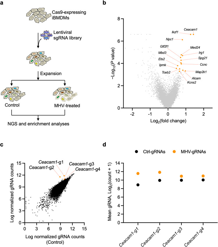
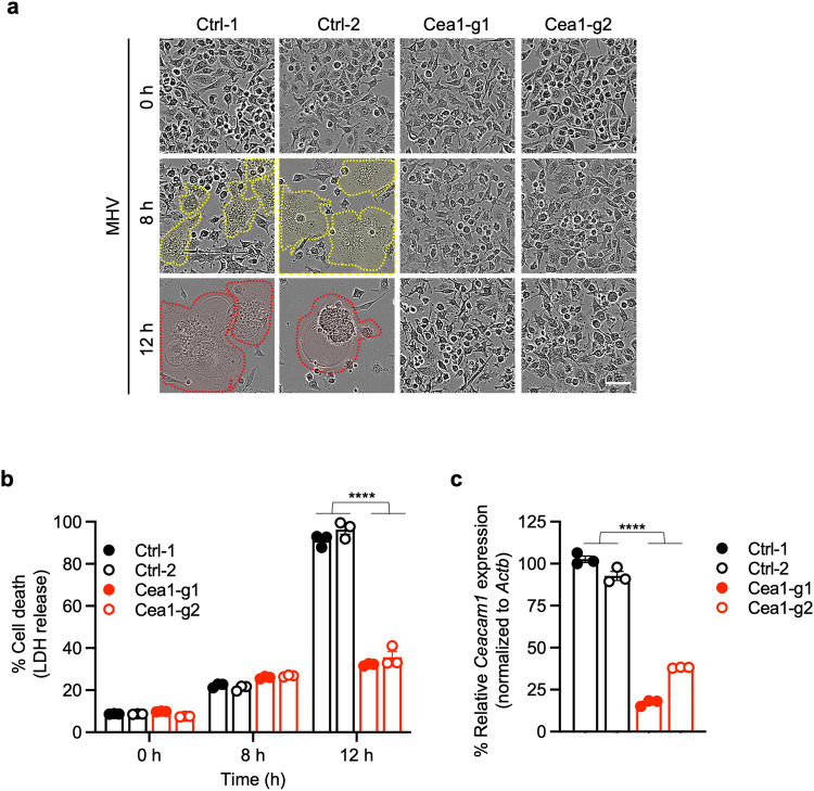
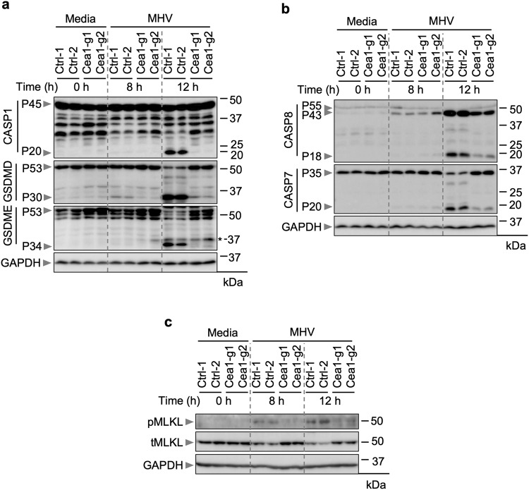
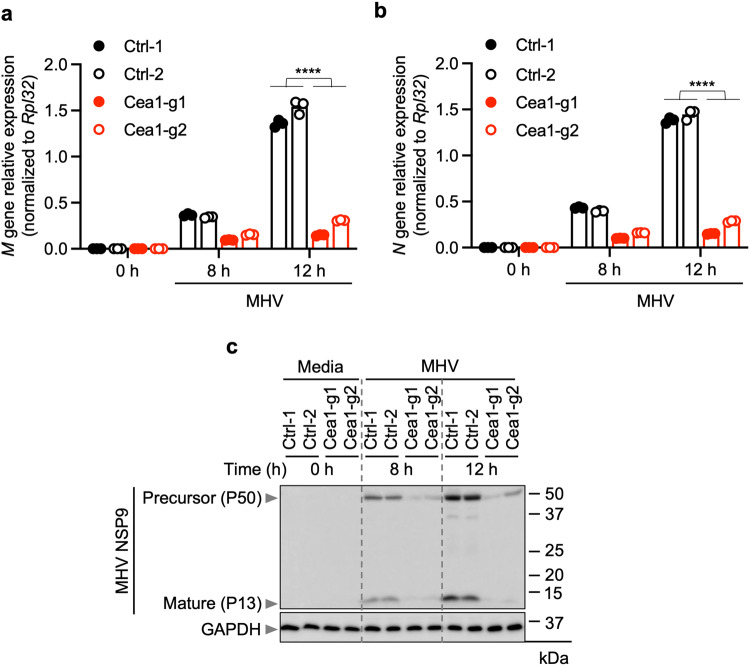
The COVID-19 pandemic, caused by the β-coronavirus (β-CoV) severe acute respiratory syndrome coronavirus 2 (SARS-CoV-2), continues to cause significant global morbidity and mortality. While vaccines have reduced the overall number of severe infections, there remains an incomplete understanding of viral entry and innate immune activation, which can drive pathology. Innate immune responses characterized by positive feedback between cell death and cytokine release can amplify the inflammatory cytokine storm during β-CoV-mediated infection to drive pathology. Therefore, there remains an unmet need to understand innate immune processes in response to β-CoV infections to identify therapeutic strategies. To address this gap, here we used an MHV model and developed a whole genome CRISPR-Cas9 screening approach to elucidate host molecules required for β-CoV infection and inflammatory cell death, PANoptosis, in macrophages, a sentinel innate immune cell. Our screen was validated through the identification of the known MHV receptor Ceacam1 as the top hit, and its deletion significantly reduced viral replication due to loss of viral entry, resulting in a downstream reduction in MHV-induced cell death. Moreover, this screen identified several other host factors required for MHV infection-induced macrophage cell death. Overall, these findings demonstrate the feasibility and power of using genome-wide PANoptosis screens in macrophage cell lines to accelerate the discovery of key host factors in innate immune processes and suggest new targets for therapeutic development to prevent β-CoV-induced pathology.


Conflict of interest statement

T.-D.K. was a consultant for Pfizer. All other authors declare no competing interests.

Figures

Fig. 1
Fig. 1. CRISPR screen identifies host factors required for β-CoV-induced cell death.
a Schematic of the CRISPR screen workflow in mouse hepatitis virus (MHV)-infected immortalized bone marrow-derived macrophages (iBMDMs). b Representative volcano plot showing the log2 mean fold change for the gRNAs in the CRISPR screen following infection of iBMDMs carrying gRNAs with MHV (MOI 0.1) for 24 h. The top 15 CRISPR screen hits are labeled. c Scatter plot highlighting the enrichment of all four gRNAs targeting Ceacam1 in the pool of iBMDMs carrying gRNAs from the whole genome CRISPR screen following infection with MHV (MOI 0.1) for 24 h. d Scatter plot depicting the distribution of the normalized gRNA counts in log scale for the individual Ceacam1 gRNAs, represented in panels (b) and (c). Ctrl-gRNAs denotes the count in the control pool of cells that were not infected; MHV-gRNAs denotes the count in the pool of cells following infection with MHV (MOI 0.1) for 24 h.
Fig. 2
Fig. 2. Ceacam1 is required for MHV-induced cell death.
a Cell death analysis in mouse hepatitis virus (MHV; MOI 0.1)-infected immortalized bone marrow-derived macrophages (iBMDMs) with and without Ceacam1 gRNA treatment with two different guides (Cea1-g1 and Cea1-g2). The yellow dotted lines denote syncytia, and the red dotted lines denote ballooning and dying syncytia. b Percentage of cell death at indicated time points after MHV infection in iBMDMs with and without Ceacam1 gRNA treatment in terms of LDH release. c Knockdown efficiency of the two different Caecam1 gRNAs, Cea1-g1 and Cea1-g2, employed in this study. Actb was used to normalize the Caecam1 expression. The data presented are representative of three independent experiments (ac). Data are shown as mean ± SEM (b, c). Analysis was performed using the Student’s t-test; ****P < 0.0001. Ctrl: Control with no gRNA. The scale bar is representative of 50 μm.
Fig. 3
Fig. 3. Ceacam1 modulates MHV-induced PANoptosis.
ac Immunoblot analysis of pro- (P45) and cleaved caspase-1 (P20; CASP1), pro- (P53) and activated (P30) gasdermin D (GSDMD), and pro- (P53) and activated (P34) gasdermin E (GSDME) (a); pro- (P55) and cleaved caspase-8 (P43, P18; CASP8) and pro- (P35) and cleaved caspase-7 (P20; CASP7) (b); and phospho- (pMLKL) and total MLKL (tMLKL) (c) from mouse hepatitis virus (MHV; MOI 0.1)-infected immortalized bone marrow-derived macrophages (iBMDMs) with and without Ceacam1 gRNA treatment with two different guides (Cea1-g1 and Cea1-g2) at the indicated time points. Asterisk denotes a non-specific band. Blots were reprobed for GAPDH to serve as the internal control. The data presented are representative of three independent experiments (ac). Ctrl: Control with no gRNA.
Fig. 4
Fig. 4. Loss of Ceacam1 reduces MHV replication.
a, b Quantification of mRNA levels of the mouse hepatitis virus (MHV) M (a) and N (b) structural proteins at the indicated time points after MHV infection (MOI 0.1) in immortalized bone marrow-derived macrophages (iBMDMs) with and without Ceacam1 gRNA treatment with two different guides (Cea1-g1 and Cea1-g2). c Immunoblot analysis of non-structural protein 9 (NSP9) precursor (P50 and P37) and mature forms (P13) in iBMDMs following the indicated treatment. Blots were reprobed for GAPDH to serve as the internal control. The data presented are representative of three independent experiments (ac). Data are shown as mean ± SEM (a, b). Analysis was performed using the Student’s t-test; ****P < 0.0001. Ctrl: Control with no gRNA.

Similar articles

Cited by

References

    1. Dong E, Du H, Gardner L. An interactive web-based dashboard to track COVID-19 in real time. Lancet Infect. Dis. 2020;20:533–534. doi: 10.1016/S1473-3099(20)30120-1. - DOI - PMC - PubMed
    1. Tan W, et al. A novel coronavirus genome identified in a cluster of pneumonia cases - Wuhan, China 2019–2020. China CDC Wkly. 2020;2:61–62. doi: 10.46234/ccdcw2020.017. - DOI - PMC - PubMed
    1. Hartenian E, et al. The molecular virology of coronaviruses. J. Biol. Chem. 2020;295:12910–12934. doi: 10.1074/jbc.REV120.013930. - DOI - PMC - PubMed
    1. Li F. Structure, function, and evolution of coronavirus spike proteins. Annu. Rev. Virol. 2016;3:237–261. doi: 10.1146/annurev-virology-110615-042301. - DOI - PMC - PubMed
    1. Cui J, Li F, Shi ZL. Origin and evolution of pathogenic coronaviruses. Nat. Rev. Microbiol. 2019;17:181–192. doi: 10.1038/s41579-018-0118-9. - DOI - PMC - PubMed

Publication types