

Using novel porous metal pillars for tibial bone defects in primary total knee arthroplasty
- PMID: 37864175
- PMCID: PMC10588064
- DOI: 10.1186/s12891-023-06962-1
Using novel porous metal pillars for tibial bone defects in primary total knee arthroplasty
Abstract
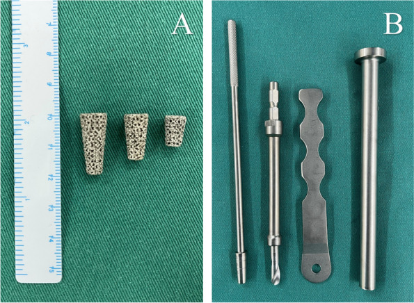
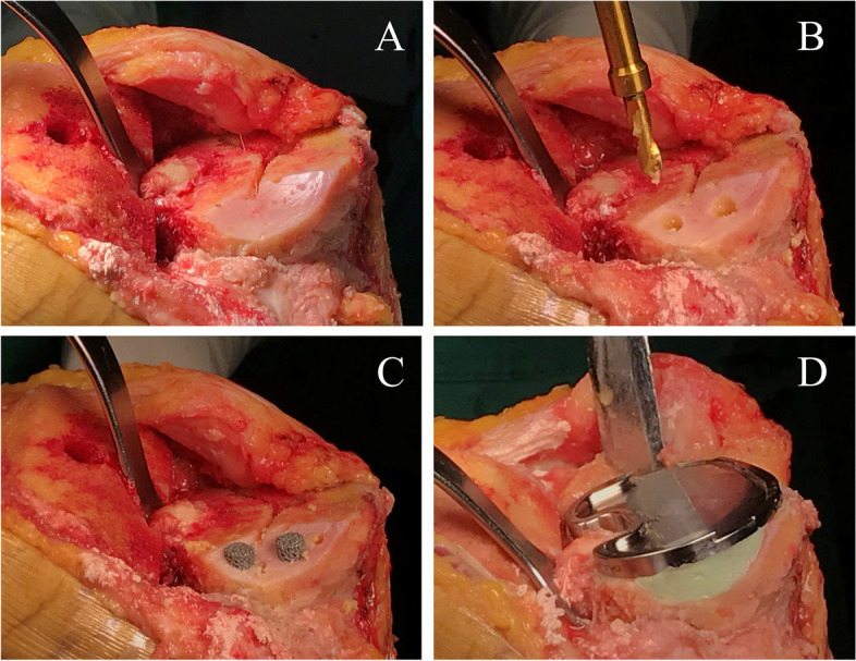
Background: The optimal method to treat tibial bone defects during primary total knee arthroplasty (TKA) is still unclear. A novel technique of porous metal pillar augmentation has been applied recently. This study aimed to assess the short-term outcomes of primary TKA with the use of novel porous metal pillars for tibial bone defects.
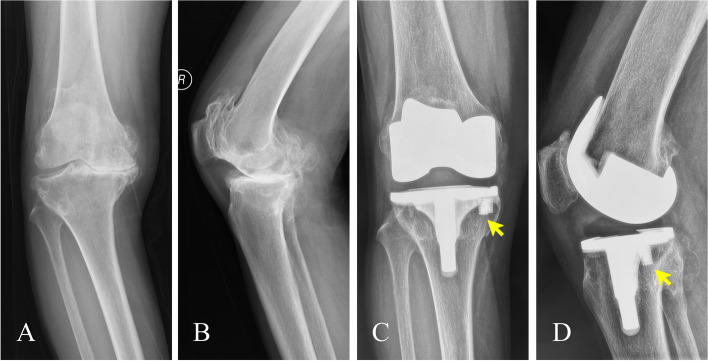
Methods: A total of 24 cases (22 patients) of primary TKA between January 2019 and December 2020 using porous metal pillars for tibial bone defects were reviewed. Clinical results were evaluated using the Knee Society knee score (KSKS) and function score (KSFS), the Western Ontario and McMaster Universities Osteoarthritis Index (WOMAC), and range of motion (ROM). Hip-knee-ankle angle (HKAA), femorotibial angle (FTA), and radiolucent lines were assessed radiologically.
Results: The median follow-up period was 36.0 months (interquartile range: 31-37 months). The KSKS, KSFS, WOMAC score, and ROM improved significantly at the final follow-up assessment compared with the preoperative evaluation. Both of the HKAA and FTA were corrected after surgery. Only one knee had a nonprogressive radiolucent line at the bone-cement interface. No radiolucent lines were detected around the pillar in any of the cases. There were no cases of prosthesis loosening and revision.
Conclusions: The use of novel porous metal pillars yielded satisfactory clinical outcomes and reliable radiological evidence of fixation in this study with a minimum 2-year follow-up. Porous metal pillar augmentation can be considered as a valuable and easy-to-use method for the management of tibial bone defects in primary TKA.
Keywords: Augmentation; Bone defect; Porous metal pillar; Total knee arthroplasty.
© 2023. BioMed Central Ltd., part of Springer Nature.
Conflict of interest statement
The authors declare no competing interests.
Figures
References
MeSH terms
Substances
Grants and funding
LinkOut - more resources
Full Text Sources
Medical
