. 2023 Oct 20;42(1):274.
doi: 10.1186/s13046-023-02834-7.

ERRα promotes glycolytic metabolism and targets the NLRP3/caspase-1/GSDMD pathway to regulate pyroptosis in endometrial cancer

Affiliations

ERRα promotes glycolytic metabolism and targets the NLRP3/caspase-1/GSDMD pathway to regulate pyroptosis in endometrial cancer

Pingping Su et al. J Exp Clin Cancer Res. .

Erratum in

Abstract

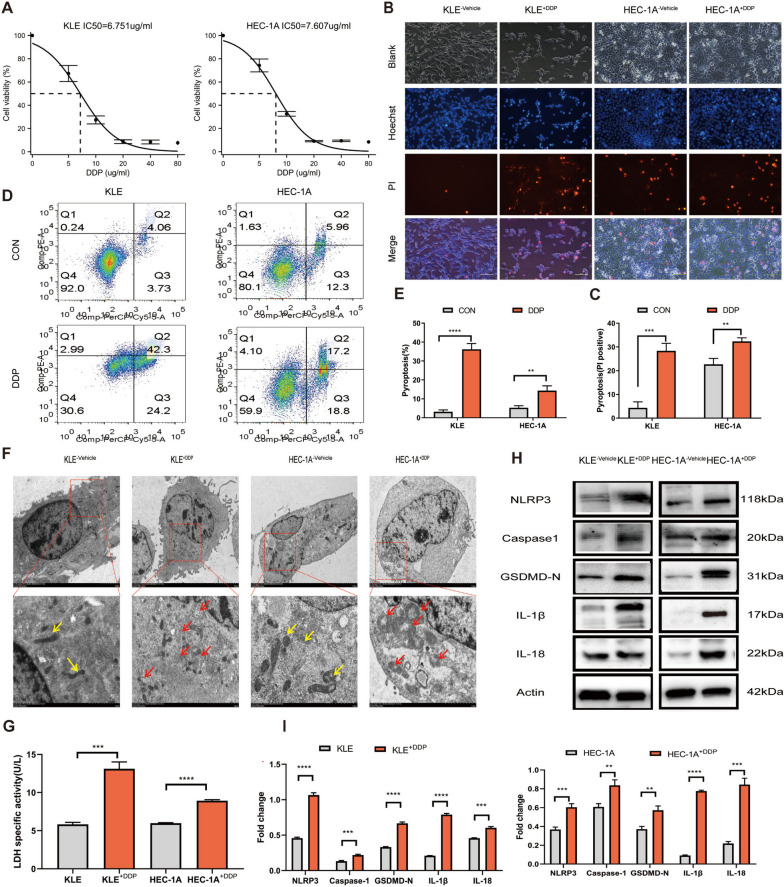
Background: Tumor cells can resist chemotherapy-induced pyroptosis through glycolytic reprogramming. Estrogen-related receptor alpha (ERRα) is a central regulator of cellular energy metabolism associated with poor cancer prognosis. Herein, we refine the oncogenic role of ERRα in the pyroptosis pathway and glycolytic metabolism.

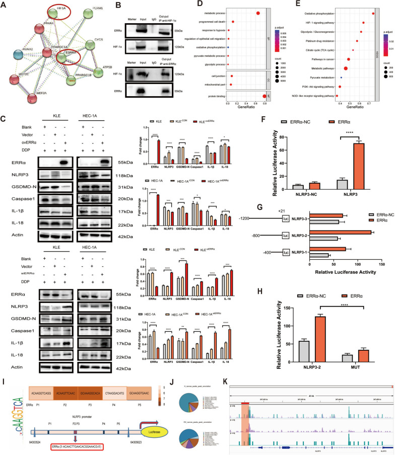
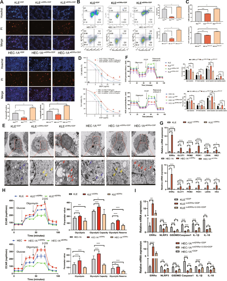
Methods: The interaction between ERRα and HIF-1α was verified using co-immunoprecipitation. The transcriptional binding sites of ERRα and NLRP3 were confirmed using dual-luciferase reporter assay and cleavage under targets and tagmentation (CUT&Tag). Flow cytometry, transmission electron microscopy, scanning electron microscopy, cell mito stress test, and extracellular acidification rate analysis were performed to investigate the effects of ERRα on the pyroptosis pathway and glycolytic metabolism. The results of these experiments were further confirmed in endometrial cancer (EC)-derived organoids and nude mice. In addition, the expression of ERRα-related pyroptosis genes was analyzed using The Cancer Genome Atlas and Gene Expression Omnibus database.

Results: Triggered by a hypoxic microenvironment, highly expressed ERRα could bind to the promoter of NLRP3 and inhibit caspase-1/GSDMD signaling, which reduced inflammasome activation and increased pyroptosis resistance, thereby resulting in the resistance of cancer cells to cisplatin. Moreover, ERRα activated glycolytic rate-limiting enzyme to bridge glycolytic metabolism and pyroptosis in EC. This phenomenon was further confirmed in EC-derived organoids and nude mice. CUT & Tag sequencing and The Cancer Genome Atlas database analysis showed that ERRα participated in glycolysis and programmed cell death, which resulted in EC progression.

Conclusions: ERRα inhibits pyroptosis in an NLRP3-dependent manner and induces glycolytic metabolism, resulting in cisplatin resistance in EC cells.

Keywords: Cisplatin resistance; ERRα; Endometrial cancer; Metabolic reprogramming; Pyroptosis.


Conflict of interest statement

The authors declare no potential conflicts of interest.

Figures

Fig. 1
Fig. 1
Pyroptosis is induced via the NLRP3/caspase-1/GSDMD pathway when KLE and HEC-1A cells are treated with DDP. A KLE and HEC-1A cells were treated with DDP at various concentration gradients (0, 5, 10, 20, 40, and 80 µg/mL) for 48 h; then, cell viability was assessed using the CCK8 assay. B, C The pyroptotic cells (PI-positive) were imaged using fluorescence microscopy after DDP treatment for 12 h (the values in each graph represent the average of three random fields per sample); scale bar: 100 µm. The KLE and HEC-1A cells from the control group were treated with the vehicle 0.9% NaCl. D, E The percentages of pyroptotic Annexin V-PE- and 7-AAD-positive cells within the second quadrant (Q2) were determined using flow cytometry after treating KLE and HEC-1A cells with DDP for 24 h. CON indicates the control group of KLE and HEC-1A cells treated with the vehicle 0.9% NaCl. F Representative TEM images of KLE and HEC-1A cells treated with DDP for 12 h. The mitochondrial membrane was disrupted, the matrix was dissolved, and the mitochondrial cristae were broken, as indicated with red arrows. The control group KLE and HEC-1A cells were treated with the vehicle 0.9% NaCl; the yellow arrows indicate the mitochondria structures that were more complete. Scale bar: 2 μm. G LDH activity in the culture supernatants of KLE and HEC-1A cells treated with DDP for 12 h was measured. H, I Whole-cell lysates of KLE and HEC-1A cells treated with DDP for 12 h or left untreated were analyzed using WB. The control group KLE−vehicle and HEC-1A−vehicle cells were treated with 0.9% NaCl. β-Actin was included as a loading control. The dose of DDP in the above-mentioned in vitro experiments was 7 µg. Data are representative of three independent experiments and shown as the mean ± SD. *p < 0.05; **p < 0. 01; ***p < 0.001; ****p < 0.0001. Abbreviations: CCK8, Cell Counting Kit-8; PI, propidium iodide; TEM, transmission electron microscopy; LDH, lactate dehydrogenase; WB, western blotting
Fig. 2
Fig. 2
ERRα inhibits caspase-1/GSDMD signaling via transcriptional binding with NLRP3. A Interaction between HIF-1α and ERRα was found in the STRING protein interaction database. B Interaction between HIF-1α and ERRα was verified using the co-immunoprecipitation (Co-IP) analysis of HEC-1A−ovERRα cell lysates. The corresponding antibodies (out-put IP: anti-HIF-1α-antibody and anti-ERRα-antibody) could precipitate both ERRα and HIF-1α, respectively, in the HEC-1A−ovERRα cell lysates. C Effects of ERRα regulation on the levels of pyroptosis-related proteins (NLRP3, GSDMD-N, caspase-1, IL-1β, and IL-18) in EC cells was analyzed using WB; β-actin was used as the loading control. Untreated KLE and HEC-1A cells and the empty vector-transfected KLE and HEC-1A cells treated with DDP were used as the control groups. D GO analysis showing the enriched biological process in EC cells overexpressing ERRα, HEC-1A cells were used as the control group. E KEGG pathway analysis for the genes that showed ERRα-binding peaks in their promoter regions. FH Dual-luciferase reporter assay to assess the interaction between ERRα and NLRP3. (F) 293 T cells were co-transfected with ERRα transcription factor, NLRP3 promoter labeled with luciferase reporter, and the Renilla luciferase control. The negative-control (NC) group was NLRP3-NC with ERRα-NC and NLRP3 with ERRα-NC. (G) Three NLRP3 promoter fragments ligated to the pGL3-basic plasmid, named NLRP3-1 (-400 to + 21), NLRP3-2 (-800 to + 21), and NLRP3-3 (-1200 to + 21), were co-transfected with ERRα transcription factor; the three NLRP3 promoter fragments with ERRα-NC were used as the control. (H) NLRP3-2-MUT and wild-type NLRP3-2 and their empty vectors were co-transfected with ERRα transcription factor. The vector-control group was NLRP3-2 with ERRα-NC and MUT with ERRα-NC. The luciferase activity was measured and normalized to that of the Renilla luciferase control 48 h post-transfection and the relative luciferase activity was determined. I Schematic diagram of putative ERRα-binding sites, as predicted by the online program JASPAR, located in the NLRP3 promoter region (P1–P5). The sequence TCAAGGTCA of ERRα is placed on the left. J CUT&Tag and sequencing analysis of the distribution map of ERRα peaks in gene functional regions. K NLRP3 promoter occupancy in HEC-1A cells was evaluated. ERRα was immunoprecipitated using an anti-Flag antibody. Data are representative of three independent experiments and shown as the mean ± SD. *p < 0.05; **p < 0. 01; ***p < 0.001; ****p < 0.0001. Abbreviations: ERRα, estrogen-related receptor alpha; HIF-1α, hypoxia-inducible factor-1 alpha. Co-IP, co-immunoprecipitation. CON; control empty vector. NC: negative control. MUT: mutation
Fig. 3
Fig. 3
Overexpression of estrogen-related receptor alpha (ERRα) enhances pyroptosis resistance accompanied by glycolytic metabolism, leading to cisplatin (DDP) resistance of EC cells. A Pyroptotic cells (PI-positive) in each well were imaged using fluorescence microscopy after DDP treatment for 12 h in the ovERRα and siERRα groups. KLE and HEC-1A cells treated with DDP for 12 h were included as the control group in the experiment. The values in each graph represent the average of three random fields per sample. Scale: 100 µm. B Percentage of AnnexinV-PE- and 7AAD-positive pyroptotic cells within the second quadrant (Q2) from different ERRα expression groups was analyzed using flow cytometry after 24 h of DDP treatment. KLE and HEC-1A cells treated with DDP for 24 h were used as the control group. C LDH activity of cell culture supernatants was measured in different ERRα expression groups after DDP treatment for 12 h. KLE and HEC-1A cells treated with DDP for 12 h were included as the control group in the experiment. D KLE and HEC-1A cells were treated with DDP at various concentration gradients for 48 h, and the IC50 of DDP in different ERRα expression groups was determined using the CCK8 assay. E Representative TEM images of KLE and HEC-1A cells in different ERRα expression groups treated with 7 µg DDP for 12 h. The mitochondrial membrane structure was indistinctly dissolved, the matrix was dissolved in a large area, and the crest was broken in siERRα cells, as indicated with the red arrows; the yellow arrows indicate ERRα-overexpressing EC cells whose mitochondrial membrane structure was relatively clear, the matrix was partially dissolved, and the cristae were slightly broken. The blue arrows indicate the mitochondria in the KLE and HEC-1A cells of control group treated with DDP for 12 h. Scale: 2 μm. F Basal, ATP-linked, and maximal respiration and spare respiratory capacities were assessed to evaluate the mitochondrial function in different ERRα expression groups. KLE+DDP and HEC-1A+DDP cells were used as the control groups. G Total RNA in KLE and HEC-1A cells was assessed using RT-qPCR to measure the expression of glycolysis-related genes in the ovERRα and siERRα groups. KLE and HEC-1A cells were used as the control group. H Extracellular acidification rate, glycolysis, glycolytic capacity, and glycolytic reserve in different ERRα expression groups are shown. KLE and HEC-1A cells were used as the control group. I Total RNA in KLE and HEC-1A cells was assessed using RT-qPCR to measure the expression of pyroptosis-related genes in EC cells. KLE−ovERRα+DDP and HEC-1A−ovERRα+DDP cells were used as the control groups. The results are presented as the average of three experimental replicates. Data are shown as the mean ± SD. Statistical tests: Student’s t-test. *p < 0.05; **p < 0.01; ***p < 0.001; ****p < 0.0001. Abbreviations: EC, endometrial cancer; IC50, inhibitory concentration; PI, propidium iodide; TEM, transmission electron microscopy; 2-DG, 2-deoxy-glucose
Fig. 4
Fig. 4
Estrogen-related receptor alpha (ERRα) interacts with HIF-1α and enhances pyroptosis resistance in an NLRP3-dependent manner. A Western blot (WB) analysis of pyroptosis-related proteins in KLE and HEC-1A cell lysates of the ovERRα group treated with CoCl2 (200 µM) for 12 h; the groups not treated with CoCl2 were used as controls. Each target protein was exposed on the same gel to ensure the same exposure conditions. B Representative SEM images of KLE and HEC-1A cells and the ovERRα group treated with CoCl2 (200 µM) for 12 h; the groups not treated with CoCl2 were used as controls. Scale: 5–30 μm. C Representative images of immunofluorescence staining of HIF-1α- and GSDMD-expressing cells per well, obtained using fluorescence microscopy after cisplatin (DDP) treatment of the ovERRα and control groups for 12 h. The groups not treated with CoCl2 were used as controls. The results represent the average of three random fields per sample. Scale: 10 µm. D-F Effect of MCC950 (10 µM) treatment on the siERRα group of KLE and HEC-1A cells measured using WB (12 h), flow-cytometry analyses (24 h), and the LDH release assay (12 h). The siERRα groups treated with DDP were included as controls. G Effect of nigericin (10 µM) treatment on the ovERRα group of KLE and HEC-1A cells measured using WB (12 h). KLE and HEC-1A cells treated with DDP were included as control groups. The dose of DDP in the above-mentioned in vitro experiments was 7 µg. The results represent the average of three experimental replicates. Data are shown as the mean ± SD. Statistical tests: Student’s t-test. *p < 0.05; **p < 0. 01; ***p < 0.001; ****p < 0.0001. Abbreviations: SEM, scanning electron microscopy
Fig. 5
Fig. 5
Knockdown of ERRα inhibits tumor growth and promotes DDP sensitivity in EC organoids and a xenograft animal model. A Cell morphology of organoids at different magnifications was recorded using an optical microscope. Scale: 50, 100, and 500 µm. B The histological structure and morphology of EC organoids were verified using H&E staining. C Fraction of organoids of different sizes after treatment with DDP at different concentrations is shown. The amount of organoid debris following treatment with DDP at different concentrations increased, and the shape and size of the debris were recorded under a microscope. D Inhibitory effect of DDP (20 µM) and XCT790 (10 µM) alone or in combination with EC organoids was recorded using an optical microscope. The group treated with DMSO was used as the control group. Scale: 100 µm. E Evaluation of cell viability following treatment with DDP (20 µM) and XCT790 (10 µM) alone or in combination in each experimental group using the CellTiter-Glo assay. Cells treated with DMSO were used as the control group. F Representative images of the IHC of ERRα, NLRP3, and caspase-1 in EC organoids are shown (the values in each graph represent the average of three random fields per sample). The groups treated with DMSO and DDP alone were used as the control groups. G After pretreatment with or without XCT790 (10 µM), the IC50 of DDP in EC organoids was determined using the CCK8 assay. H Representative images of tumors captured at the end of the study and tumor size in BALB/c nude mice of the four treatment groups. I Tumor growth curves of the four treatment groups are shown. The KLE+DMSO and KLE+DDP treatment groups were used as control groups (day 0 was defined as the day when different cell groups were implanted). J Representative images of the IHC of ERRα, NLRP3, and GSDMD in tumor sections from xenograft mice are shown (each point represents the average of three random fields per sample). The KLE+DMSO and KLE+DDP treatment groups were used as control groups. K-L WB analysis of ERRα, NLRP3, caspase-1, GSDMD-N, IL-18, and IL-1β in tumor specimens of xenograft mice. Tubulin was used as a loading control. The KLE+DMSO and KLE+DDP treatment groups were used as control groups. Data are shown as the mean ± SD. Statistical tests: Student’s t-test. *p < 0.05; **p < 0.01; ***p < 0.001; ****p < 0.0001. Abbreviations: H&E, hematoxylin and eosin
Fig. 6
Fig. 6
Estrogen-related receptor alpha (ERRα) is involved in cell pyroptosis and participates in the reprogramming of energy metabolism based on TCGA data analysis. A Differential expression map of ERRα. ERRα expression in GTEx (78 cases) combined with TCGA paracancerous (23 cases) and TCGA endometrial cancer (181 cases) samples. Wilcoxon rank sum test was used for statistical analysis. B ERRα expression in normal (35 cases) and endometrial cancer (EC) tissues (552 cases) from TCGA. Wilcoxon rank sum test was used for statistical analysis. C ERRα expression in EC cancer tissues (23 cases) and the corresponding normal tissues from TCGA database. Paired sample t-test was used for statistical analysis. D ROC curves shows that ERRα (known as ESRRA) has high diagnostic accuracy in EC. EF Associations between ERRα expression and residual tumor and histologic grade. G Using the Cox proportional hazard regression model, clinical stage, histologic grade, radiation therapy, primary treatment outcome, and residual tumor were included in the nomogram for predicting the probability of 1-, 3-, and 5-year OS in patients with EC. H Calibration plot of the nomogram for predicting the probability of OS in patients with EC at 1, 3, and 5 years. I GO analysis of TCGA data showed that ERRα-related genes in EC were enriched in glucose metabolism, pyruvate metabolism, programmed necrotic cell death, and glycolysis/gluconeogenesis. The subset of representative terms from this cluster was visualized with the p-value set to 0.05. J) Differential expression map of pyroptosis-related genes. The expression of pyroptosis-related genes in GTEx (78 cases) combined with TCGA paracancerous (23 cases) and TCGA endometrial cancer (181 cases) samples. Wilcoxon rank sum test was used for statistical analysis. K Pyroptosis-related gene expression in normal (35 cases) and EC tissues (552 cases) from TCGA. Wilcoxon rank sum test was used for statistical analysis. L Expression of pyroptosis-related genes in EC tissues (23 cases) and the corresponding normal tissues in TCGA database. Paired sample t-test was used for statistical analysis. M Analysis of GEO database data to show the correlation between the expression of ERRα and the pyroptosis pathway. N-P Survival analysis using ERRα,NLRP3, and GSDMD expression in EC. OS of patients with high versus low ERRα, NLRP3, and GSDMD expression levels. *p < 0.05; **p < 0.01; ***p < 0.001. Abbreviations: ERRα, estrogen-related receptor alpha; TCGA, The Cancer Genome Atlas; ROC, receiver operating characteristic; GO, Gene Ontology; OS, overall survival; ns, no statistical significance
Fig. 7
Fig. 7
Molecular mechanisms underlying the regulation of estrogen-related receptor alpha (ERRα) expression in pyroptosis. ERRα promotes the resistance of EC cells to pyroptosis by regulating NLRP3 inflammasomes. Platinum activates the inflammasome components, followed by the cleavage of CASP1 (Caspase 1) and production of inflammatory factors such as IL-1β and IL-18, and then triggers the cleavage of GSDMD and promotes cell membrane rupture by forming GSDMD-N. In addition, HIF-1α interacts with ERRα and enhances glycolytic metabolism in cancer cells to resist chemotherapy. Moreover, this effect might be triggered and enhanced by the hypoxic tumor microenvironment

References

    1. Miranda-Galvis M, Teng Y. Targeting hypoxia-driven metabolic reprogramming to constrain tumor progression and metastasis[J]. Int J Mol Sci. 2020;21(15):5487. - PMC - PubMed
    1. Semenza GL. HIF-1 mediates metabolic responses to intratumoral hypoxia and oncogenic mutations[J]. J Clin Investig. 2013;123(9):3664–71. - PMC - PubMed
    1. Vaupel P, Schmidberger H, Mayer A. The Warburg effect: essential part of metabolic reprogramming and central contributor to cancer progression[J]. Int J Radiat Biol. 2019;95(7):912–9. - PubMed
    1. Sun L, Suo C, Li S, et al. Metabolic reprogramming for cancer cells and their microenvironment: beyond the Warburg effect[J]. Biochim Biophys Acta Rev Cancer. 2018;1870(1):51–66. - PubMed
    1. Desbats MA, Giacomini I, Prayer-Galetti T, et al. Metabolic plasticity in chemotherapy resistance[J]. Front Oncol. 2020;10:281. - PMC - PubMed

MeSH terms

LinkOut - more resources