. 2024 Apr;30(4):e14493.
doi: 10.1111/cns.14493. Epub 2023 Oct 21.

Truncated Dyrk1A aggravates neuronal apoptosis by inhibiting ASF-mediated Bcl-x exon 2b inclusion

Affiliations

Truncated Dyrk1A aggravates neuronal apoptosis by inhibiting ASF-mediated Bcl-x exon 2b inclusion

Shuqiang Zhang et al. CNS Neurosci Ther. 2024 Apr.

Abstract

Aim: Aggravated neuronal loss, caused mainly by neuronal apoptosis, is observed in the brain of patients with Alzheimer's disease (AD) and animal models of AD. A truncated form of Dual-specific and tyrosine phosphorylation-regulated protein kinase 1A (Dyrk1A) plays a vital role in AD pathogenesis. Downregulation of anti-apoptotic Bcl-xL is tightly correlated with neuronal loss in AD. However, the molecular regulation of neuronal apoptosis and Bcl-x expression by Dyrk1A in AD remains largely elusive. Here, we aimed to explore the role and molecular mechanism of Dyrk1A in apoptosis.

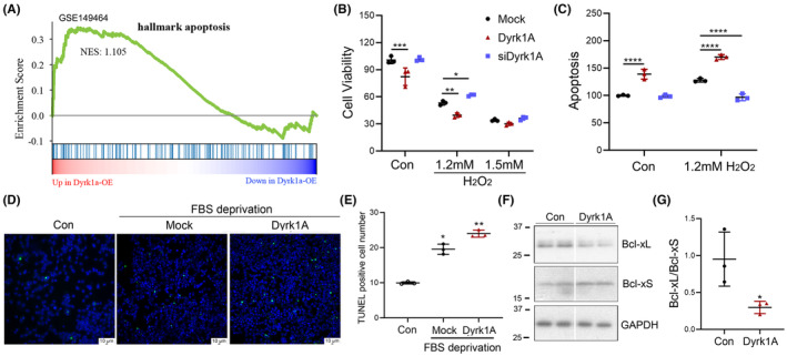
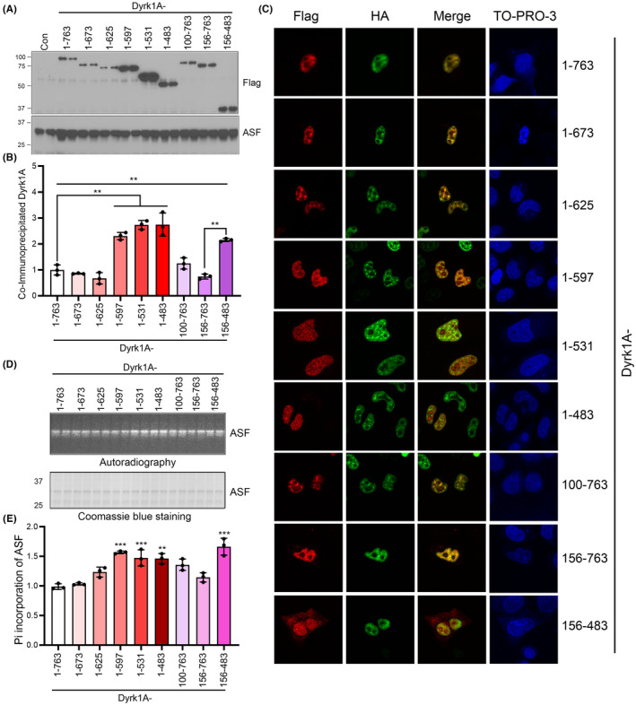
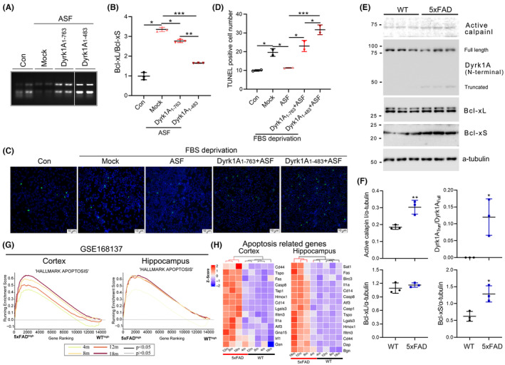
Methods: Cell Counting Kit-8 (CCK8), flow cytometry, and TdT-mediated dUTP Nick-End Labeling (TUNEL) were used to check apoptosis. The cells, transfected with Dyrk1A or/and ASF with Bcl-x minigene, were used to assay Bcl-x expression by RT-PCR and Western blots. Co-immunoprecipitation, autoradiography, and immunofluorescence were conducted to check the interaction of ASF and Dyrk1A. Gene set enrichment analysis (GSEA) of apoptosis-related genes was performed in mice overexpressing Dyrk1A (TgDyrk1A) and AD model 5xFAD mice.

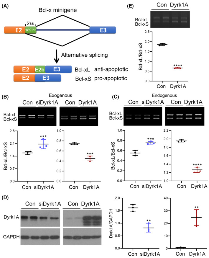
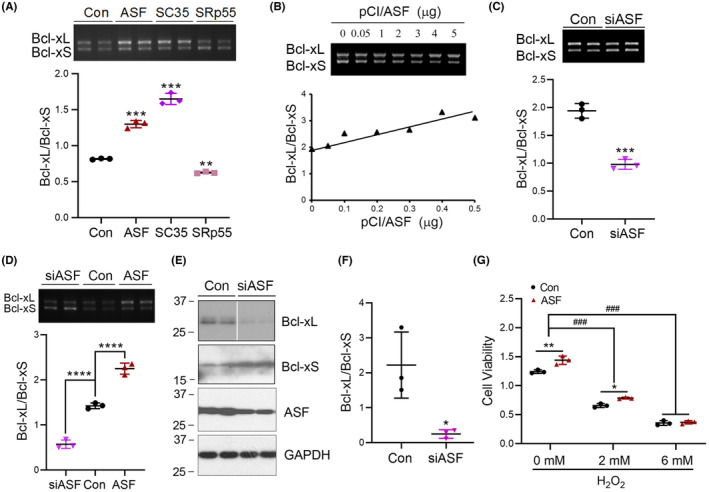
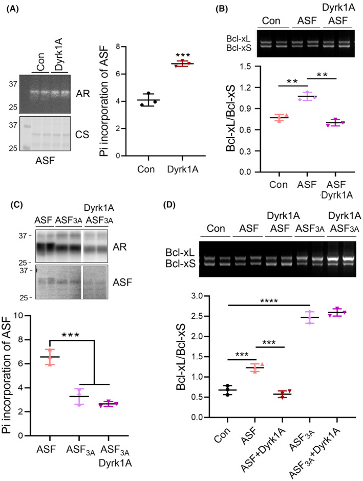
Results: Dyrk1A promoted Bcl-xS expression and apoptosis. Splicing factor ASF promoted Bcl-x exon 2b inclusion, leading to increased Bcl-xL expression. Dyrk1A suppressed ASF-mediated Bcl-x exon 2b inclusion via phosphorylation. The C-terminus deletion of Dyrk1A facilitated its binding and kinase activity to ASF. Moreover, Dyrk1a1-483 further suppressed the ASF-mediated Bcl-x exon 2b inclusion and aggravated apoptosis. The truncated Dyrk1A, increased Bcl-xS, and enrichment of apoptosis-related genes was observed in the brain of 5xFAD mice.

Conclusions: We speculate that increased Dyrk1A and truncated Dyrk1A may aggravate neuronal apoptosis by decreasing the ratio of Bcl-xL/Bcl-xS via phosphorylating ASF in AD.

Keywords: ASF; Bcl‐x; Dyrk1A; alternative splicing; apoptosis.


Conflict of interest statement

The authors declare that they have no competing interests.

Figures

FIGURE 1
FIGURE 1
Dyrk1A promotes cell apoptosis and increases expression of pro‐apoptotic protein Bcl‐xS. (A) GESA analysis of the apoptosis gene enrichment of hippocampal RNA sequencing data from Dyrk1A overexpressed (TgDyrk1A) mice at 16 weeks. (B,C) Dyrk1A was overexpressed or silenced in U87 cells. The cells were treated with H2O2 at the indicated concentration for 2 h. Cell viability was measured by CCK8 (B) and flow cytometry (C). (D,E) HEK‐293 T cells were transfected with Dyrk1A and were deprived of FBS for 24 h. The apoptosis was determined by TUNEL staining (D). The TUNEL positive cells were counted in a total of 1000 cells (E). (F,G) Dyrk1A was overexpressed in HEK‐293A cells for 48 h. The cells were harvested and analyzed by western blot to detect Bcl‐xL, Bcl‐xS, and GAPDH (F). The Bcl‐xL/Bcl‐xS ratio was calculated after densitometry (G). Data are presented as mean ± SD; n = 3; *, p < 0.05; **, p < 0.01.
FIGURE 2
FIGURE 2
Dyrk1A promotes Bcl‐x exon 2b exclusion leading to enhanced Bcl‐xS expression. (A) A schematic depicting the alternative splicing of Bcl‐x exon 2b. (B–D) siRNA of Dyrk1A or Dyrk1A were co‐transfected with Bcl‐x mini‐gene into HEK‐293A cells. The splicing products of exogenous (B) and endogenous (C) of Bcl‐x exon 2b were analyzed by RT‐PCR using two sets of primers corresponding to endogenous and exogenous Bcl‐x. The Bcl‐xL/Bcl‐xS ratio was calculated following densitometry. The level of Dyrk1A was analyzed by immunoblotting (D). (E) Dyrk1A was co‐transfected with Bcl‐x mini‐gene into U87 cells. Data are presented as mean ± SD; n = 3; *, p < 0.05; **, p < 0.01.
FIGURE 3
FIGURE 3
ASF promotes Bcl‐xL expression and increases cell viability. (A) The splicing factors ASF, SC35, and SRp55 were co‐transfected with Bcl‐x mini‐gene into HEK‐293A cells. The splicing products of Bcl‐x exon 2b were analyzed by RT‐PCR. (B,C) ASF was overexpressed or knocked down with siRNA in Bcl‐x mini‐gene transfected HEK‐293A cells. The alternative splicing products of Bcl‐x exon 2b was analyzed by RT‐PCR. The Bcl‐xL/Bcl‐xS ratio was calculated and plotted against the concentration of pCI/ASF (B). (D) U87 cells were co‐transfected with siASF or ASF and Bcl‐x mini‐gene. The splicing products were analyzed by RT‐PCR. The ratio of Bcl‐xL to Bcl‐xS was calculated. (E,F) The expression of ASF was knocked down by siASF. Western blotting was performed to quantify the expression of Bcl‐xL, Bcl‐xS, ASF, and GAPDH. The ratio of Bcl‐xL/Bcl‐xS was calculated (F). (G) The cells were transfected with ASF and treated with different concentration H2O2 for 2 h. Cell viability was analyzed by CCK8. Data are presented as mean ± SD, n = 3; *, p < 0.05; **, p < 0.01, ***, p < 0.001, ****, p < 0.0001.
FIGURE 4
FIGURE 4
Suppression of ASF‐mediated Bcl‐x exon 2b inclusion by Dyrk1A requires Ser227, Ser234, and Ser238 phosphorylation. (A) HEK‐293FT cells were transfected with Dyrk1A for 45 h and labeled with 32Pi for 3 h. The cells were lysed with RIPA buffer. The ASF was immunoprecipitated and separated by SDS‐PAGE. Incorporation of 32Pi into ASF was analyzed by autoradiography and normalized by total ASF showed by Coomassie Blue Staining. (B) Dyrk1A and ASF were co‐expressed with Bcl‐x mini‐gene in U87 cells. The splicing products of Bcl‐x exon 2b were analyzed by RT‐PCR. (C) Dyrk1A and mutated ASF, ASF3A, were co‐transfected to HEK‐293 T cells for 45 h, 32Pi was added to the culture medium for another 3 h. The ASF was immunoprecipitated and separated by SDS‐PAGE. 32Pi incorporation was analyzed by autoradiography and normalized with total ASF showed by Coomassie Blue Staining. (D) Dyrk1A and ASF or ASF3A were co‐transfected with Bcl‐x mini‐gene in HEK‐293A cells. The splicing products of Bcl‐x exon 2b were analyzed by RT‐PCR. The Bcl‐xL/Bcl‐xS ratio was calculated. CS, Coomassie Blue Staining; AR, autoradiography; ASF3A, Ser227, Ser234, and Ser238 of ASF mutated to Ala. *, compared with Con; #, compared with ASF. Data are presented as mean ± SD, n = 3; *, p < 0.05; **, p < 0.01.
FIGURE 5
FIGURE 5
Truncated Dyrk1As without the C‐terminus enhanced its interaction with ASF and the phosphorylation level of ASF. (A,B) Dyrk1A and its series deletions were transfected to HEK‐293FT cells for 48 h. The cells were lysed and incubated with ASF antibody to immunoprecipitate endogenous ASF. The co‐immunoprecipitated Dyrk1A was detected by western blotting using anti‐FLAG antibody (A). The co‐immunoprecipitated Dyrk1A was normalized with total Dyrk1A (showed in Figure S1B) (B). (C) Dyrk1A or Dyrk1A truncation mutants and ASF were co‐transfected into Hela cells. Polyclonal anti‐FLAG and monoclonal anti‐HA were used to immunostain Dyrk1A and ASF respectively. The fluorescent‐labeled anti‐mouse (green) and anti‐rabbit (red) antibodies were used to check the overexpressed protein. TO‐PRO‐3 was used for nuclear staining. (D,E) The HEK‐293FT cells were transfected with Dyrk1A and its deletion mutants and treated with 32Pi medium for 3 h. The cells were lysed with RIPA buffer. The monoclonal ASF antibody was used to immunoprecipitate endogenous ASF. The phosphorylation levels of ASF were checked by autoradiography (D) and normalized with total ASF (showed by Coomassie Blue Staining) (E). CS, Coomassie Blue Staining; AR, autoradiography; Data are presented as mean ± SD, n = 3; *, p < 0.05; **, p < 0.01.
FIGURE 6
FIGURE 6
Truncation of Dyrk1A may cause decreased Bcl‐xL and increased apoptosis gene enrichment in 5xFAD mice. (A,B) Dyrk1A1–763 or Dyrk1A1–483 and ASF were co‐transfected to HEK‐293 T cells. The total RNA was extracted to check the expression of Bcl‐xL and Bcl‐xS (A). The Bcl‐xL/Bcl‐xS ratio was calculated (B). (C,D) HEK‐293 T cells were co‐transfected with ASF, Dyrk1A1–763, or Dyrk1A1–483 for 24 h. The cells were deprived of FBS to induce apoptosis and were then subjected TUNEL staining (C). The TUNEL positive cells were counted in every 1000 cells (D). (E,F) The protein level of activated calpain I, full length and truncated Dyrk1A, Bcl‐xL, and Bcl‐xS in 5xFAD cortex at 10 m were checked by western blotting using calpain I, Dyrk1A (N‐terminal), Bcl‐xL, and Bcl‐xS antibodies (E). The densities of activated calpain I, Bcl‐xL, and Bcl‐xS bands were normalized to a‐tubulin, and the truncated Dyrk1A was normalized to the full length Dyrk1A (F). (G) Apoptosis gene enrichment was analyzed by GESA from hippocampal (right) and cortex (left) RNA sequencing data of 5xFAD mice at 4 m, 8 m, 12 m, and 18 m (GSE168137). (H) Apoptosis‐related 14 or 15 gene expression patterns were increased in 5xFAD mice, as represented by Z scores in heatmap. Thin curve, p > 0.05, thick curve, p < 0.05. Data are presented as mean ± SD, for cell experiment n = 3, for animal experiment n = 2; *, p < 0.05; **, p < 0.01; ***, p < 0.001.

Similar articles

Cited by

References

    1. Bennett DA, Schneider JA, Wilson RS, Bienias JL, Arnold SE. Neurofibrillary tangles mediate the association of amyloid load with clinical Alzheimer disease and level of cognitive function. Arch Neurol. 2004;61(3):378‐384. - PubMed
    1. Braak H, Braak E, Grundke‐Iqbal I, Iqbal K. Occurrence of neuropil threads in the senile human brain and in Alzheimer's disease: a third location of paired helical filaments outside of neurofibrillary tangles and neuritic plaques. Neurosci Lett. 1986;65(3):351‐355. - PubMed
    1. Shimohama S. Apoptosis in Alzheimer's disease‐‐an update. Apoptosis. 2000;5(1):9‐16. - PubMed
    1. Gomez‐Isla T, Price JL, McKeel DW Jr, Morris JC, Growdon JH, Hyman BT. Profound loss of layer II entorhinal cortex neurons occurs in very mild Alzheimer's disease. J Neurosci. 1996;16(14):4491‐4500. - PMC - PubMed
    1. Martinez‐Pinilla E, Ordóñez C, Del Valle E, Navarro A, Tolivia J. Regional and gender study of neuronal density in brain during aging and in Alzheimer's disease. Front Aging Neurosci. 2016;8:213. - PMC - PubMed

Publication types

MeSH terms