. 2023 Nov:97:104834.
doi: 10.1016/j.ebiom.2023.104834. Epub 2023 Oct 20.

The microbial metabolite desaminotyrosine enhances T-cell priming and cancer immunotherapy with immune checkpoint inhibitors

Affiliations

The microbial metabolite desaminotyrosine enhances T-cell priming and cancer immunotherapy with immune checkpoint inhibitors

Laura Joachim et al. EBioMedicine. 2023 Nov.

Abstract

Background: Inter-individual differences in response to immune checkpoint inhibitors (ICI) remain a major challenge in cancer treatment. The composition of the gut microbiome has been associated with differential ICI outcome, but the underlying molecular mechanisms remain unclear, and therapeutic modulation challenging.

Methods: We established an in vivo model to treat C57Bl/6j mice with the type-I interferon (IFN-I)-modulating, bacterial-derived metabolite desaminotyrosine (DAT) to improve ICI therapy. Broad spectrum antibiotics were used to mimic gut microbial dysbiosis and associated ICI resistance. We utilized genetic mouse models to address the role of host IFN-I in DAT-modulated antitumour immunity. Changes in gut microbiota were assessed using 16S-rRNA sequencing analyses.

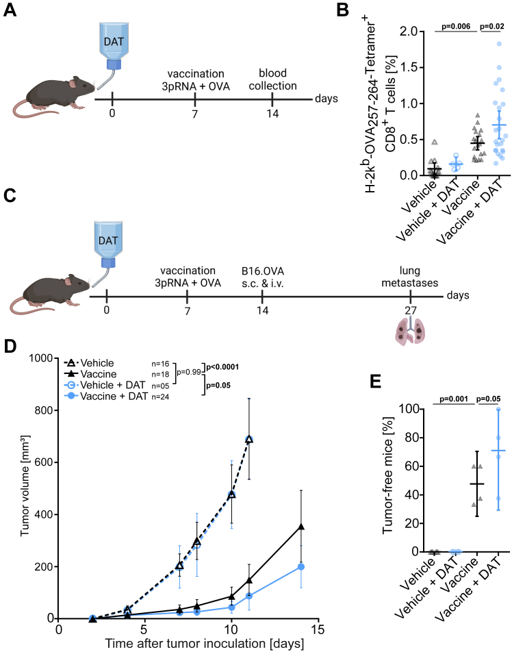
Findings: We found that oral supplementation of mice with the microbial metabolite DAT delays tumour growth and promotes ICI immunotherapy with anti-CTLA-4 or anti-PD-1. DAT-enhanced antitumour immunity was associated with more activated T cells and natural killer cells in the tumour microenvironment and was dependent on host IFN-I signalling. Consistent with this, DAT potently enhanced expansion of antigen-specific T cells following vaccination with an IFN-I-inducing adjuvant. DAT supplementation in mice compensated for the negative effects of broad-spectrum antibiotic-induced dysbiosis on anti-CTLA-4-mediated antitumour immunity. Oral administration of DAT altered the gut microbial composition in mice with increased abundance of bacterial taxa that are associated with beneficial response to ICI immunotherapy.

Interpretation: We introduce the therapeutic use of an IFN-I-modulating bacterial-derived metabolite to overcome resistance to ICI. This approach is a promising strategy particularly for patients with a history of broad-spectrum antibiotic use and associated loss of gut microbial diversity.

Funding: Melanoma Research Alliance, Deutsche Forschungsgemeinschaft, German Cancer Aid, Wilhelm Sander Foundation, Novartis Foundation.

Keywords: Antibiotics; Desaminotyrosine; Gut microbiome; Immune checkpoint inhibitors; Melanoma; Microbial metabolites.


Conflict of interest statement

Declaration of interests S.H. is a consultant for Bristol Myers-Squibb, Novartis, Merck, Abbvie, and Roche. S.H. has received research funding from Bristol Myers-Squibb and Novartis. H.P. is a consultant for Gilead, Abbvie, Pfizer, Novartis, Servier, and Bristol Myers-Squibb. K.S. is consultant for TRIMT GmbH and has filed a patent for radiopharmaceutical target. H.P. and S.H. have received research funding from Bristol Myers-Squibb. E.T.O. has received honoraria from BeiGene. C.P. received research funding from Almirall and honoraria from MSD, BMS, Pierre-Fabre, Sanofi, Novartis, Almirall, Pelpharma, Pritzer, Merck, Leo Pharma, Sun Pharma, Janssen, Abbvie, Amazentis, and Scarletred. S.H. is an employee of and holds equity interest in Roche/Genentech. The remaining authors declare no financial conflict of interest.

Figures

Fig. 1
Fig. 1
Treatment with bacterial-derived metabolite DAT delays tumour growth and enhances anti-CTLA-4 immunotherapy via increased T cell activation. (A) Experimental setup: Mice were provided with DAT via the drinking water ad libitum beginning on the day of tumour cell inoculation (day 0) until the end of the experiment. Anti-CTLA-4 or isotype control antibodies were administered i.p. on days 7 (200 μg), 10 (100 μg), and 13 (100 μg). Tumours were extracted on day 15. (B) Percentage of mice without a palpable tumour on day 7 before treatment onset from n = 6 independent experiments [p-values correspond to an unpaired t-test with Welch's correction]. (C) Mean tumour growth from n = 3–6 independent experiments [p-values correspond to a two-way ANOVA with Tukey's multiple comparison test on day 14]. (D) Overall survival of n = 3 independent experiments [p-values correspond to a Log-rank (Mantel–Cox) test]. In n = 4 independent experiments, tumours were extracted for analysis of immune cells in the tumour microenvironment (TME) via flow cytometry (E) Representative dot plots and (F) scatter plots of the frequency of IFNγ+ CD4+ cells, (G) mean fluorescence intensity (MFI) of IFNγ expression in CD4+ cells normalized to ‘Ctrl + isotype’ group, (H) frequency of IFNγ+ CD8+ cells, (I) MFI of IFNγ expression in CD8+ cells normalized to ‘control + isotype’ group, and (J) frequency of IFNγ+ natural killer (NK) cells in the TME [p-values correspond to the respective one-way ANOVA with Tukey's multiple comparison test]. All graphs show mean with 95% confidence interval (CI). For an analysis of area under the curve (AUC) of tumour volume growth see Fig. S8.
Fig. 2
Fig. 2
DAT enhances T cell and DC activation in vitro. (AC) Splenic T cells were stimulated with CD3/CD28-coated beads and IL-2 for 24 h, and were then exposed to different concentrations of DAT. T-cell activation and differentiation were analysed via flow cytometry from n = 4 independent experiments. Frequency of (A) IFNγ+ CD8+ cells, (B) CD25+ CD8+ cells, and (C) T-bet+ CD4+ cells. (DE) Bone marrow-derived DCs were incubated with different concentrations of DAT, and in some cases additionally with LPS. After 24 h, surface expression of (D) CD80 or (E) CD86 on DCs from n = 3 independent experiments was analysed via flow cytometry. Data show CD80 or CD86 MFI. [p-values correspond to the respective one-way ANOVA with Tukey's multiple comparison test]. All graphs show mean with 95% CI.
Fig. 3
Fig. 3
The additive effect of DAT and ICI immunotherapy is time and dose dependent. (A) Scheme of experimental setup of B-C: Mice were inoculated s.c. with B16.OVA cells on day 0, and anti-CTLA-4 or isotype control antibodies were administered i.p. on days 7 (200 μg), 10 (100 μg), and 13 (100 μg) as described for Fig. 1, and DAT was continuously provided via the drinking water beginning on day 5. (B) Tumour volume on day 7 before onset of treatment with anti-CTLA-4 from n = 3 independent [the reported p-values correspond to a one-way ANOVA with Tukey's multiple comparison test]. (C) Continuous tumour growth from n = 2–3 independent experiments over time [p-values correspond to a two-way ANOVA with Tukey's multiple comparison test on day 14]. (D) Scheme of experimental setup of E–G: Mice were inoculated s.c. with B16.OVA cells on day 0, and anti-CTLA-4 or isotype control antibodies were administered i.p. on days 7, 10 and 13 as described for Fig. 1, and DAT was provided by daily oral gavage beginning on the day of tumour cell inoculation until the end of the experiment. (E) Tumour volume on day 7 before onset of treatment with anti-CTLA-4 [p-values correspond to an unpaired t-test]. (F) Continuous tumour growth over time from n = 3 independent experiments [p-values correspond to a two-way ANOVA with Tukey's multiple comparison test on day 12]. (G) Overall survival of tumour-bearing animals [p-values correspond to a Log-rank (Mantel–Cox) test]. All graphs show mean with 95% CI. For an integrated analysis of AUC of tumour volume growth see Fig. S8.
Fig. 4
Fig. 4
DAT enhances antigen-specific priming and expansion of cytotoxic T cells. Mice were provided with DAT via the drinking water from day 0 until the end of the experiment. On day 7 mice were injected s.c. with a vaccine/adjuvant mix of 50 μg OVA protein and 10 μg 3pRNA into the medial part of the upper tight. (A) Scheme for experimental setup I: For some mice, blood samples were taken on day 14, and (B) the frequency of OVA-specific CD8+ T cells in the peripheral blood was analysed from n = 5 independent experiments [p-values correspond to a one-way ANOVA with Bonferroni's multiple comparison test]. (C) Scheme for experimental setup II: Some mice were challenged with B16.OVA cells injected s.c. and i.v. on day 14. (D) Continuous growth of s.c. tumours from n = 4 independent experiments [p-values correspond to a two-way ANOVA with Tukey's multiple comparison test on day 11 and an additional unpaired t-test with Welch's correction for ‘vaccine’ and ‘vaccine + DAT’ group on day 14]. (E) The formation of lung pseudo-metastases on day 27 was analysed. Lungs were extracted on day 27, pseudo-metastases per lung were counted and the percentage of tumour-free mice per experiment (defined as animal with less than 5 metastases per lung) were determined [p-values correspond to a one-way ANOVA with Bonferroni's multiple comparison test]. All graphs show mean with 95% CI. For an integrated analysis of AUC of tumour volume growth see Fig. S8.
Fig. 5
Fig. 5
Host IFN-I signalling is required for the additive effect of DAT and ICI. Mice with genetic deficiency for the interferon-alpha receptor 1 were (Ifnar1−/−) were provided with DAT via the drinking water, inoculated with B16 melanomas and were treated with anti-CTLA-4 or isotype control antibodies as described for Fig. 1. (A) Mean tumour volume on day 7 before onset of ICI treatment from n = 6 independent experiments [the reported p-values correspond to an unpaired t-test.] (B) Percentage of mice without a palpable tumour on day 7 before treatment onset of ICI treatment from 6 independent experiments [the reported p-values correspond to an unpaired t-test]. (C) Mean tumour growth per experimental group from n = 6 independent experiments [p-values correspond to a two-way ANOVA with Tukey's multiple comparison test on day 13]. (D) Overall survival from n = 3 independents [p-values correspond to a Log-rank (Mantel–Cox) test]. In n = 3 independent experiments, tumours were extracted for analysis of immune cells in the TME via flow cytometry on day 15: (E) IFNγ+ CD4+ cells, (F) IFNγ+ CD8+ cells, (G) OVA antigen-specific T cells, and (H) IFNγ+ NK cells in the TME [p-values correspond to the respective one-way ANOVA with Tukey's multiple comparison test]. All graphs show mean with 95% CI. For an integrated analysis of AUC of tumour volume growth see Fig. S8.
Fig. 6
Fig. 6
DAT compensates for the adverse effects that result from antibiotic treatment during anti-CTLA-4 immunotherapy. Mice were treated with a mixture of broad-spectrum antibiotics (ampicillin, neomycin, vancomycin, metronidazole) and amphotericin B via the drinking water starting on day −4 before s.c. B16.OVA cell inoculation on day 0. The Abx-containing drinking water was provided until day 21. Some mice were additionally supplemented with DAT via the drinking water starting on day 0. Anti-CTLA-4 or isotype control antibodies were administered i.p. on days 7 (200 μg), 10 (100 μg), and 13 (100 μg). (A) Mean tumour growth per experimental group from at least n = 3 independent experiments [p-values correspond to a two-way ANOVA with Tukey's multiple comparison test on day 12 for comparisons of ‘Ctrl + isotype’ and other groups and a two-way ANOVA with Tukey's multiple comparison test on day 14 excluding ‘Ctrl + isotype’ for all other comparisons; the statistical analysis was performed on the whole data set from Fig. S6C]. (B) Overall survival in Abx-treated tumour-bearing mice from at least n = 3 independent experiments [p-values correspond to a Log-rank (Mantel–Cox) test; the statistical analysis was performed on the whole data set from Fig. S6D]. (C) Expansion of OVA antigen-specific T cells in the peripheral blood on day 15 with Ctrl + Abx + isotype n = 25, Ctrl + Abx + anti-CTLA-4 n = 15, DAT + Abx + isotype n = 12, DAT + Abx + anti-CTLA-4 n = 17, Ctrl + isotype n = 15, Ctrl + anti-CTLA-4 n = 17, DAT + isotype n = 16, and DAT + anti-CTLA-4 n = 17 [p-values correspond to a Kruskal–Wallis test with Dunn's multiple comparison test]. Graph (A) shows mean with 95% CI and graph (C) shows median with its corresponding CI at a requested confidence level of 95%. For an integrated analysis of AUC of tumour volume growth see Fig. S8.
Fig. 7
Fig. 7
Oral DAT supplementation alters the microbial composition of the gut in mice. Mice were provided with DAT via the drinking water ad libitum for 7 days. Stool samples of DAT treated (n = 10) or untreated (n = 10) mice were collected on day 7 and were analysed via NGS for 16S-rRNA. (A) Effective richness [p-values correspond to an unpaired t-test] and (B) Shannon effective representing alpha-diversity [p-values correspond to an unpaired t-test]. (C) Non-metric multidimensional scaling (NMDS) biplot showing distance between DAT and Ctrl groups. Taxonomic binning on (D) class and (E) order level [p-values correspond to an unpaired t-test]. (F) Taxonomic binning on order level of mice on day 0 which were retrospectively categorized into responder and non-responder to anti-CTLA-4 therapy [p-values correspond to an unpaired t-test with Welch's correction]. (G) Taxonomic binning on family level on day 7 after DAT treatment [p-values correspond to an unpaired t-test with Welch's correction]. All graphs show mean with 95% CI.

References

    1. Callahan M.K., Wolchok J.D. At the Bedside: CTLA-4- and PD-1-blocking antibodies in cancer immunotherapy. J Leukoc Biol. 2013;94:41–53. doi: 10.1189/jlb.1212631. - DOI - PMC - PubMed
    1. Hodi F.S., O'Day S.J., McDermott D.F., et al. Improved survival with ipilimumab in patients with metastatic melanoma. N Engl J Med. 2010;363:711–723. doi: 10.1056/NEJMoa1003466. - DOI - PMC - PubMed
    1. Atkins M.B., Larkin J. Immunotherapy combined or sequenced with targeted therapy in the treatment of solid tumors: current perspectives. J Natl Cancer Inst. 2016;108 doi: 10.1093/jnci/djv414. - DOI - PubMed
    1. Pires da Silva I., Ahmed T., Reijers I.L.M., et al. Ipilimumab alone or ipilimumab plus anti-PD-1 therapy in patients with metastatic melanoma resistant to anti-PD-(L)1 monotherapy: a multicentre, retrospective, cohort study. Lancet Oncol. 2021;22:836–847. doi: 10.1016/S1470-2045(21)00097-8. - DOI - PubMed
    1. Jessurun C.A.C., Vos J.A.M., Limpens J., Luiten R.M. Biomarkers for response of melanoma patients to immune checkpoint inhibitors: a systematic review. Front Oncol. 2017;7:233. doi: 10.3389/fonc.2017.00233. - DOI - PMC - PubMed