. 2023 Nov 9;186(23):5041-5053.e19.
doi: 10.1016/j.cell.2023.09.017. Epub 2023 Oct 20.

De novo protein identification in mammalian sperm using in situ cryoelectron tomography and AlphaFold2 docking

Affiliations

De novo protein identification in mammalian sperm using in situ cryoelectron tomography and AlphaFold2 docking

Zhen Chen et al. Cell. .

Abstract

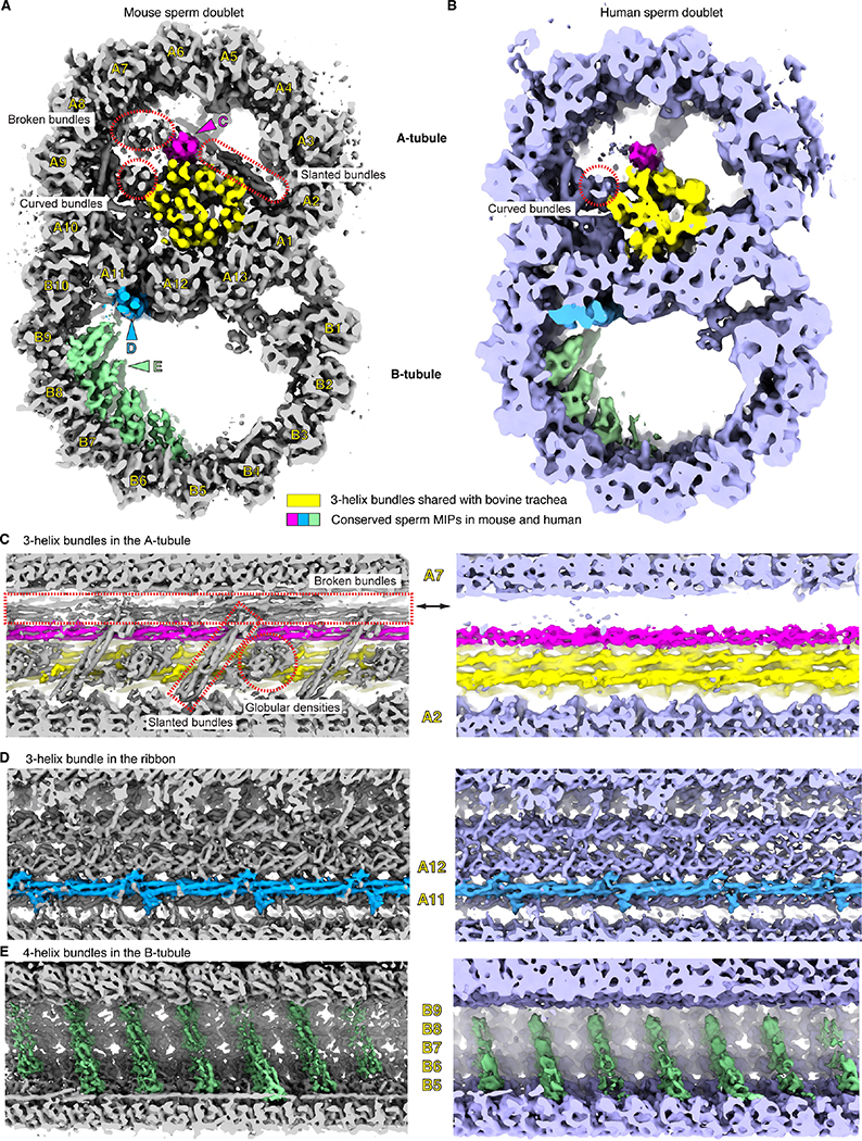
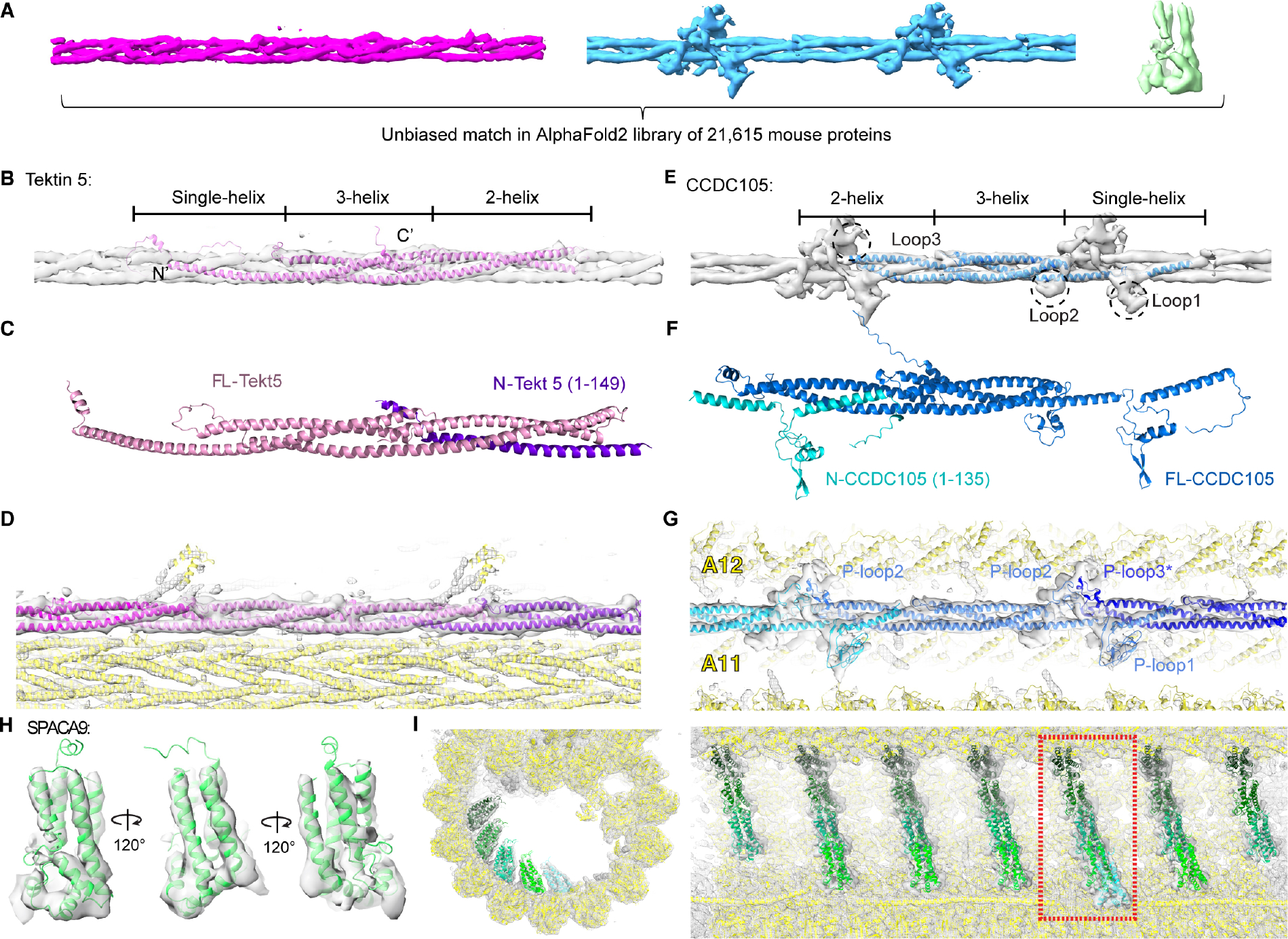
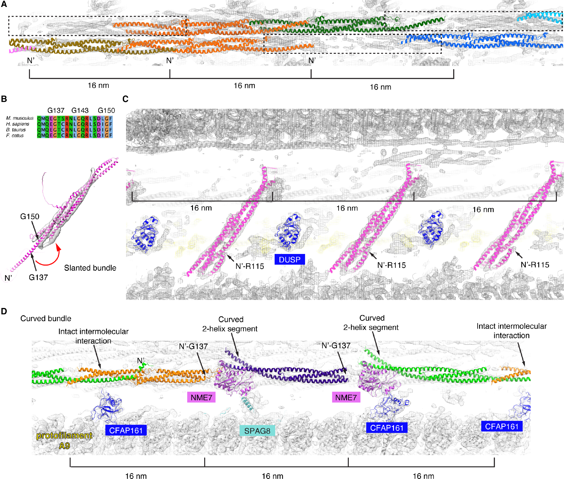
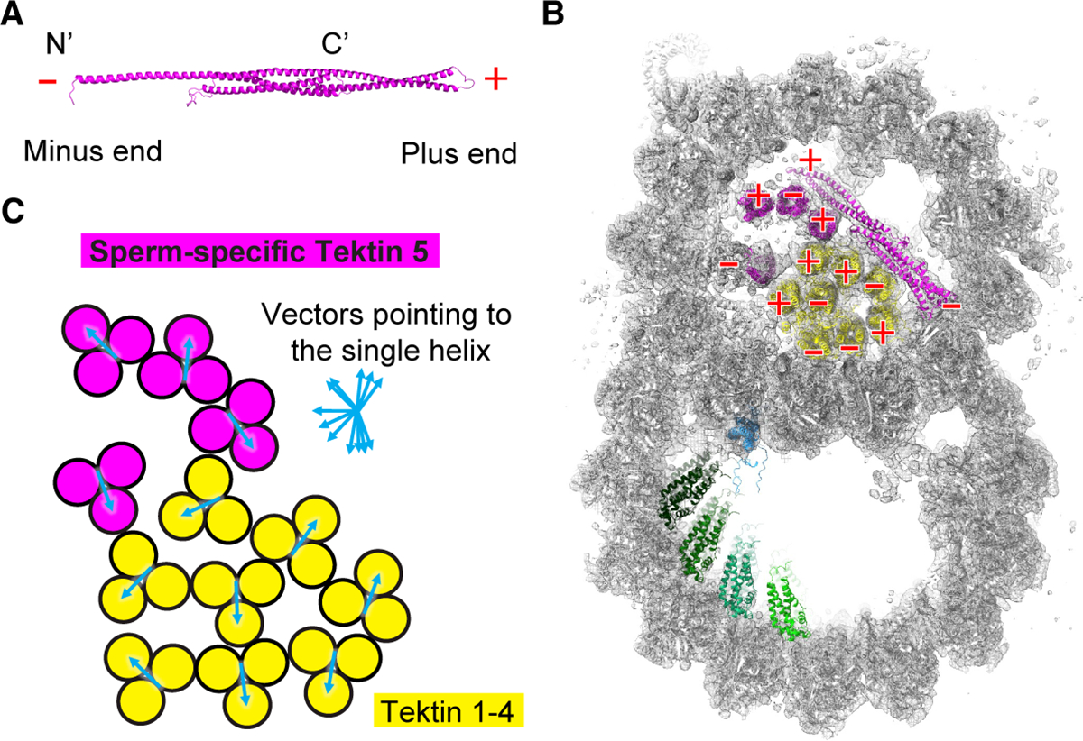
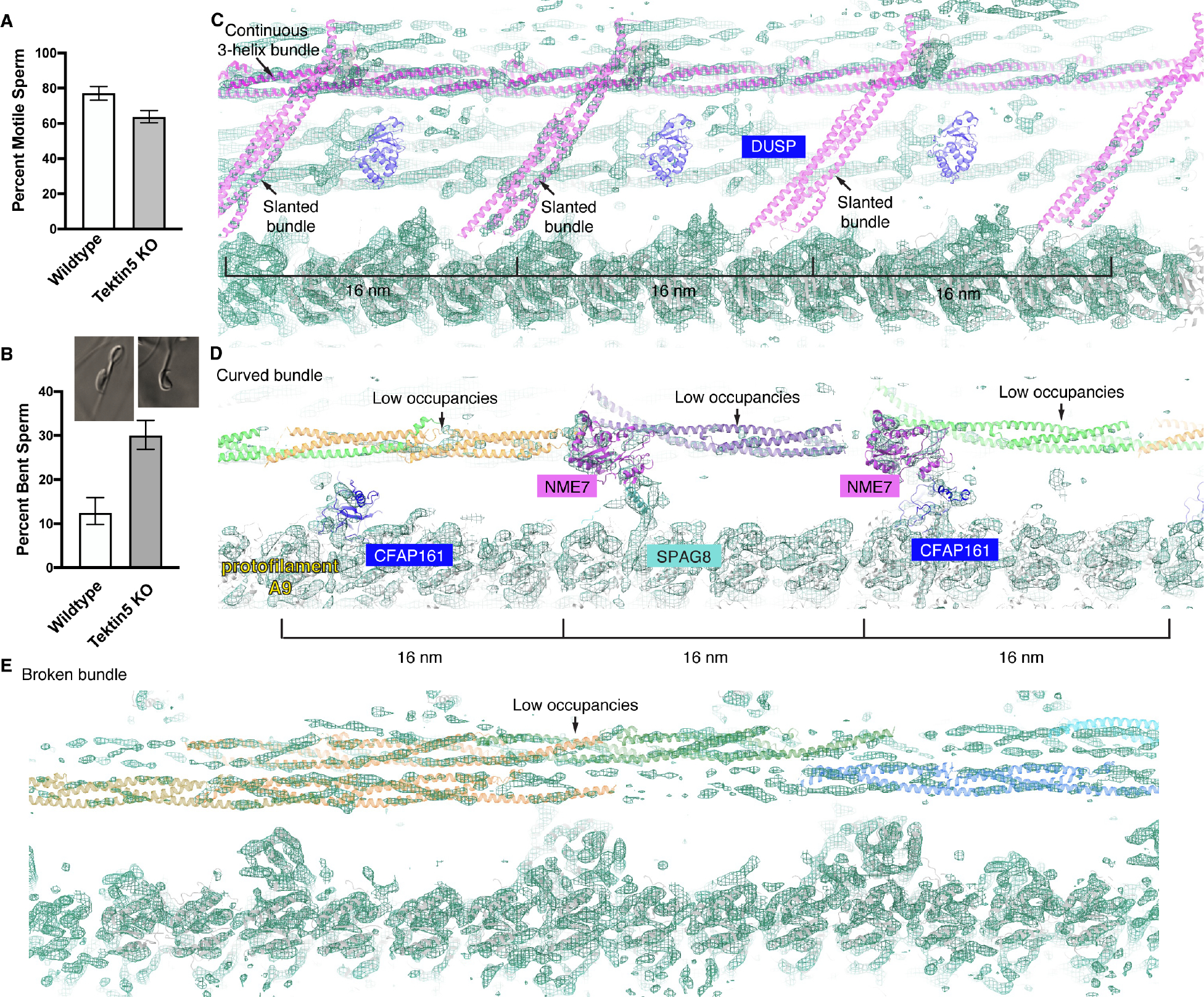
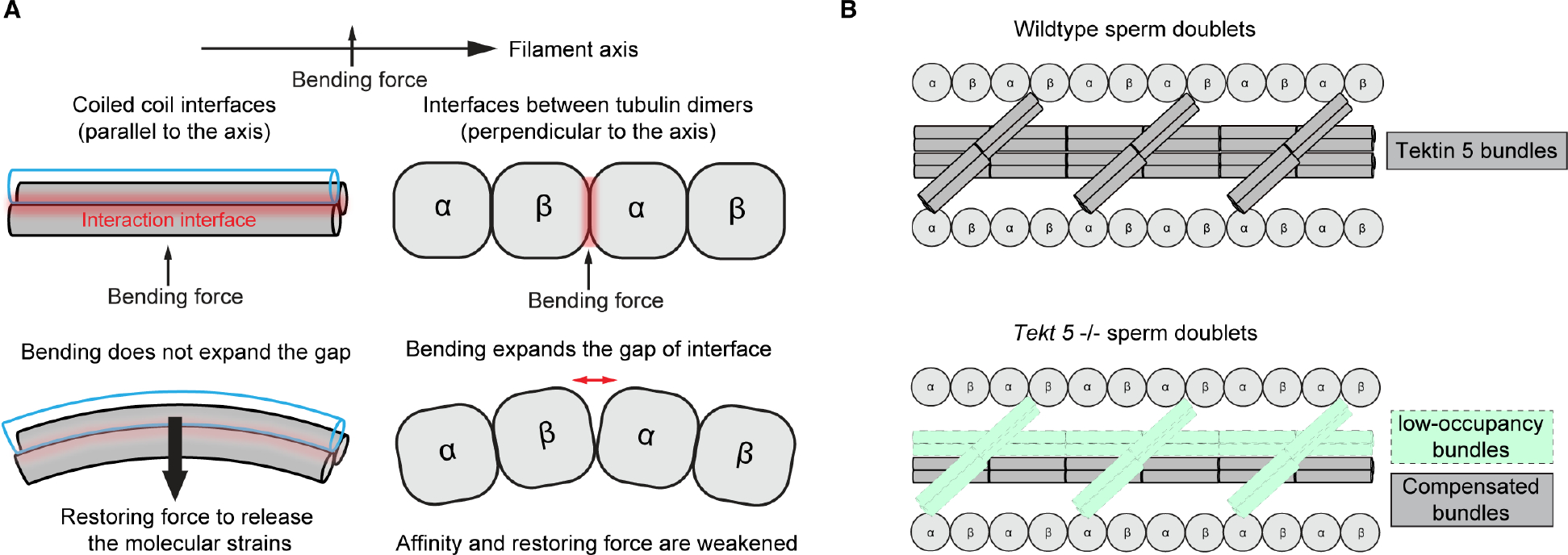
To understand the molecular mechanisms of cellular pathways, contemporary workflows typically require multiple techniques to identify proteins, track their localization, and determine their structures in vitro. Here, we combined cellular cryoelectron tomography (cryo-ET) and AlphaFold2 modeling to address these questions and understand how mammalian sperm are built in situ. Our cellular cryo-ET and subtomogram averaging provided 6.0-Å reconstructions of axonemal microtubule structures. The well-resolved tertiary structures allowed us to unbiasedly match sperm-specific densities with 21,615 AlphaFold2-predicted protein models of the mouse proteome. We identified Tektin 5, CCDC105, and SPACA9 as novel microtubule-associated proteins. These proteins form an extensive interaction network crosslinking the lumen of axonemal doublet microtubules, suggesting their roles in modulating the mechanical properties of the filaments. Indeed, Tekt5 -/- sperm possess more deformed flagella with 180° bends. Together, our studies presented a cellular visual proteomics workflow and shed light on the in vivo functions of Tektin 5.

Keywords: AlphaFold2 modeling; axoneme; cellular cryo-ET; doublet; microtubule; microtubule inner protein; sperm; visual proteomics.


Conflict of interest statement

Declaration of interests We declare that one or more authors have a competing interest as defined by Nature Portfolio. The Krogan Laboratory has received research support from Vir Biotechnology, F. Hoffmann-La Roche, and Rezo Therapeutics. N.J.K. has previously held financially compensated consulting agreements with the Icahn School of Medicine at Mount Sinai, New York, and Twist Bioscience Corp. He currently has financially compensated consulting agreements with Maze Therapeutics, Interline Therapeutics, Rezo Therapeutics, and GEn1E Lifesciences, Inc. He is on the Board of Directors of Rezo Therapeutics and is a shareholder in Tenaya Therapeutics, Maze Therapeutics, Rezo Therapeutics, and Interline Therapeutics.

Figures

Figure 1.
Figure 1.. The 3D reconstructions of mouse and human sperm doublets revealed novel MIPs.
(A), (B) Transverse cross-section views of the doublets of mouse (A) and human (B) sperm. Conserved sperm MIP densities are highlighted (pink, blue and green) and the corresponding viewing angles of (C)-(E) are indicated (colored arrowheads). The 3-helix densities in A-tubule shared with Bovine trachea doublets (EMD-24664) are colored (yellow) . Divergent sperm densities are also indicated (red dashed shapes). Individual protofilaments of the doublets are labeled as A1–13 and B1–10. (C)-(E). Zoom-in views of the conserved sperm MIP densities along the longitudinal axis. In (C), mouse sperm-specific densities are indicated and labeled (red dashed shapes, see more in Figures S2 and S3). In (E), although the striations are 8 nm apart from one another, the overall periodicity is 48 nm.
Figure 2.
Figure 2.. De novo protein identification of sperm MIPs assisted by AlphaFold2.
(A) Conserved densities in mouse and human sperm were segmented from the averages of 16-nm repeats of mouse sperm doublets and searched in the AlphaFold2 library of the mouse proteome (21,615 proteins). (B) The predicted structure of Tektin 5 based on AlphaFold2 was fitted into the continuous 3-helix bundle. (C) Modeling of a complex formed by a full-length Tektin 5 and a truncated one (N-Tekt 5: a.a. 1–149) using Colabfold . (D) Fitting and modeling of Tektin 5s into the 3-helix bundle densities in the A-tubule. The nearby densities accounted for by other proteins are also shown (yellow ribbon). (E) An unbiased search in the AlphaFold2 library identified CCDC105 as the candidate for the continuous 3-helix density at the ribbon. The three conserved proline-rich loops among CCDC105 orthologs could account for the protrusion densities but were not modeled (See Figures S6C–D). (F) Modeling of a complex formed by a full-length CCDC105 and a truncated one (N-CCDC105: a.a. 1–135) using Colabfold . (G) Fitting and modeling of CCDC105 into the 3-helix bundle density at the ribbon. The nearby densities are accounted for by other proteins (yellow ribbon). (H) The AlphaFold2 model for SPACA9 was directly fitted into the density and viewed from different angles. (I) Two orthogonal views of the striations of SPAC9 in the B-tubule. Different SPACA9 molecules are colored with different shades of green. The left panel showed a particular striation indicated in the right panel (the dashed rectangle).
Figure 3.
Figure 3.. Conformational plasticity of Tektin 5.
(A) The two broken 3-helix bundles could be explained by two complete and a third partial copies of Tektin 5 (dashed rectangles) per 48-nm repeat, instead of three Tektins in the continuous 3-helix bundle. (B) The AlphaFold2 model of mouse Tektin 5 was fitted into the slanted helical densities. Sequence alignment of Tektin 5 from M. musculus, H. sapiens, B. taurus and F. catus is shown from Q133-F151 (the numbering of amino acids is based on M. musculus Tektin 5). The conserved Gly137, Gly143 and Gly150 are near the turning point of the bent α-helix. (C) The fitting of Tektin 5 and DUSP3 protein (its homologs are also possible candidates) into the 16-nm repeating features, see the same view of the map in Figure 1C. (D) Three modified Tektin 5 were fitted into the densities of curved bundles in the mouse sperm doublet (as indicated in Figure 1A). The intact intermolecular interaction interface, N-termini of the Tektin 5s and curved 2-helix segments are indicated (arrows). Nearby MIPs shared between mouse sperm flagella and bovine trachea cilia are also colored and labeled (NME7, CFAP161, SPAG8). (E) The cross-section schematic is shown. The highlighted models of panels A-D are indicated using arrows.
Figure 4.
Figure 4.. Sperm doublets are composed of microtubules and extensive coiled-coil bundles.
(A) The plus and minus ends of Tektin 5 were named based on the N- and C-termini of the protein. (B) The cross-section view of the mouse sperm doublets shows the polarities of 3-helix bundles pointing toward the readers. (C) The orientations for each 3-helix bundle were represented by a vector starting from the middle point of the 2-helix segment and pointing toward the single-helix segment of the other Tektin molecule.
Figure 5.
Figure 5.. Characterization of mutant Tekt 5 −/− sperm.
(A) The percentages of motile sperm from wild-type and Tekt 5 knockout mice (> 200 cells were counted for each mouse and three knockout −/− mice and two wild-type mice were analyzed, the pool percentage and 95% Confidence Intervals (Wilson/Brown method) were shown). (B) The percentages of bent sperm from wild-type and Tekt 5 knockout mice (> 200 cells were counted for each mouse and three knockout −/− mice and two wild-type mice were analyzed, the pool percentage and 95% Confidence Intervals by Wilson/Brown method were shown). Two examples of bent sperm are shown. (C) An overlay of wild-type models with the densities of Tekt 5 −/− sperm around the slanted bundles. The continuous 3-helix bundle assigned as Tektin 5 (high occupancies) and slanted helical bundles (low occupancies) are shown. The densities corresponding to the DUSP proteins are barely resolved. Note there are substantially less densities for these models compared to Figure 3C. (D) An overlay of wildtype models with the densities of Tekt 5 −/− sperm around the curved bundles. The occupancies of the curved bundles are lower than the other MIPs and tubulins. Note there are substantially less densities for these models compared to Figure 3D. (E) The two broken 3-helix bundles have lower occupancies compared to the surrounding MIPs and tubulins. Note there are substantially less densities for these models compared to Figure 3A.
Figure 6.
Figure 6.. Coiled-coil interfaces are suitable to withstand mechanical stress from orthogonal directions.
(A) A model of how 3-helix bundles would be able to bear mechanical stress differently compared to the microtubules. The bending curvatures and gaps are exaggerated for illustration purposes. (B) A schematic of wild-type and mutant sperm doublets structures highlighting the Tektin 5 bundles and the partial redundancy.

Similar articles

Cited by

References

    1. Sironen A, Shoemark A, Patel M, Loebinger MR, and Mitchison HM (2020). Sperm defects in primary ciliary dyskinesia and related causes of male infertility. Cell Mol Life Sci 77, 2029–2048. 10.1007/s00018-019-03389-7. - DOI - PMC - PubMed
    1. Fernandez-Lopez P, Garriga J, Casas I, Yeste M, and Bartumeus F (2022). Predicting fertility from sperm motility landscapes. Commun Biol 5, 1027. 10.1038/s42003-022-03954-0. - DOI - PMC - PubMed
    1. Ishikawa T (2017). Axoneme Structure from Motile Cilia. Cold Spring Harbor perspectives in biology 9. 10.1101/cshperspect.a028076. - DOI - PMC - PubMed
    1. Linck RW, Chemes H, and Albertini DF (2016). The axoneme: the propulsive engine of spermatozoa and cilia and associated ciliopathies leading to infertility. J Assist Reprod Genet 33, 141–156. 10.1007/s10815-016-0652-1. - DOI - PMC - PubMed
    1. Fawcett DW (1975). The mammalian spermatozoon. Dev Biol 44, 394–436. 10.1016/0012-1606(75)90411-x. - DOI - PubMed

Publication types